PKC alpha (Protein kinase C alpha) is an 80 kDa member of the PKC subfamily, AGC Ser/Thr protein kinase family of enzymes. It is widely expressed, and serves multiple cell-specific functions. PKC alpha is activated by increased diacylglycerol and intracellular calcium, and translocates to several sites such as the Golgi, nucleus and plasma membrane. Human PKC alpha is 672 amino acids (aa) in length. It contains two zinc finger regions (aa 36‑151), a protein kinase domain (aa 339‑557), and an AGC kinase region (aa 598‑668). Phosphorylations on Thr497, Thr638 and Ser657 are necessary for kinase activity. Over aa 604‑672, human PKC alpha shows 100% aa identity to mouse PKC alpha.

Key Product Details
Species Reactivity
Validated:
Human, Mouse, Rat
Cited:
Mouse, Rat
Applications
Validated:
Immunohistochemistry, Western Blot, Simple Western
Cited:
Immunohistochemistry, Western Blot
Label
Unconjugated
Antibody Source
Polyclonal Goat IgG
Loading...
Product Specifications
Immunogen
E. coli-derived recombinant human PKC alpha
Lys604-Val672
Accession # P17252
Lys604-Val672
Accession # P17252
Specificity
Detects endogenous human, mouse, and rat PKC alpha in Western blots.
Clonality
Polyclonal
Host
Goat
Isotype
IgG
Scientific Data Images for PKC alpha Antibody
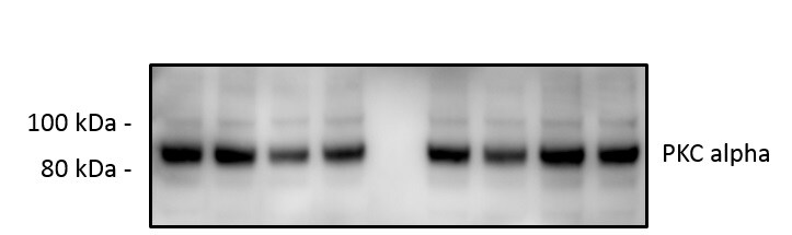
Detection of Human/Mouse/Rat PKC alpha by Western Blot.
Western blot shows lysates of HeLa human cervical epithelial carcinoma cell line, Saos-2 human osteosarcoma cell line, Balb/3T3 mouse embryonic fibroblast cell line, and NRK rat normal kidney cell line. PVDF membrane was probed with 1 µg/mL of Human/Mouse/Rat PKCa Antigen Affinity-purified Polyclonal Antibody (Catalog # AF5340) followed by HRP-conjugated Anti-Goat IgG Secondary Antibody (Catalog # HAF109). A specific band was detected for PKCa at approximately 90 kDa (as indicated). This experiment was conducted under reducing conditions and using Immunoblot Buffer Group 1.
PKC alpha in Human Breast Cancer Tissue.
PKCa was detected in immersion fixed paraffin-embedded sections of human breast cancer tissue using Goat Anti-Human/Mouse/Rat PKCa Antigen Affinity-purified Polyclonal Antibody (Catalog # AF5340) at 10 µg/mL overnight at 4 °C. Tissue was stained using the Anti-Goat HRP-DAB Cell & Tissue Staining Kit (brown; Catalog # CTS008) and counterstained with hematoxylin (blue). Specific staining was localized to cancer cell cytoplasm. View our protocol for Chromogenic IHC Staining of Paraffin-embedded Tissue Sections.
Detection of Human PKC alpha by Simple WesternTM.
Simple Western shows lysates of Saos‑2 human osteosarcoma cell line, loaded at 0.5 mg/ml. A specific band was detected for PKC alpha at approximately 84 kDa (as indicated) using 10 µg/mL of Goat Anti-Human/Mouse/Rat PKC alpha Antigen Affinity-purified Polyclonal Antibody (Catalog # AF5340). This experiment was conducted under reducing conditions and using the 12-230kDa separation system.Applications for PKC alpha Antibody
Application
Recommended Usage
Immunohistochemistry
5-15 µg/mL
Sample: Immersion fixed paraffin-embedded sections of human breast cancer tissue
Sample: Immersion fixed paraffin-embedded sections of human breast cancer tissue
Simple Western
10 µg/mL
Sample: Saos-2 human osteosarcoma cell line
Sample: Saos-2 human osteosarcoma cell line
Western Blot
1 µg/mL
Sample: HeLa human cervical epithelial carcinoma cell line, Saos-2 human osteosarcoma cell line, Balb/3T3 mouse embryonic fibroblast cell line, and NRK rat normal kidney cells
Sample: HeLa human cervical epithelial carcinoma cell line, Saos-2 human osteosarcoma cell line, Balb/3T3 mouse embryonic fibroblast cell line, and NRK rat normal kidney cells
Reviewed Applications
Read 3 reviews rated 5 using AF5340 in the following applications:
Formulation, Preparation, and Storage
Purification
Antigen Affinity-purified
Reconstitution
Reconstitute at 0.2 mg/mL in sterile PBS. For liquid material, refer to CoA for concentration.
Loading...
Formulation
Lyophilized from a 0.2 μm filtered solution in PBS with Trehalose. See Certificate of Analysis for details.
*Small pack size (-SP) is supplied either lyophilized or as a 0.2 µm filtered solution in PBS.
*Small pack size (-SP) is supplied either lyophilized or as a 0.2 µm filtered solution in PBS.
Shipping
Lyophilized product is shipped at ambient temperature. Liquid small pack size (-SP) is shipped with polar packs. Upon receipt, store immediately at the temperature recommended below.
Stability & Storage
Use a manual defrost freezer and avoid repeated freeze-thaw cycles.
- 12 months from date of receipt, -20 to -70 °C as supplied.
- 1 month, 2 to 8 °C under sterile conditions after reconstitution.
- 6 months, -20 to -70 °C under sterile conditions after reconstitution.
Calculators
Background: PKC alpha
Long Name
Protein Kinase C alpha
Alternate Names
PKCA, PRKCA
Gene Symbol
PRKCA
UniProt
Additional PKC alpha Products
Product Documents for PKC alpha Antibody
Certificate of Analysis
To download a Certificate of Analysis, please enter a lot or batch number in the search box below.
Note: Certificate of Analysis not available for kit components.
Product Specific Notices for PKC alpha Antibody
For research use only
Related Research Areas
Citations for PKC alpha Antibody
 Powered by Bioz
Customer Reviews for PKC alpha Antibody (3)
5 out of 5
3 Customer Ratings
Have you used PKC alpha Antibody?
Submit a review and receive an Amazon gift card!
$25/€18/£15/$25CAN/¥2500 Yen for a review with an image
$10/€7/£6/$10CAN/¥1110 Yen for a review without an image
Submit a review
Customer Images
Showing
1
-
3 of
3 reviews
Showing All
Filter By:
-
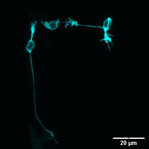
Application: ImmunocytochemistrySample Tested: Mouse Retinal CellsSpecies: MouseVerified Customer | Posted 01/21/2021Isolated retinal cells stained with gt-PKCalpha (AF5340, 1:1000; cyan); Labelling conforms to rod bipolar cell morphology, comparable to PKCalpha antibodies from mouse and rabbit.Papain dissociation of retina tissue, PFA fixation and staining for isolated rod bipolar cells with gt-PKCalpha (AF5340, 1:1000) + dk-a-gt DyLight594 (1:250); PKCalpha labelling conforms to rod bipolar cell morphology.
 Bio-Techne ResponseThis review was submitted through the legacy Novus Innovators Program, reflecting a new species or application tested on a primary antibody.
 -
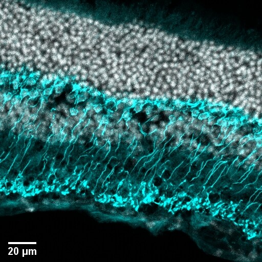
Application: Immunohistochemistry-FrozenSample Tested: C57BL/6 mouse retinaSpecies: MouseVerified Customer | Posted 01/21/2021Mouse retina cryosection stained with gt-PKCalpha (AF5340, 1:250; cyan) and counterstained with DAPI (grey). PKCalpha labelling conforms to the rod bipolar cell morphology, comparable to established PKCalpha marker antibodies.gt-PKCalpha (AF5340, 1:250) + dk-a-gt DyLight594 (1:250) PFA-fixed tissue
-
Application: Western BlotSample Tested: EndothelialSpecies: MouseVerified Customer | Posted 10/11/2019
There are no reviews that match your criteria.
Protocols
Find general support by application which include: protocols, troubleshooting, illustrated assays, videos and webinars.
- Antigen Retrieval Protocol (PIER)
- Antigen Retrieval for Frozen Sections Protocol
- Appropriate Fixation of IHC/ICC Samples
- Cellular Response to Hypoxia Protocols
- Chromogenic IHC Staining of Formalin-Fixed Paraffin-Embedded (FFPE) Tissue Protocol
- Chromogenic Immunohistochemistry Staining of Frozen Tissue
- ClariTSA™ Fluorophore Kits
- Detection & Visualization of Antibody Binding
- Fluorescent IHC Staining of Frozen Tissue Protocol
- Graphic Protocol for Heat-induced Epitope Retrieval
- Graphic Protocol for the Preparation and Fluorescent IHC Staining of Frozen Tissue Sections
- Graphic Protocol for the Preparation and Fluorescent IHC Staining of Paraffin-embedded Tissue Sections
- Graphic Protocol for the Preparation of Gelatin-coated Slides for Histological Tissue Sections
- IHC Sample Preparation (Frozen sections vs Paraffin)
- Immunofluorescent IHC Staining of Formalin-Fixed Paraffin-Embedded (FFPE) Tissue Protocol
- Immunohistochemistry (IHC) and Immunocytochemistry (ICC) Protocols
- Immunohistochemistry Frozen Troubleshooting
- Immunohistochemistry Paraffin Troubleshooting
- Preparing Samples for IHC/ICC Experiments
- Preventing Non-Specific Staining (Non-Specific Binding)
- Primary Antibody Selection & Optimization
- Protocol for Heat-Induced Epitope Retrieval (HIER)
- Protocol for Making a 4% Formaldehyde Solution in PBS
- Protocol for VisUCyte™ HRP Polymer Detection Reagent
- Protocol for the Preparation & Fixation of Cells on Coverslips
- Protocol for the Preparation and Chromogenic IHC Staining of Frozen Tissue Sections
- Protocol for the Preparation and Chromogenic IHC Staining of Frozen Tissue Sections - Graphic
- Protocol for the Preparation and Chromogenic IHC Staining of Paraffin-embedded Tissue Sections
- Protocol for the Preparation and Chromogenic IHC Staining of Paraffin-embedded Tissue Sections - Graphic
- Protocol for the Preparation and Fluorescent IHC Staining of Frozen Tissue Sections
- Protocol for the Preparation and Fluorescent IHC Staining of Paraffin-embedded Tissue Sections
- Protocol for the Preparation of Gelatin-coated Slides for Histological Tissue Sections
- R&D Systems Quality Control Western Blot Protocol
- TUNEL and Active Caspase-3 Detection by IHC/ICC Protocol
- The Importance of IHC/ICC Controls
- Troubleshooting Guide: Immunohistochemistry
- Troubleshooting Guide: Western Blot Figures
- Western Blot Conditions
- Western Blot Protocol
- Western Blot Protocol for Cell Lysates
- Western Blot Troubleshooting
- Western Blot Troubleshooting Guide
- View all Protocols, Troubleshooting, Illustrated assays and Webinars
Loading...
Associated Pathways


mTOR Signaling Pathway
 VEGF - VEGF R2 Signaling Pathways
 Wnt Signaling Pathways: beta-Catenin-dependent Wnt Signaling
