
Key Product Details
Validated by
Species Reactivity
Validated:
Cited:
Applications
Validated:
Cited:
Label
Antibody Source
Product Specifications
Immunogen
Met1-Asn175
Accession # P40763
Specificity
Clonality
Host
Isotype
Scientific Data Images for STAT3 Antibody (232209)
Detection of Human, Mouse and Rat STAT3 by Western Blot.
Western Blot shows lysates of HepG2 human hepatocellular carcinoma cell line, NCI‑H1703 human non-small cell lung carcinoma cell line, C6 rat glioma cell line and NIH‑3T3 mouse embryonic fibroblast cell line. PVDF membrane was probed with 1 µg/ml of Mouse Anti-Human/Mouse/Rat STAT3 Monoclonal Antibody (Catalog # MAB1799) followed by HRP-conjugated Anti-Mouse IgG Secondary Antibody (Catalog # HAF018). A specific band was detected for STAT3 at approximately 86 kDa (as indicated). This experiment was conducted under reducing conditions and using Western Blot Buffer Group 1.
STAT3 in HeLa Human Cell Line.
STAT3 was detected in immersion fixed HeLa human cervical epithelial carcinoma cell line using Mouse Anti-Human/Mouse/Rat STAT3 Monoclonal Antibody (Catalog # MAB1799) at 3 µg/mL for 3 hours at room temperature. Cells were stained using the NorthernLights™ 557-conjugated Anti-Mouse IgG Secondary Antibody (red; Catalog # NL007) and counterstained with DAPI (blue). Specific staining was localized to cytoplasm and nuclei. View our protocol for Fluorescent ICC Staining of Cells on Coverslips.
Detection of STAT3 in Jurkat Human Cell Line by Flow Cytometry.
Jurkat human acute T cell leukemia cell line was stained with Mouse Anti-Human STAT3 Monoclonal Antibody (Catalog # MAB1799, filled histogram) or isotype control antibody (Catalog # MAB0041, open histogram), followed by Allophycocyanin-conjugated Anti-Mouse IgG F(ab')2Secondary Antibody (Catalog # F0101B). To facilitate intracellular staining, cells were fixed with paraformaldehyde and permeabilized with methanol.
Detection of STAT3 in HeLa Human Cell Line by Flow Cytometry.
HeLa human cell line was stained with Mouse Anti-Human/Mouse/Rat STAT3 Monoclonal Antibody (Catalog # MAB1799, filled histogram) or isotype control antibody (Catalog # MAB0041, open histogram) followed by anti-Mouse IgG PE-conjugated secondary antibody (Catalog # F0102B). To facilitate intracellular staining, cells were fixed with Flow Cytometry Fixation Buffer (Catalog # FC004) and permeabilized with Flow Cytometry Permeabilization/Wash Buffer I (Catalog # FC005). View our protocol for Staining Intracellular Molecules.
STAT3 Specificity is Shown by Immunocytochemistry in Knockout Cell Line.
STAT3 was detected in immersion fixed HeLa human cervical epithelial carcinoma cell line treated with IFN-alpha 1, but is not detected in STAT3 knockout (KO) HeLa cell line using Mouse Anti-Human/ Mouse/Rat STAT3 Monoclonal Antibody (Catalog # MAB1799) at 1 µg/mL for 3 hours at room temperature. Cells were stained using the NorthernLights™ 557-conjugated Anti-Mouse IgG Secondary Antibody (red; Catalog # NL007) and counterstained with DAPI (blue). Specific staining was localized to cytoplasm and nuclei. View our protocol for Fluorescent ICC Staining of Cells on Coverslips.
Western Blot Shows Human STAT3 Specificity by Using Knockout Cell Line.
Western blot shows lysates of HeLa human cervical epithelial carcinoma parental cell line, STAT1 knockout (KO) HeLa cell line, STAT2 KO HeLa cell line, STAT3 KO HeLa cell line, STAT5a KO HeLa cell line, STAT5b KO HeLa cell line, STAT6 KO HeLa cell line,. PVDF membrane was probed with 0.5 µg/mL of Mouse Anti-Human/Mouse/Rat STAT3 Monoclonal Antibody (Catalog # MAB1799) followed by HRP-conjugated Anti-Mouse IgG Secondary Antibody (Catalog # HAF018). A specific band was detected for STAT3 at approximately 80 kDa (as indicated) in the parental HeLa cell line, but is not detectable in the STAT3 knockout HeLa cell line. GAPDH (Catalog # AF5718) is shown as a loading control. This experiment was conducted under reducing conditions and using Immunoblot Buffer Group 1.
STAT3 Specificity is Shown by Flow Cytometry in Knockout Cell Line.
STAT3 knockout HeLa human cervical epithelial cell line was stained with Mouse Anti-Human/Mouse STAT3 Monoclonal Antibody (Catalog # MAB1799, filled histogram) or isotype control antibody (Catalog # MAB0041, open histogram) followed by anti-Mouse IgG PE-conjugated secondary antibody (Catalog # F0102B). No staining in the STAT3 knockout HeLa cell line was observed. To facilitate intracellular staining, cells were fixed with Flow Cytometry Fixation Buffer (Catalog # FC004) and permeabilized with Flow Cytometry Permeabilization/Wash Buffer I (Catalog # FC005). View our protocol for Staining Intracellular Molecules.
Detection of Mouse STAT3 by Western Blot
Effects of kurarinone on the phosphorylation of p-STAT1 and p-STAT3 in lymph nodes. Single cell suspensions were collected from ILNs on day 42, the protein expression levels of p-STAT1, STAT1, p-STAT3, and STAT3 were measured using Western blots. (A) Representative images of Western blot and (B) Densitometric analysis for protein expressions was performed using ImageJ software. Data are presented as mean ± SEM of 6 mice from one of three experiments. (*) p < 0.05, (**) p < 0.01, (***) p < 0.001 versus vehicle-treated CIA mice group (One Way ANOVA followed by Tukey’s multiple comparison test). Image collected and cropped by CiteAb from the following open publication (https://pubmed.ncbi.nlm.nih.gov/33924467), licensed under a CC-BY license. Not internally tested by R&D Systems.
Detection of STAT3 by Western Blot
Galectin-3 expression induces activation of PYK2, STAT1 and GSK3 alpha / beta signalling. Expression of 37 protein kinases in SW620 cells in response to 10 µg/ml galectin-3 or BSA for 0.5 h was assessed by Proteome Profiler Human Phospho-Kinase Array (A, Percentage changes of the kinases in cell response to galectin-3 in comparison to control are shown at the bottom panel). The presence of galectin-3 increases the phosphorylation of PYK2, GSK3 alpha / beta, and STAT1 and decreases phosphorylation of STAT3. SW620 cells treated with 10 µg/ml galectin-3 for different times were assessed by immunoblotting using antibodies against p-PYK2, p-STAT-1, p-GSK3 alpha / beta or p-STAT-3 (B). The blots were striped and reprobed with antibodies against PYK2, STAT-1, GSK3 alpha / beta or STAT-3. The band density was quantified and expressed as percentages of phospho-/non-phosphorylated proteins (C). In D and E, SW620 cells were treated with 10 µg/ml galectin-3 or BSA followed by introduction of GSK3 alpha / beta inhibitor SB 216763 (SB) or PKY2 inhibitor PF-431396 (PF) for 15 min and the levels of phosphorylated PYK2, STAT-1, GSK3 alpha / beta or STAT-3 were analysed by immunoblotting. The blots were striped and reprobed with antibodies against PYK2, STAT-1, GSK3 alpha / beta or STAT-3. The densities of the blots from three independent experiments were quantified and are expressed as the percentage of phosphorylated/non-phosphorylated levels of each protein. ***P < 0.001, **P < 0.01, *P < 0.05 (ANOVA). Image collected and cropped by CiteAb from the following open publication (https://pubmed.ncbi.nlm.nih.gov/37055381), licensed under a CC-BY license. Not internally tested by R&D Systems.
Detection of STAT3 by Western Blot
Aspirin modulated the JAK/p-STAT3 signaling pathway in atypical hyperplastic intestinal mucosal cells of UC mice (n = 4 for each group). (A) Western blotting images for JAK/p-STAT3 signaling pathway-associated molecules expression. (B) Statistical analysis and comparison for p-STAT3 expression. (C) Statistical analysis and comparison for STAT3 expression. (D) Statistical analysis and comparison for cyclin D1 expression. (E) Statistical analysis and comparison for SOCS3 expression. *P <.05 versus control group. #P <.05 versus UC model group. JAK, Janus kinase; UC, ulcerative colitis; p-STAT3, phosphorylated-STAT3; STAT3, signal transducer and activator of transcription 3; SOCS3, suppressor of cytokine signaling 3. Image collected and cropped by CiteAb from the following open publication (https://pubmed.ncbi.nlm.nih.gov/35946886), licensed under a CC-BY license. Not internally tested by R&D Systems.
Detection of STAT3 by Western Blot
Aspirin modulated the JAK/p-STAT3 signaling pathway in atypical hyperplastic intestinal mucosal cells of UC mice (n = 4 for each group). (A) Western blotting images for JAK/p-STAT3 signaling pathway-associated molecules expression. (B) Statistical analysis and comparison for p-STAT3 expression. (C) Statistical analysis and comparison for STAT3 expression. (D) Statistical analysis and comparison for cyclin D1 expression. (E) Statistical analysis and comparison for SOCS3 expression. *P <.05 versus control group. #P <.05 versus UC model group. JAK, Janus kinase; UC, ulcerative colitis; p-STAT3, phosphorylated-STAT3; STAT3, signal transducer and activator of transcription 3; SOCS3, suppressor of cytokine signaling 3. Image collected and cropped by CiteAb from the following open publication (https://pubmed.ncbi.nlm.nih.gov/35946886), licensed under a CC-BY license. Not internally tested by R&D Systems.Applications for STAT3 Antibody (232209)
Immunocytochemistry
Sample: Immersion fixed human peripheral blood mononuclear cells and HeLa human cervical epithelial carcinoma cell line
Immunoprecipitation
Sample: Daudi human Burkitt's lymphoma cell line, see our available Western blot detection antibodies
Intracellular Staining by Flow Cytometry
Sample: HeLa or Jurkat human acute T cell leukemia cell line fixed with paraformaldehyde and permeabilized with methanol
Knockout Validated
STAT3 is specifically detected in HeLa human cervical epithelial carcinoma parental cell line but is not detectable in STAT3 knockout HeLa cell line.
Western Blot
Sample: HepG2 human hepatocellular carcinoma cell line, NCI-H1703 human non-small cell lung carcinoma cell line, NIH-3T3 mouse embryonic fibroblast cell line, and PC-12 rat adrenal pheochromocytoma cell line
Reviewed Applications
Read 1 review rated 5 using MAB1799 in the following applications:
Flow Cytometry Panel Builder
Bio-Techne Knows Flow Cytometry
Save time and reduce costly mistakes by quickly finding compatible reagents using the Panel Builder Tool.
Advanced Features
- Spectra Viewer - Custom analysis of spectra from multiple fluorochromes
- Spillover Popups - Visualize the spectra of individual fluorochromes
- Antigen Density Selector - Match fluorochrome brightness with antigen density

Formulation, Preparation, and Storage
Purification
Reconstitution
Reconstitute at 0.5 mg/mL in sterile PBS. For liquid material, refer to CoA for concentration.
Formulation
*Small pack size (-SP) is supplied either lyophilized or as a 0.2 µm filtered solution in PBS.
Shipping
Stability & Storage
- 12 months from date of receipt, -20 to -70 °C as supplied.
- 1 month, 2 to 8 °C under sterile conditions after reconstitution.
- 6 months, -20 to -70 °C under sterile conditions after reconstitution.
Calculators
Background: STAT3
Long Name
Alternate Names
Gene Symbol
UniProt
Additional STAT3 Products
Product Documents for STAT3 Antibody (232209)
Certificate of Analysis
To download a Certificate of Analysis, please enter a lot or batch number in the search box below.
Note: Certificate of Analysis not available for kit components.
Product Specific Notices for STAT3 Antibody (232209)
For research use only
Citations for STAT3 Antibody (232209)
 Powered by Bioz
Customer Reviews for STAT3 Antibody (232209) (1)
Have you used STAT3 Antibody (232209)?
Submit a review and receive an Amazon gift card!
$25/€18/£15/$25CAN/¥2500 Yen for a review with an image
$10/€7/£6/$10CAN/¥1110 Yen for a review without an image
Submit a review
Customer Images
-
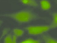
Application: Immunocytochemistry/ImmunofluorescenceSample Tested: Hepatocellular carcinoma cell lineSpecies: HumanVerified Customer | Posted 08/27/2021

There are no reviews that match your criteria.
Protocols
Find general support by application which include: protocols, troubleshooting, illustrated assays, videos and webinars.
- 7-Amino Actinomycin D (7-AAD) Cell Viability Flow Cytometry Protocol
- Appropriate Fixation of IHC/ICC Samples
- Cellular Response to Hypoxia Protocols
- ClariTSA™ Fluorophore Kits
- Detection & Visualization of Antibody Binding
- Extracellular Membrane Flow Cytometry Protocol
- Flow Cytometry Protocol for Cell Surface Markers
- Flow Cytometry Protocol for Staining Membrane Associated Proteins
- Flow Cytometry Staining Protocols
- Flow Cytometry Troubleshooting Guide
- ICC Cell Smear Protocol for Suspension Cells
- ICC Immunocytochemistry Protocol Videos
- ICC for Adherent Cells
- Immunocytochemistry (ICC) Protocol
- Immunocytochemistry Troubleshooting
- Immunofluorescence of Organoids Embedded in Cultrex Basement Membrane Extract
- Immunohistochemistry (IHC) and Immunocytochemistry (ICC) Protocols
- Immunoprecipitation Protocol
- Intracellular Flow Cytometry Protocol Using Alcohol (Methanol)
- Intracellular Flow Cytometry Protocol Using Detergents
- Intracellular Nuclear Staining Flow Cytometry Protocol Using Detergents
- Intracellular Staining Flow Cytometry Protocol Using Alcohol Permeabilization
- Intracellular Staining Flow Cytometry Protocol Using Detergents to Permeabilize Cells
- Preparing Samples for IHC/ICC Experiments
- Preventing Non-Specific Staining (Non-Specific Binding)
- Primary Antibody Selection & Optimization
- Propidium Iodide Cell Viability Flow Cytometry Protocol
- Protocol for Liperfluo
- Protocol for VisUCyte™ HRP Polymer Detection Reagent
- Protocol for the Characterization of Human Th22 Cells
- Protocol for the Characterization of Human Th9 Cells
- Protocol for the Fluorescent ICC Staining of Cell Smears - Graphic
- Protocol for the Fluorescent ICC Staining of Cultured Cells on Coverslips - Graphic
- Protocol for the Preparation and Fluorescent ICC Staining of Cells on Coverslips
- Protocol for the Preparation and Fluorescent ICC Staining of Non-adherent Cells
- Protocol for the Preparation and Fluorescent ICC Staining of Stem Cells on Coverslips
- Protocol for the Preparation of a Cell Smear for Non-adherent Cell ICC - Graphic
- Protocol: Annexin V and PI Staining by Flow Cytometry
- Protocol: Annexin V and PI Staining for Apoptosis by Flow Cytometry
- R&D Systems Quality Control Western Blot Protocol
- TUNEL and Active Caspase-3 Detection by IHC/ICC Protocol
- The Importance of IHC/ICC Controls
- Troubleshooting Guide: Fluorokine Flow Cytometry Kits
- Troubleshooting Guide: Western Blot Figures
- Western Blot Conditions
- Western Blot Protocol
- Western Blot Protocol for Cell Lysates
- Western Blot Troubleshooting
- Western Blot Troubleshooting Guide
- View all Protocols, Troubleshooting, Illustrated assays and Webinars
Associated Pathways


