TLR9 (Toll receptor 9; also CD289) is a 145-150 kDa member of the Toll-like receptor family of molecules. It is expressed by colonic epithelium, CD123+ plasmacytoid dendritic cells, and transitional B cells, and responds to unmethylated DNA CpG motifs that contain either a GTCGTT sequence (in human), or a GACGTT sequence (in mouse). TLR9 is found in the ER and translocates to either the cell membrane or to lysosomes where it binds bacterial DNA. Precursor human TLR9 is a type I transmembrane protein 1032 amino acids (aa) in length. It possesses a 793 aa extracellular region that contains 26 LRRs (aa 26-818) plus a 193 aa cytoplasmic domain. The full-length 150 kDa form is suggested to be ligand-binding but nonsignaling. The active form is believed to be an 80 kDa cleavage product found in the endosome compartment. There are multiple splice forms. One contains a deletion of aa 2-16, a second possesses an alternate start site at Met58, while a third and fourth show alternative start sites aa 23 and 24 upstream of the standard site. Over aa 64-189, human TLR9 shares 76% aa identity with mouse TLR9.

Key Product Details
Species Reactivity
Human, Mouse
Applications
Immunohistochemistry, Immunocytochemistry
Label
Unconjugated
Antibody Source
Monoclonal Mouse IgG1 Clone # 980915
Loading...
Product Specifications
Immunogen
Human TLR9 synthetic peptide
Accession # Q9NR96
Accession # Q9NR96
Specificity
Detects human TLR9 in direct ELISAs.
Clonality
Monoclonal
Host
Mouse
Isotype
IgG1
Scientific Data Images for TLR9 Antibody (980915)
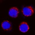
TLR9 in Mouse Splenocytes.
TLR9 was detected in immersion fixed mouse splenocytes using Mouse Anti-Human TLR9 Monoclonal Antibody (Catalog # MAB36581) at 3 µg/mL for 3 hours at room temperature. Cells were stained using the NorthernLights™ 557-conjugated Anti-Mouse IgG Secondary Antibody (red; NL007) and counterstained with DAPI (blue). Specific staining was localized to plasma membrane. View our protocol for Fluorescent ICC Staining of Non-adherent Cells.
TLR9 in Mouse Spleen.
TLR9 was detected in perfusion fixed frozen sections of mouse spleen using Mouse Anti-Human TLR9 Monoclonal Antibody (Catalog # MAB36581) at 1.7 µg/mL for 1 hour at room temperature followed by incubation with the Anti-Mouse IgG VisUCyte™ HRP Polymer Antibody (VC001). Tissue was stained using DAB (brown) and counterstained with hematoxylin (blue). Specific staining was localized to plasma membrane. View our protocol for IHC Staining with VisUCyte HRP Polymer Detection Reagents. and HDLM‑2 Human Hodgkin’s Lymphoma Cell Line (negative).")
Detection of TLR9 in Daudi Human Burkitt's Lymphoma Cell Line (positive) and HDLM‑2 Human Hodgkin’s Lymphoma Cell Line (negative).
TLR9 was detected in immersion fixed Daudi Human Burkitt's Lymphoma Cell Line (positive) and absent in HDLM‑2 Human Hodgkin’s Lymphoma Cell Line (negative) Cells using Mouse Anti-Human/Mouse TLR9 Monoclonal Antibody (Catalog # MAB36581) at 5 µg/mL for 3 hours at room temperature. Cells were stained using the NorthernLights™ 557-conjugated Anti-Mouse IgG Secondary Antibody (red; Catalog # NL007) and counterstained with DAPI (blue). Specific staining was localized to cytoplasm. View our protocol for Fluorescent ICC Staining of Non-adherent Cells.
Detection of TLR9 in Human Tonsil.
TLR9 was detected in immersion fixed paraffin-embedded sections of human tonsil using Mouse Anti-Human/Mouse TLR9 Monoclonal Antibody (Catalog # MAB36581) at 5 µg/ml for 1 hour at room temperature followed by incubation with the Anti-Mouse IgG VisUCyte™ HRP Polymer Antibody (Catalog # VC001). Before incubation with the primary antibody, tissue was subjected to heat-induced epitope retrieval using VisUCyte Antigen Retrieval Reagent-Basic (Catalog # VCTS021). Tissue was stained using DAB (brown) and counterstained with hematoxylin (blue). Specific staining was localized to the cytoplasm. View our protocol for IHC Staining with VisUCyte HRP Polymer Detection Reagents.Applications for TLR9 Antibody (980915)
Application
Recommended Usage
Immunocytochemistry
3-25 µg/mL
Sample: Immersion fixed mouse splenocytes, Daudi Human Burkitt's Lymphoma Cell Line (positive) and HDLM‑2 Human Hodgkin's Lymphoma Cell Line (negative) Cells.
Sample: Immersion fixed mouse splenocytes, Daudi Human Burkitt's Lymphoma Cell Line (positive) and HDLM‑2 Human Hodgkin's Lymphoma Cell Line (negative) Cells.
Immunohistochemistry
1-25 µg/mL
Sample: Perfusion fixed frozen sections of mouse spleen and immersion fixed paraffin-embedded sections of human tonsil
Sample: Perfusion fixed frozen sections of mouse spleen and immersion fixed paraffin-embedded sections of human tonsil
Reviewed Applications
Read 1 review rated 5 using MAB36581 in the following applications:
Formulation, Preparation, and Storage
Purification
Protein A or G purified from hybridoma culture supernatant
Reconstitution
Reconstitute at 0.5 mg/mL in sterile PBS. For liquid material, refer to CoA for concentration.
Loading...
Formulation
Lyophilized from a 0.2 μm filtered solution in PBS with Trehalose. See Certificate of Analysis for details.
*Small pack size (-SP) is supplied either lyophilized or as a 0.2 µm filtered solution in PBS.
*Small pack size (-SP) is supplied either lyophilized or as a 0.2 µm filtered solution in PBS.
Shipping
Lyophilized product is shipped at ambient temperature. Liquid small pack size (-SP) is shipped with polar packs. Upon receipt, store immediately at the temperature recommended below.
Stability & Storage
Use a manual defrost freezer and avoid repeated freeze-thaw cycles.
- 12 months from date of receipt, -20 to -70 °C as supplied.
- 1 month, 2 to 8 °C under sterile conditions after reconstitution.
- 6 months, -20 to -70 °C under sterile conditions after reconstitution.
Calculators
Background: TLR9
Long Name
Toll-like Receptor 9
Alternate Names
CD289
Gene Symbol
TLR9
UniProt
Additional TLR9 Products
Product Documents for TLR9 Antibody (980915)
Certificate of Analysis
To download a Certificate of Analysis, please enter a lot or batch number in the search box below.
Note: Certificate of Analysis not available for kit components.
Product Specific Notices for TLR9 Antibody (980915)
For research use only
Customer Reviews for TLR9 Antibody (980915) (1)
5 out of 5
1 Customer Rating
Have you used TLR9 Antibody (980915)?
Submit a review and receive an Amazon gift card!
$25/€18/£15/$25CAN/¥2500 Yen for a review with an image
$10/€7/£6/$10CAN/¥1110 Yen for a review without an image
Submit a review
Customer Images
Showing
1
-
1 of
1 review
Showing All
Filter By:
-
Application: Immunocytochemistry/ImmunofluorescenceSample Tested: SplenocytesSpecies: MouseVerified Customer | Posted 07/08/2022

There are no reviews that match your criteria.
Protocols
Find general support by application which include: protocols, troubleshooting, illustrated assays, videos and webinars.
- Antigen Retrieval Protocol (PIER)
- Antigen Retrieval for Frozen Sections Protocol
- Appropriate Fixation of IHC/ICC Samples
- Cellular Response to Hypoxia Protocols
- Chromogenic IHC Staining of Formalin-Fixed Paraffin-Embedded (FFPE) Tissue Protocol
- Chromogenic Immunohistochemistry Staining of Frozen Tissue
- ClariTSA™ Fluorophore Kits
- Detection & Visualization of Antibody Binding
- Fluorescent IHC Staining of Frozen Tissue Protocol
- Graphic Protocol for Heat-induced Epitope Retrieval
- Graphic Protocol for the Preparation and Fluorescent IHC Staining of Frozen Tissue Sections
- Graphic Protocol for the Preparation and Fluorescent IHC Staining of Paraffin-embedded Tissue Sections
- Graphic Protocol for the Preparation of Gelatin-coated Slides for Histological Tissue Sections
- ICC Cell Smear Protocol for Suspension Cells
- ICC Immunocytochemistry Protocol Videos
- ICC for Adherent Cells
- IHC Sample Preparation (Frozen sections vs Paraffin)
- Immunocytochemistry (ICC) Protocol
- Immunocytochemistry Troubleshooting
- Immunofluorescence of Organoids Embedded in Cultrex Basement Membrane Extract
- Immunofluorescent IHC Staining of Formalin-Fixed Paraffin-Embedded (FFPE) Tissue Protocol
- Immunohistochemistry (IHC) and Immunocytochemistry (ICC) Protocols
- Immunohistochemistry Frozen Troubleshooting
- Immunohistochemistry Paraffin Troubleshooting
- Preparing Samples for IHC/ICC Experiments
- Preventing Non-Specific Staining (Non-Specific Binding)
- Primary Antibody Selection & Optimization
- Protocol for Heat-Induced Epitope Retrieval (HIER)
- Protocol for Making a 4% Formaldehyde Solution in PBS
- Protocol for VisUCyte™ HRP Polymer Detection Reagent
- Protocol for the Fluorescent ICC Staining of Cell Smears - Graphic
- Protocol for the Fluorescent ICC Staining of Cultured Cells on Coverslips - Graphic
- Protocol for the Preparation & Fixation of Cells on Coverslips
- Protocol for the Preparation and Chromogenic IHC Staining of Frozen Tissue Sections
- Protocol for the Preparation and Chromogenic IHC Staining of Frozen Tissue Sections - Graphic
- Protocol for the Preparation and Chromogenic IHC Staining of Paraffin-embedded Tissue Sections
- Protocol for the Preparation and Chromogenic IHC Staining of Paraffin-embedded Tissue Sections - Graphic
- Protocol for the Preparation and Fluorescent ICC Staining of Cells on Coverslips
- Protocol for the Preparation and Fluorescent ICC Staining of Non-adherent Cells
- Protocol for the Preparation and Fluorescent ICC Staining of Stem Cells on Coverslips
- Protocol for the Preparation and Fluorescent IHC Staining of Frozen Tissue Sections
- Protocol for the Preparation and Fluorescent IHC Staining of Paraffin-embedded Tissue Sections
- Protocol for the Preparation of Gelatin-coated Slides for Histological Tissue Sections
- Protocol for the Preparation of a Cell Smear for Non-adherent Cell ICC - Graphic
- TUNEL and Active Caspase-3 Detection by IHC/ICC Protocol
- The Importance of IHC/ICC Controls
- Troubleshooting Guide: Immunohistochemistry
- View all Protocols, Troubleshooting, Illustrated assays and Webinars
Loading...
Associated Pathways


