Myoglobin is a 17KDa cytoplasmic oxygen-binding protein encoded by the MB gene and expressed in myocytes of the heart and skeletal muscle. Its name derives from its structural and functional similarity to hemoglobin, the oxygen binding protein found in red blood cells. Functions of myoglobin include oxygen storage and transport, as well as scavenging of NO and reactive oxygen species. Myoglobin also serves as a sensitive marker for muscle injury resulting from cardiac infarction. Myoglobin was the first protein to have its three-dimensional structure determined by X-ray crystallography.
Human Myoglobin Antibody (2269D)
R&D Systems | Catalog # MAB97204
Recombinant Monoclonal Antibody.

Key Product Details
Species Reactivity
Human
Applications
Immunohistochemistry, Western Blot, ELISA
Label
Unconjugated
Antibody Source
Recombinant Monoclonal Rabbit IgG Clone # 2269D
Loading...
Product Specifications
Immunogen
Purified human Myoglobin antigen from human heart
Accession # P02144
Accession # P02144
Specificity
Detects human Myoglobin in direct ELISAs.
Clonality
Monoclonal
Host
Rabbit
Isotype
IgG
Scientific Data Images for Human Myoglobin Antibody (2269D)
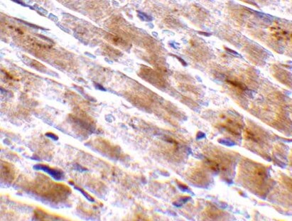
Myoglobin in Human Heart.
Myoglobin was detected in immersion fixed paraffin-embedded sections of human heart using Rabbit Anti-Human Myoglobin Monoclonal Antibody (Catalog # MAB97204) at 3 µg/mL for 1 hour at room temperature followed by incubation with the Anti-Goat IgG VisUCyte™ HRP Polymer Antibody (Catalog # VC004). Tissue was stained using DAB (brown) and counterstained with hematoxylin (blue). Specific staining was localized to cardiomyocytes. View our protocol for IHC Staining with VisUCyte HRP Polymer Detection Reagents.
Detection of Human Myoglobin by Western Blot.
Western blot shows lysates of human heart tissue and human liver tissue (negative control). PVDF membrane was probed with 0.05 µg/mL of Rabbit Anti-Human Myoglobin Monoclonal Antibody (Catalog # MAB97204) followed by HRP-conjugated Anti-Rabbit IgG Secondary Antibody (Catalog # HAF008). A specific band was detected for Myoglobin at approximately 16 kDa (as indicated). This experiment was conducted under reducing conditions and using Immunoblot Buffer Group 1.
Human Myoglobin ELISA Standard Curve.
Recombinant Human Myoglobin protein was serially diluted 2-fold and captured by Rabbit Anti-Human Myoglobin Monoclonal Antibody (Catalog # MAB97204) coated on a Clear Polystyrene Microplate (Catalog # DY990). Rabbit Anti-Human/Mouse/Rat Myoglobin Monoclonal Antibody (Catalog # MAB97203) was biotinylated and incubated with the protein captured on the plate. Detection of the standard curve was achieved by incubating Streptavidin-HRP (Catalog # DY998) followed by Substrate Solution (Catalog # DY999) and stopping the enzymatic reaction with Stop Solution (Catalog # DY994).Applications for Human Myoglobin Antibody (2269D)
Application
Recommended Usage
ELISA
This antibody functions as an ELISA capture antibody when paired with Rabbit Anti-Human/Mouse/Rat Myoglobin Monoclonal Antibody (Catalog # MAB97203).
This product is intended for assay development on various assay platforms requiring antibody pairs.
Immunohistochemistry
3-25 µg/mL
Sample: Immersion fixed paraffin-embedded sections of human heart
Sample: Immersion fixed paraffin-embedded sections of human heart
Western Blot
0.05 µg/mL
Sample: Human heart tissue
Sample: Human heart tissue
Reviewed Applications
Read 1 review rated 5 using MAB97204 in the following applications:
Formulation, Preparation, and Storage
Purification
Protein A or G purified from cell culture supernatant
Reconstitution
Reconstitute at 0.5 mg/mL in sterile PBS. For liquid material, refer to CoA for concentration.
Loading...
Formulation
Lyophilized from a 0.2 μm filtered solution in PBS with Trehalose. *Small pack size (SP) is supplied either lyophilized or as a 0.2 µm filtered solution in PBS.
Shipping
Lyophilized product is shipped at ambient temperature. Liquid small pack size (-SP) is shipped with polar packs. Upon receipt, store immediately at the temperature recommended below.
Stability & Storage
Use a manual defrost freezer and avoid repeated freeze-thaw cycles.
- 12 months from date of receipt, -20 to -70 °C as supplied.
- 1 month, 2 to 8 °C under sterile conditions after reconstitution.
- 6 months, -20 to -70 °C under sterile conditions after reconstitution.
Calculators
Background: Myoglobin
Alternate Names
MB
Gene Symbol
MB
UniProt
Additional Myoglobin Products
Product Documents for Human Myoglobin Antibody (2269D)
Certificate of Analysis
To download a Certificate of Analysis, please enter a lot or batch number in the search box below.
Note: Certificate of Analysis not available for kit components.
Product Specific Notices for Human Myoglobin Antibody (2269D)
For research use only
Customer Reviews for Human Myoglobin Antibody (2269D) (1)
5 out of 5
1 Customer Rating
Have you used Human Myoglobin Antibody (2269D)?
Submit a review and receive an Amazon gift card!
$25/€18/£15/$25CAN/¥2500 Yen for a review with an image
$10/€7/£6/$10CAN/¥1110 Yen for a review without an image
Submit a review
Customer Images
Showing
1
-
1 of
1 review
Showing All
Filter By:
-
Application: ImmunohistochemistrySample Tested: Heart tissueSpecies: HumanVerified Customer | Posted 07/04/2022

There are no reviews that match your criteria.
Protocols
Find general support by application which include: protocols, troubleshooting, illustrated assays, videos and webinars.
- Antigen Retrieval Protocol (PIER)
- Antigen Retrieval for Frozen Sections Protocol
- Appropriate Fixation of IHC/ICC Samples
- Cellular Response to Hypoxia Protocols
- Chromogenic IHC Staining of Formalin-Fixed Paraffin-Embedded (FFPE) Tissue Protocol
- Chromogenic Immunohistochemistry Staining of Frozen Tissue
- ClariTSA™ Fluorophore Kits
- Detection & Visualization of Antibody Binding
- ELISA Sample Preparation & Collection Guide
- ELISA Troubleshooting Guide
- Fluorescent IHC Staining of Frozen Tissue Protocol
- Graphic Protocol for Heat-induced Epitope Retrieval
- Graphic Protocol for the Preparation and Fluorescent IHC Staining of Frozen Tissue Sections
- Graphic Protocol for the Preparation and Fluorescent IHC Staining of Paraffin-embedded Tissue Sections
- Graphic Protocol for the Preparation of Gelatin-coated Slides for Histological Tissue Sections
- How to Run an R&D Systems DuoSet ELISA
- How to Run an R&D Systems Quantikine ELISA
- How to Run an R&D Systems Quantikine™ QuicKit™ ELISA
- IHC Sample Preparation (Frozen sections vs Paraffin)
- Immunofluorescent IHC Staining of Formalin-Fixed Paraffin-Embedded (FFPE) Tissue Protocol
- Immunohistochemistry (IHC) and Immunocytochemistry (ICC) Protocols
- Immunohistochemistry Frozen Troubleshooting
- Immunohistochemistry Paraffin Troubleshooting
- Preparing Samples for IHC/ICC Experiments
- Preventing Non-Specific Staining (Non-Specific Binding)
- Primary Antibody Selection & Optimization
- Protocol for Heat-Induced Epitope Retrieval (HIER)
- Protocol for Making a 4% Formaldehyde Solution in PBS
- Protocol for VisUCyte™ HRP Polymer Detection Reagent
- Protocol for the Preparation & Fixation of Cells on Coverslips
- Protocol for the Preparation and Chromogenic IHC Staining of Frozen Tissue Sections
- Protocol for the Preparation and Chromogenic IHC Staining of Frozen Tissue Sections - Graphic
- Protocol for the Preparation and Chromogenic IHC Staining of Paraffin-embedded Tissue Sections
- Protocol for the Preparation and Chromogenic IHC Staining of Paraffin-embedded Tissue Sections - Graphic
- Protocol for the Preparation and Fluorescent IHC Staining of Frozen Tissue Sections
- Protocol for the Preparation and Fluorescent IHC Staining of Paraffin-embedded Tissue Sections
- Protocol for the Preparation of Gelatin-coated Slides for Histological Tissue Sections
- Quantikine HS ELISA Kit Assay Principle, Alkaline Phosphatase
- Quantikine HS ELISA Kit Principle, Streptavidin-HRP Polymer
- R&D Systems Quality Control Western Blot Protocol
- Sandwich ELISA (Colorimetric) – Biotin/Streptavidin Detection Protocol
- Sandwich ELISA (Colorimetric) – Direct Detection Protocol
- TUNEL and Active Caspase-3 Detection by IHC/ICC Protocol
- The Importance of IHC/ICC Controls
- Troubleshooting Guide: ELISA
- Troubleshooting Guide: Immunohistochemistry
- Troubleshooting Guide: Western Blot Figures
- Western Blot Conditions
- Western Blot Protocol
- Western Blot Protocol for Cell Lysates
- Western Blot Troubleshooting
- Western Blot Troubleshooting Guide
- View all Protocols, Troubleshooting, Illustrated assays and Webinars
Loading...