Nuclear Factor kappa B2 (NF kappa B2 or NF kappa B p52) is a member of the NF kappa B/Rel family of transcription factors. NF kappa B2 dimerizes with other members of the NF kappa B/Rel family to regulate expression of genes that participate in immune, apoptotic, and oncogenic processes.

Key Product Details
Validated by
Biological Validation
Species Reactivity
Validated:
Human
Cited:
Human
Applications
Validated:
Western Blot, Immunocytochemistry, Chromatin Immunoprecipitation (ChIP)
Cited:
Flow Cytometry
Label
Unconjugated
Antibody Source
Monoclonal Mouse IgG1 Clone # 291319
Loading...
Product Specifications
Immunogen
E. coli-derived recombinant human NF kappa B2
Met1-Asn447
Accession # Q00653
Met1-Asn447
Accession # Q00653
Specificity
Detects human NFkB2 in direct ELISAs and Western blots. In Western blots, no cross-reactivity with endogenous mouse NFkB2 in NIH 3T3 cells is observed.
Clonality
Monoclonal
Host
Mouse
Isotype
IgG1
Scientific Data Images for Human NFkB2 Antibody (291319)
Detection of Human NF kappa B2 by Western Blot.
Western blot shows lysates of Daudi human Burkitt's lymphoma cell line untreated (-) or treated (+) with 100 ng/mL Recombinant Human CD40 Ligand/TNFSF5 aa 108-261 (Catalog # 6245-CL) for 4 hours. Gels were loaded with 20 µg of cytoplasmic (Cyto) and 10 µg of nuclear extracts (Nuc). For additional reference, lysates of Raji human Burkitt's lymphoma cell line were included. PVDF membrane was probed with 0.5 µg/mL Mouse Anti-Human NF kappa B2 Monoclonal Antibody (Catalog # MAB28881) followed by HRP-conjugated Anti-Mouse IgG Secondary Antibody (Catalog # HAF007). Specific bands for NF kappa B2 were detected at approximately 52 kDa and 100 kDa (as indicated). This experiment was conducted under reducing conditions and using Immunoblot Buffer Group 1.
Detection of NF kappa B2-regulated Genes by Chromatin Immunoprecipitation.
Jurkat human acute T cell leukemia cell line treated with 50 ng/mL PMA and 200 ng/mL calcium ionomycin overnight was fixed using formaldehyde, resuspended in lysis buffer, and sonicated to shear chromatin. NF kappa B2/DNA complexes were immunoprecipitated using 5 µg Mouse Anti-Human NF kappa B2 Monoclonal Antibody (Catalog # MAB28881) or control antibody (Catalog # MAB002) for 15 minutes in an ultrasonic bath, followed by Biotinylated Anti-Mouse IgG Secondary Antibody (Catalog # BAF007). Immunocomplexes were captured using 50 µL of MagCellect Streptavidin Ferrofluid (Catalog # MAG999) and DNA was purified using chelating resin solution. Thec-mycpromoter was detected by standard PCR.
NF kappa B2 in HeLa Human Cell Line.
NF kappa B2 was detected in immersion fixed HeLa human cervical epithelial carcinoma cell line using Mouse Anti-Human NF kappa B2 Monoclonal Antibody (Catalog # MAB28881) at 3 µg/mL for 3 hours at room temperature. Cells were stained using the NorthernLights™ 557-conjugated Anti-Mouse IgG Secondary Antibody (red; Catalog # NL007) and counterstained with DAPI (blue). Specific staining was localized to cytoplasm and nuclei. View our protocol for Fluorescent ICC Staining of Cells on Coverslips.Applications for Human NFkB2 Antibody (291319)
Application
Recommended Usage
Chromatin Immunoprecipitation (ChIP)
5 µg/5 x 106 cells
Sample: PMA and calcium ionomycin treated Jurkat human acute T cell leukemia cell line chromatin, c-myc promoter detected by standard PCR
Sample: PMA and calcium ionomycin treated Jurkat human acute T cell leukemia cell line chromatin, c-myc promoter detected by standard PCR
Immunocytochemistry
3-25 µg/mL
Sample: Immersion fixed human peripheral blood mononuclear cells and HeLa human cervical epithelial carcinoma cell line
Sample: Immersion fixed human peripheral blood mononuclear cells and HeLa human cervical epithelial carcinoma cell line
Western Blot
0.5 µg/mL
Sample: Daudi human Burkitt's lymphoma cell line treated with Recombinant Human CD40 Ligand/TNFSF5 (Catalog # 6245-CL)
Sample: Daudi human Burkitt's lymphoma cell line treated with Recombinant Human CD40 Ligand/TNFSF5 (Catalog # 6245-CL)
Reviewed Applications
Read 6 reviews rated 4.3 using MAB28881 in the following applications:
Formulation, Preparation, and Storage
Purification
Protein A or G purified from hybridoma culture supernatant
Reconstitution
Reconstitute at 0.5 mg/mL in sterile PBS. For liquid material, refer to CoA for concentration.
Loading...
Formulation
Lyophilized from a 0.2 μm filtered solution in PBS with Trehalose. *Small pack size (SP) is supplied either lyophilized or as a 0.2 µm filtered solution in PBS.
Shipping
Lyophilized product is shipped at ambient temperature. Liquid small pack size (-SP) is shipped with polar packs. Upon receipt, store immediately at the temperature recommended below.
Stability & Storage
Use a manual defrost freezer and avoid repeated freeze-thaw cycles.
- 12 months from date of receipt, -20 to -70 °C as supplied.
- 1 month, 2 to 8 °C under sterile conditions after reconstitution.
- 6 months, -20 to -70 °C under sterile conditions after reconstitution.
Calculators
Background: NFkB2
Long Name
Nuclear Factor Kappa-B, Subunit 2
Alternate Names
LYT10, NF-kB2
Gene Symbol
NFKB2
UniProt
Additional NFkB2 Products
Product Documents for Human NFkB2 Antibody (291319)
Certificate of Analysis
To download a Certificate of Analysis, please enter a lot or batch number in the search box below.
Note: Certificate of Analysis not available for kit components.
Product Specific Notices for Human NFkB2 Antibody (291319)
For research use only
Related Research Areas
Citations for Human NFkB2 Antibody (291319)
 Powered by Bioz
Customer Reviews for Human NFkB2 Antibody (291319) (6)
4.3 out of 5
6 Customer Ratings
Have you used Human NFkB2 Antibody (291319)?
Submit a review and receive an Amazon gift card!
$25/€18/£15/$25CAN/¥2500 Yen for a review with an image
$10/€7/£6/$10CAN/¥1110 Yen for a review without an image
Submit a review
Customer Images

Showing
1
-
5 of
6 reviews
Showing All
Filter By:
-
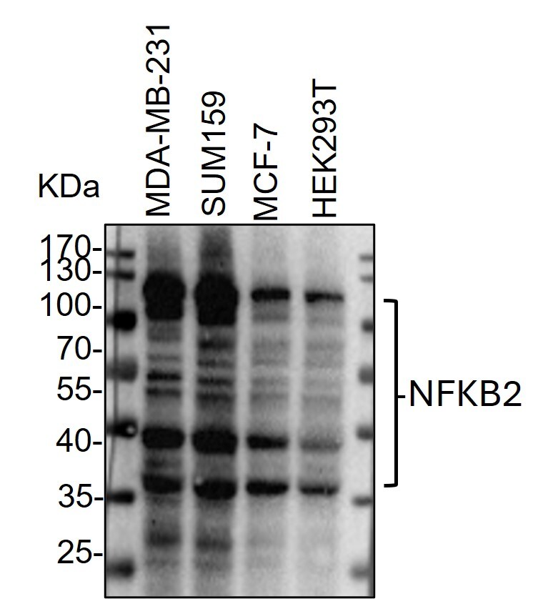
Application: Western BlotVerified Customer | Posted 12/31/2023Western Blot: whole cell lysates from MDA-MB-231, SUM159, MCF-7 and HEK293T cells were loaded with 50 ug/lane. 10% SDS-PAGE. NFKB2 Antibody (MAB28881-100) was used for primary antibody: 1:1000, 4℃, overnight.

-
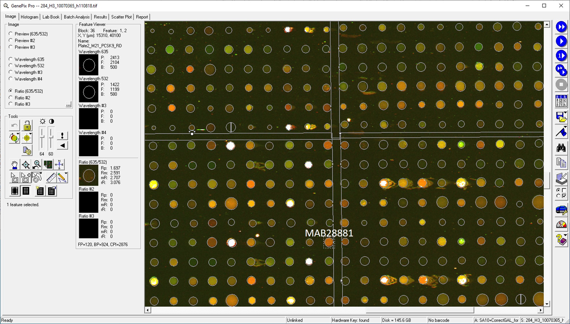
Application: MicroarraysSample Tested: EDTA PlasmaSpecies: HumanVerified Customer | Posted 01/14/2021
-
Application: MicroarraySample Tested: EDTA PlasmaSpecies: HumanVerified Customer | Posted 02/07/2020Antibody was printed on custom arrays and incubated with fluorescently labeled human EDTA plasma
-
Application: ELISASample Tested: PDX samplesSpecies: HumanVerified Customer | Posted 01/16/2020
-
Application: MicroarraysSample Tested: EDTA PlasmaSpecies: HumanVerified Customer | Posted 11/14/2018
-
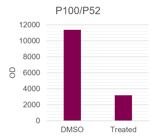
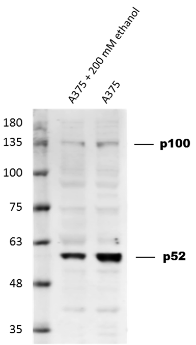
Application: Western BlotSample Tested: A375 human melanoma cell lineSpecies: HumanVerified Customer | Posted 03/01/2017Down-regulation of NFKB-2 p100/p52 in Human melanoma A375 cell line upon treatment with ethanol. Dilution: 1:1,000 in PBS with 5% BSA. Secondary Ab: anti-Mouse IgG 1:5,000.
There are no reviews that match your criteria.
Protocols
Find general support by application which include: protocols, troubleshooting, illustrated assays, videos and webinars.
- Appropriate Fixation of IHC/ICC Samples
- Cellular Response to Hypoxia Protocols
- ChIP Protocol Video
- Chromatin Immunoprecipitation (ChIP) Protocol
- Chromatin Immunoprecipitation Protocol
- ClariTSA™ Fluorophore Kits
- Detection & Visualization of Antibody Binding
- ICC Cell Smear Protocol for Suspension Cells
- ICC Immunocytochemistry Protocol Videos
- ICC for Adherent Cells
- Immunocytochemistry (ICC) Protocol
- Immunocytochemistry Troubleshooting
- Immunofluorescence of Organoids Embedded in Cultrex Basement Membrane Extract
- Immunohistochemistry (IHC) and Immunocytochemistry (ICC) Protocols
- Preparing Samples for IHC/ICC Experiments
- Preventing Non-Specific Staining (Non-Specific Binding)
- Primary Antibody Selection & Optimization
- Protocol for VisUCyte™ HRP Polymer Detection Reagent
- Protocol for the Fluorescent ICC Staining of Cell Smears - Graphic
- Protocol for the Fluorescent ICC Staining of Cultured Cells on Coverslips - Graphic
- Protocol for the Preparation and Fluorescent ICC Staining of Cells on Coverslips
- Protocol for the Preparation and Fluorescent ICC Staining of Non-adherent Cells
- Protocol for the Preparation and Fluorescent ICC Staining of Stem Cells on Coverslips
- Protocol for the Preparation of a Cell Smear for Non-adherent Cell ICC - Graphic
- R&D Systems Quality Control Western Blot Protocol
- TUNEL and Active Caspase-3 Detection by IHC/ICC Protocol
- The Importance of IHC/ICC Controls
- Troubleshooting Guide: Western Blot Figures
- Western Blot Conditions
- Western Blot Protocol
- Western Blot Protocol for Cell Lysates
- Western Blot Troubleshooting
- Western Blot Troubleshooting Guide
- View all Protocols, Troubleshooting, Illustrated assays and Webinars
Loading...
Associated Pathways


Pathogen or Damage-activated C-Type Lectin Receptor Signaling Pathways
 TGF-beta Signaling Pathways
 Toll-Like Receptor Signaling Pathways
