Human NG2/MCSP Antibody (LHM-2)
R&D Systems | Catalog # MAB2585

Key Product Details
Species Reactivity
Validated:
Human
Cited:
Human, Mouse, Equine
Applications
Validated:
Immunohistochemistry, Western Blot, Flow Cytometry, CyTOF-ready
Cited:
Immunohistochemistry, Immunohistochemistry-Paraffin, Western Blot, Flow Cytometry, Immunocytochemistry
Label
Unconjugated
Antibody Source
Monoclonal Mouse IgG1 Clone # LHM-2
Loading...
Product Specifications
Immunogen
Human melanoma cells
Specificity
Detects human NG2/MCSP in Western blots.
Clonality
Monoclonal
Host
Mouse
Isotype
IgG1
Scientific Data Images for Human NG2/MCSP Antibody (LHM-2)
Detection of Human NG2/MCSP by Western Blot.
Western blot shows lysates of A375 human melanoma cell line and SK-Mel-28 human malignant melanoma cell line. PVDF membrane was probed with 1 µg/mL of Mouse Anti-Human NG2/MCSP Monoclonal Antibody (Catalog # MAB2585) followed by HRP-conjugated Anti-Mouse IgG Secondary Antibody (Catalog # HAF018). A specific band was detected for NG2/MCSP at approximately 300 kDa (as indicated). This experiment was conducted under reducing conditions and using Immunoblot Buffer Group 1.
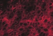
NG2/MCSP in Human Skin Cancer Tissue.
NG2/MCSP was detected in immersion fixed paraffin-embedded sections of human skin cancer tissue (melanoma) using 10 µg/mL Mouse Anti-Human NG2/MCSP Monoclonal Antibody (Catalog # MAB2585) overnight at 4 °C. Before incubation with the primary antibody tissue was subjected to heat-induced epitope retrieval using Antigen Retrieval Reagent-Basic (Catalog # CTS013). Tissue was stained with the Anti-Mouse HRP-DAB Cell & Tissue Staining Kit (brown; Catalog # CTS002) and counterstained with hematoxylin (blue). View our protocol for Chromogenic IHC Staining of Paraffin-embedded Tissue Sections.
Detection of NG2/MCSP in A375 cells by Flow Cytometry
A375 cells were stained with Mouse Anti-Human NG2/MCSP Monoclonal Antibody (Catalog # MAB2585, filled histogram) or isotype control antibody (Catalog # MAB002, open histogram) followed by Allophycocyanin-conjugated Anti-Mouse IgG Secondary Antibody (Catalog # F0101B). View our protocol for Staining Membrane-associated Proteins.Applications for Human NG2/MCSP Antibody (LHM-2)
Application
Recommended Usage
CyTOF-ready
Ready to be labeled using established conjugation methods. No BSA or other carrier proteins that could interfere with conjugation.
Flow Cytometry
0.25 µg/106 cells
Sample: see below
Sample: see below
Immunohistochemistry
8-25 µg/mL
Sample: Immersion fixed paraffin-embedded sections of human skin cancer tissue (melanoma) subjected to Antigen Retrieval Reagent-Basic (Catalog # CTS013)
Sample: Immersion fixed paraffin-embedded sections of human skin cancer tissue (melanoma) subjected to Antigen Retrieval Reagent-Basic (Catalog # CTS013)
Western Blot
1 µg/mL
Sample: A375 human melanoma cell line and SK‑Mel‑28 human malignant melanoma cell line
Sample: A375 human melanoma cell line and SK‑Mel‑28 human malignant melanoma cell line
Reviewed Applications
Read 2 reviews rated 5 using MAB2585 in the following applications:
Flow Cytometry Panel Builder
Bio-Techne Knows Flow Cytometry
Save time and reduce costly mistakes by quickly finding compatible reagents using the Panel Builder Tool.
Advanced Features
- Spectra Viewer - Custom analysis of spectra from multiple fluorochromes
- Spillover Popups - Visualize the spectra of individual fluorochromes
- Antigen Density Selector - Match fluorochrome brightness with antigen density

Formulation, Preparation, and Storage
Purification
Protein A or G purified from hybridoma culture supernatant
Reconstitution
Reconstitute at 0.5 mg/mL in sterile PBS. For liquid material, refer to CoA for concentration.
Loading...
Formulation
Lyophilized from a 0.2 μm filtered solution in PBS with Trehalose. See Certificate of Analysis for details.
*Small pack size (-SP) is supplied either lyophilized or as a 0.2 µm filtered solution in PBS.
*Small pack size (-SP) is supplied either lyophilized or as a 0.2 µm filtered solution in PBS.
Shipping
Lyophilized product is shipped at ambient temperature. Liquid small pack size (-SP) is shipped with polar packs. Upon receipt, store immediately at the temperature recommended below.
Stability & Storage
Use a manual defrost freezer and avoid repeated freeze-thaw cycles.
- 12 months from date of receipt, -20 to -70 °C as supplied.
- 1 month, 2 to 8 °C under sterile conditions after reconstitution.
- 6 months, -20 to -70 °C under sterile conditions after reconstitution.
Calculators
Background: NG2/MCSP
References
- Kupsch, J.M. et al. (1995) Melanoma Res. 5:403.
- Stegmuller, J. et al. (2002) J. Neurocytol. 31:497.
- Legg, J. et al. (2003) Development 130:6049.
Long Name
Chondroitin Sulfate Proteoglycan NG2/Melanoma Associated Chondroitin Sulfate Proteoglycan
Alternate Names
AN2, CSPG4, HMW-MAA, MCSP, MCSPG, MEL-CSPG
Gene Symbol
CSPG4
Additional NG2/MCSP Products
Product Documents for Human NG2/MCSP Antibody (LHM-2)
Certificate of Analysis
To download a Certificate of Analysis, please enter a lot or batch number in the search box below.
Note: Certificate of Analysis not available for kit components.
Product Specific Notices for Human NG2/MCSP Antibody (LHM-2)
For research use only
Related Research Areas
Citations for Human NG2/MCSP Antibody (LHM-2)
 Powered by Bioz
Customer Reviews for Human NG2/MCSP Antibody (LHM-2) (2)
5 out of 5
2 Customer Ratings
Have you used Human NG2/MCSP Antibody (LHM-2)?
Submit a review and receive an Amazon gift card!
$25/€18/£15/$25CAN/¥2500 Yen for a review with an image
$10/€7/£6/$10CAN/¥1110 Yen for a review without an image
Submit a review
Customer Images
Showing
1
-
2 of
2 reviews
Showing All
Filter By:
-
Application: ImmunohistochemistrySample Tested: brain tumorSpecies: HumanVerified Customer | Posted 10/15/2021

-
Application: ImmunohistochemistrySample Tested: White matter (brain)Species: HumanVerified Customer | Posted 07/15/2021
There are no reviews that match your criteria.
Protocols
Find general support by application which include: protocols, troubleshooting, illustrated assays, videos and webinars.
- 7-Amino Actinomycin D (7-AAD) Cell Viability Flow Cytometry Protocol
- Antigen Retrieval Protocol (PIER)
- Antigen Retrieval for Frozen Sections Protocol
- Appropriate Fixation of IHC/ICC Samples
- Cellular Response to Hypoxia Protocols
- Chromogenic IHC Staining of Formalin-Fixed Paraffin-Embedded (FFPE) Tissue Protocol
- Chromogenic Immunohistochemistry Staining of Frozen Tissue
- ClariTSA™ Fluorophore Kits
- Detection & Visualization of Antibody Binding
- Extracellular Membrane Flow Cytometry Protocol
- Flow Cytometry Protocol for Cell Surface Markers
- Flow Cytometry Protocol for Staining Membrane Associated Proteins
- Flow Cytometry Staining Protocols
- Flow Cytometry Troubleshooting Guide
- Fluorescent IHC Staining of Frozen Tissue Protocol
- Graphic Protocol for Heat-induced Epitope Retrieval
- Graphic Protocol for the Preparation and Fluorescent IHC Staining of Frozen Tissue Sections
- Graphic Protocol for the Preparation and Fluorescent IHC Staining of Paraffin-embedded Tissue Sections
- Graphic Protocol for the Preparation of Gelatin-coated Slides for Histological Tissue Sections
- IHC Sample Preparation (Frozen sections vs Paraffin)
- Immunofluorescent IHC Staining of Formalin-Fixed Paraffin-Embedded (FFPE) Tissue Protocol
- Immunohistochemistry (IHC) and Immunocytochemistry (ICC) Protocols
- Immunohistochemistry Frozen Troubleshooting
- Immunohistochemistry Paraffin Troubleshooting
- Intracellular Flow Cytometry Protocol Using Alcohol (Methanol)
- Intracellular Flow Cytometry Protocol Using Detergents
- Intracellular Nuclear Staining Flow Cytometry Protocol Using Detergents
- Intracellular Staining Flow Cytometry Protocol Using Alcohol Permeabilization
- Intracellular Staining Flow Cytometry Protocol Using Detergents to Permeabilize Cells
- Preparing Samples for IHC/ICC Experiments
- Preventing Non-Specific Staining (Non-Specific Binding)
- Primary Antibody Selection & Optimization
- Propidium Iodide Cell Viability Flow Cytometry Protocol
- Protocol for Heat-Induced Epitope Retrieval (HIER)
- Protocol for Liperfluo
- Protocol for Making a 4% Formaldehyde Solution in PBS
- Protocol for VisUCyte™ HRP Polymer Detection Reagent
- Protocol for the Characterization of Human Th22 Cells
- Protocol for the Characterization of Human Th9 Cells
- Protocol for the Preparation & Fixation of Cells on Coverslips
- Protocol for the Preparation and Chromogenic IHC Staining of Frozen Tissue Sections
- Protocol for the Preparation and Chromogenic IHC Staining of Frozen Tissue Sections - Graphic
- Protocol for the Preparation and Chromogenic IHC Staining of Paraffin-embedded Tissue Sections
- Protocol for the Preparation and Chromogenic IHC Staining of Paraffin-embedded Tissue Sections - Graphic
- Protocol for the Preparation and Fluorescent IHC Staining of Frozen Tissue Sections
- Protocol for the Preparation and Fluorescent IHC Staining of Paraffin-embedded Tissue Sections
- Protocol for the Preparation of Gelatin-coated Slides for Histological Tissue Sections
- Protocol: Annexin V and PI Staining by Flow Cytometry
- Protocol: Annexin V and PI Staining for Apoptosis by Flow Cytometry
- R&D Systems Quality Control Western Blot Protocol
- TUNEL and Active Caspase-3 Detection by IHC/ICC Protocol
- The Importance of IHC/ICC Controls
- Troubleshooting Guide: Fluorokine Flow Cytometry Kits
- Troubleshooting Guide: Immunohistochemistry
- Troubleshooting Guide: Western Blot Figures
- Western Blot Conditions
- Western Blot Protocol
- Western Blot Protocol for Cell Lysates
- Western Blot Troubleshooting
- Western Blot Troubleshooting Guide
- View all Protocols, Troubleshooting, Illustrated assays and Webinars
Loading...
Associated Pathways
