

Scientific images may be resized, cropped, or adjusted for brightness or contrast for presentation purposes. These updates are limited to presentation and do not affect the underlying scientific interpretation of the data. Where available, additional source data may be provided upon request.
Key Product Details
Validated by
Species Reactivity
Validated:
Cited:
Applications
Validated:
Cited:
Label
Antibody Source
Product Specifications
Immunogen
Gln24-Arg137
Accession # P52952
Specificity
Clonality
Host
Isotype
Scientific Data Images for Human NKX2.5 Antibody
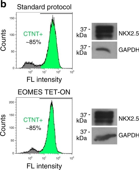
Detection of Human NKX2.5 by Western Blot
EOMES programs hESCs into functional CMs at high efficiency. a Illustration of growth factor-mediated and optimized EOMES induction-based cardiac differentiation protocols. Bottom right: Immunoblot validating doxycycline-dependent EOMES expression in a transgenic EOMESKO/E.TET-ON hESC line. b Typical yields of hESC-CMs (left, flow cytometry) and NKX2.5 expression (right, immunoblot) obtained with the two protocols (day 10). c Immunostainings 21 days after the initiation of EOMES induction. Weak perinuclear ANP staining is typical in overall MLC2v-positive hESC-CMs. Scale bars: 25 (top) and 50 µm (bottom). d Acceleration and slowdown of spontaneous beat rates in pCMs following exposure to 10 µM isoprenaline and 10 µM propranolol, respectively, on multielectrode arrays. e Microarray-based time course analysis comparing the indicated protocols and cell lines. RESCUE cells carry an inducible EOMES transgene on EOMESKO HuES6 background. Underlying data are from Supplementary Data 2 Image collected and cropped by CiteAb from the following publication (https://pubmed.ncbi.nlm.nih.gov/29382828), licensed under a CC-BY license. Not internally tested by R&D Systems.
Detection of Human NKX2.5 by Immunocytochemistry/Immunofluorescence
Contextual requirements of EOMES-mediated CM programming. a DOX dose dependency of the EOMES TET-ON protocol using the WTE.TET-ON hESC line. Top panel: Immunostains at 1.5 wk. Scale bar: 100 µm. Bottom: Flow cytometry analysis. Numbers indicate average percentages of cTnT-positive CMs from 3–6 experiments per condition. b CM programming by EOMES necessitates suppression of autocrine WNT signaling from the third day of transgene induction (qPCR data, n = 2). c DOX dose-dependent CM programming using an independent WTE.TET-ON hiPSC line. The data shows immunostaining (scale bar: 100 µm), RT-qPCR (n = 4), and FACS analysis (n = 3) performed at 1 wk of differentiation. The asymmetrical shape of the DOX titration data with this line is in part due to the incomplete repression of SOX2 at 0.05 µg/ml, which caused overgrowth of the cultures with neural precursors (also see Supplementary Fig. 3f). Error bars: s.e.m. Image collected and cropped by CiteAb from the following publication (https://pubmed.ncbi.nlm.nih.gov/29382828), licensed under a CC-BY license. Not internally tested by R&D Systems.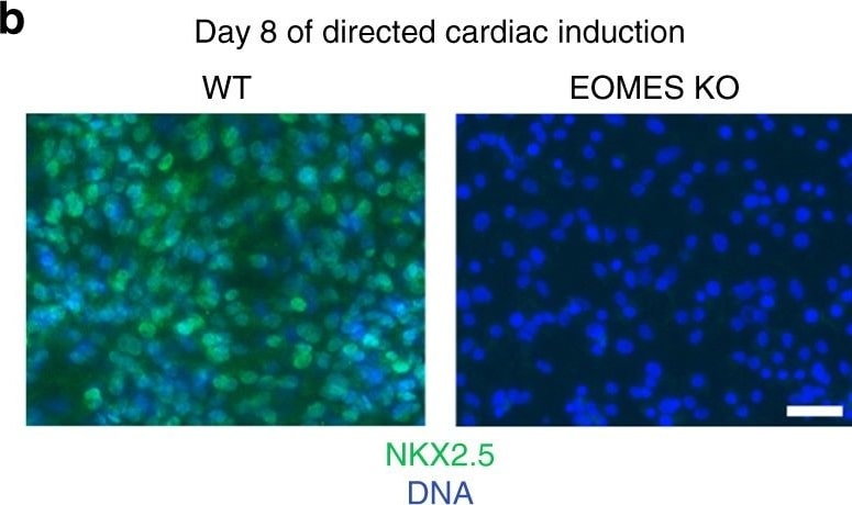
Detection of Human NKX2.5 by Immunocytochemistry/Immunofluorescence
EOMES knockout hESCs fail to differentiate into cardiomyocytes. a Immunoblot confirming EOMES expression and its absence in WT and KO cells, respectively, at the cardiac mesoderm stage of directed differentiation. b EOMES KO cells fail to express the early cardiomyocyte marker NKX2.5 following exposure to a directed differentiation protocol. Scale bar: 50 µm. c EOMES KO cells show a general failure in markedly upregulating essential pan-cardiac genes (qPCR data, n = 3; error bars: s.e.m.). For each gene, the expression level in the highest expressing sample (“max” = WT in all three cases) is set to 1. d Differentiation of EOMES KO hESCs under signaling factor-assisted, non-cardiac mesoderm/endoderm-permissive culture conditions. Top: EOMES KO cells fail to express alpha -fetoprotein following Activin A-assisted endodermal induction. Scale bar: 50 µm. Bottom: Non-cardiac mesodermal differentiation competence of EOMES KO hESCs. KO cells were differentiated using varied meso and endoderm-permissive induction conditions. The data denote transformed p-values of annotation terms enriched in gene sets upregulated in EOMES KO samples compared to undifferentiated hESCs or differentiated WT controls (cutoffs: 10 or 3-fold, respectively). Underlying data: Supplementary Data 1 Image collected and cropped by CiteAb from the following publication (https://pubmed.ncbi.nlm.nih.gov/29382828), licensed under a CC-BY license. Not internally tested by R&D Systems.Applications for Human NKX2.5 Antibody
Immunohistochemistry
Sample: Immersion fixed paraffin-embedded sections of human heart
Western Blot
Sample: Recombinant Human NKX2.5
Reviewed Applications
Read 2 reviews rated 4 using AF2444 in the following applications:
Formulation, Preparation, and Storage
Purification
Reconstitution
Reconstitute at 0.2 mg/mL in sterile PBS. For liquid material, refer to CoA for concentration.
Formulation
Shipping
Stability & Storage
- 12 months from date of receipt, -20 to -70 °C as supplied.
- 1 month, 2 to 8 °C under sterile conditions after reconstitution.
- 6 months, -20 to -70 °C under sterile conditions after reconstitution.
Calculators
Background: NKX2.5
Long Name
Alternate Names
Entrez Gene IDs
Gene Symbol
UniProt
Additional NKX2.5 Products
Product Documents for Human NKX2.5 Antibody
Certificate of Analysis
To download a Certificate of Analysis, please enter a lot or batch number in the search box below.
Note: Certificate of Analysis not available for kit components.
Product Specific Notices for Human NKX2.5 Antibody
For research use only
Citations for Human NKX2.5 Antibody
 Powered by Bioz
Customer Reviews for Human NKX2.5 Antibody (2)
Have you used Human NKX2.5 Antibody?
Submit a review and receive an Amazon gift card!
$25/€18/£15/$25CAN/¥2500 for a review with an image
$10/€7/£6/$10CAN/¥1110 for a review without an image
Submit a review
-
Application: ImmunofluorescenceSample Tested: See PMID 24123507Species: MouseVerified Customer | Posted 01/07/2015
-
Application: ImmunofluorescenceSample Tested: See PMID 24123507Species: OtherVerified Customer | Posted 01/06/2015
There are no reviews that match your criteria.
Protocols
Find general support by application which include: protocols, troubleshooting, illustrated assays, videos and webinars.
- Antigen Retrieval Protocol (PIER)
- Antigen Retrieval for Frozen Sections Protocol
- Appropriate Fixation of IHC/ICC Samples
- Cellular Response to Hypoxia Protocols
- Chromogenic IHC Staining of Formalin-Fixed Paraffin-Embedded (FFPE) Tissue Protocol
- Chromogenic Immunohistochemistry Staining of Frozen Tissue
- ClariTSA™ Fluorophore Kits
- Detection & Visualization of Antibody Binding
- Fluorescent IHC Staining of Frozen Tissue Protocol
- Graphic Protocol for Heat-induced Epitope Retrieval
- Graphic Protocol for the Preparation and Fluorescent IHC Staining of Frozen Tissue Sections
- Graphic Protocol for the Preparation and Fluorescent IHC Staining of Paraffin-embedded Tissue Sections
- Graphic Protocol for the Preparation of Gelatin-coated Slides for Histological Tissue Sections
- IHC Sample Preparation (Frozen sections vs Paraffin)
- Immunofluorescent IHC Staining of Formalin-Fixed Paraffin-Embedded (FFPE) Tissue Protocol
- Immunohistochemistry (IHC) and Immunocytochemistry (ICC) Protocols
- Immunohistochemistry Frozen Troubleshooting
- Immunohistochemistry Paraffin Troubleshooting
- Preparing Samples for IHC/ICC Experiments
- Preventing Non-Specific Staining (Non-Specific Binding)
- Primary Antibody Selection & Optimization
- Protocol for Heat-Induced Epitope Retrieval (HIER)
- Protocol for Making a 4% Formaldehyde Solution in PBS
- Protocol for VisUCyte™ HRP Polymer Detection Reagent
- Protocol for the Preparation & Fixation of Cells on Coverslips
- Protocol for the Preparation and Chromogenic IHC Staining of Frozen Tissue Sections
- Protocol for the Preparation and Chromogenic IHC Staining of Frozen Tissue Sections - Graphic
- Protocol for the Preparation and Chromogenic IHC Staining of Paraffin-embedded Tissue Sections
- Protocol for the Preparation and Chromogenic IHC Staining of Paraffin-embedded Tissue Sections - Graphic
- Protocol for the Preparation and Fluorescent IHC Staining of Frozen Tissue Sections
- Protocol for the Preparation and Fluorescent IHC Staining of Paraffin-embedded Tissue Sections
- Protocol for the Preparation of Gelatin-coated Slides for Histological Tissue Sections
- R&D Systems Quality Control Western Blot Protocol
- TUNEL and Active Caspase-3 Detection by IHC/ICC Protocol
- The Importance of IHC/ICC Controls
- Troubleshooting Guide: Immunohistochemistry
- Troubleshooting Guide: Western Blot Figures
- Western Blot Conditions
- Western Blot Protocol
- Western Blot Protocol for Cell Lysates
- Western Blot Troubleshooting
- Western Blot Troubleshooting Guide
- View all Protocols, Troubleshooting, Illustrated assays and Webinars

