p70 S6 kinase is a Ser/Thr kinase activated by such mitogens as EGF, IGF-I, and insulin. The S6 protein of the 40S ribosomal subunit is a major substrate of p70 S6 kinase, and its phosphorylation upregulates the translation of mRNAs containing an oligopyrimidine tract at their transcriptional start site. The activity of p70 S6 kinase is controlled by multiple phosphorylation events, with phosphorylation at T229 by PDK1 and T389 by mTOR most critical for kinase function.

Key Product Details
Species Reactivity
Validated:
Human
Cited:
Human, Mouse
Applications
Validated:
Immunohistochemistry, Western Blot, Intracellular Staining by Flow Cytometry, Simple Western, CyTOF-ready
Cited:
Western Blot
Label
Unconjugated
Antibody Source
Polyclonal Rabbit IgG
Loading...
Product Specifications
Immunogen
E. coli-derived recombinant human p70 S6K
Accession # M60725
Accession # M60725
Specificity
Detects human p70 S6K and p85 S6K (also known as p70 S6K alpha I), an isoform with 23 extra residues at the N-terminus. Reactivity with beta isoforms of p70 S6K is unknown.
Clonality
Polyclonal
Host
Rabbit
Isotype
IgG
Scientific Data Images for Human p70 S6 Kinase Antibody
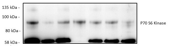
Detection of Human p70 S6 Kinase by Western Blot.
Western blot shows lysates of MCF-7 human breast cancer cell line. PVDF membrane was probed with 0.2 µg/mL of Rabbit Anti-Human p70 S6 Kinase Antigen Affinity-purified Polyclonal Antibody (Catalog # AF8962) followed by HRP-conjugated Anti-Rabbit IgG Secondary Antibody (HAF008). A specific band was detected for p70 S6 Kinase at approximately 70 and 90 kDa (as indicated). This experiment was conducted under reducing conditions and using Immunoblot Buffer Group 1.
Detection of p70 S6 Kinase in HeLa Human Cell Line by Flow Cytometry.
HeLa human cervical epithelial carcinoma cell line was stained with Rabbit Anti-Human p70 S6 Kinase Antigen Affinity-purified Polyclonal Antibody (Catalog # AF8962, filled histogram) or control antibody (AB-105-C, open histogram), followed by Phycoerythrin-conjugated Anti-Rabbit IgG Secondary Antibody (F0110). To facilitate intracellular staining, cells were fixed with paraformaldehyde and permeabilized with methanol.
p70 S6 Kinase in Human Breast Cancer Tissue.
p70 S6 Kinase was detected in immersion fixed paraffin-embedded sections of human breast cancer tissue using Rabbit Anti-Human p70 S6 Kinase Antigen Affinity-purified Polyclonal Antibody (Catalog # AF8962) at 1.7 µg/mL overnight at 4 °C. Tissue was stained using the Anti-Rabbit HRP-DAB Cell & Tissue Staining Kit (brown; CTS005) and counterstained with hematoxylin (blue). Specific labeling was localized to the nuclei of epithelial cells. View our protocol for Chromogenic IHC Staining of Paraffin-embedded Tissue Sections.
p70 S6 Kinase in Human Breast Cancer Tissue.
p70 S6 Kinase was detected in immersion fixed paraffin-embedded sections of human breast cancer tissue using Rabbit Anti-Human p70 S6 Kinase Antigen Affinity-purified Polyclonal Antibody (Catalog # AF8962) at 15 µg/mL overnight at 4 °C. Tissue was stained using the Anti-Rabbit HRP-DAB Cell & Tissue Staining Kit (brown; CTS005) and counterstained with hematoxylin (blue). Lower panel shows a lack of labeling if primary antibodies are omitted and tissue is stained only with secondary antibody followed by incubation with detection reagents. View our protocol for Chromogenic IHC Staining of Paraffin-embedded Tissue Sections.
Detection of p70 S6 Kinase by Simple WesternTM.
Simple Western lane view shows lysates of MCF-7 human breast cancer cell line, loaded at 0.2 mg/mL. A specific band was detected for p70 S6 Kinase at approximately 66 kDa (as indicated) using 2 µg/mL of Rabbit Anti-Human p70 S6 Kinase Antigen Affinity-purified Polyclonal Antibody (Catalog # AF8962). This experiment was conducted under reducing conditions and using the 12-230 kDa separation system.
Detection of Human p70 S6 Kinase/S6K by Western Blot
Time-dependent changes in autophagic induction with mild MPP+ exposure.(a) SH-SY5Y cells were exposed to 10 and 200 μM MPP+ for up to 48 h. Time-dependent changes in the phosphorylation levels of various proteins were estimated by western blotting. (b,c,d) SH-SY5Y cells were exposed to 10 and 200 μM MPP+ for 24 h (b), 36 h (c) or 48 h (d) with or without 200 nM bafilomycin A1 (Baf) for the last 4 h; LC3-II turnover was estimated by western blotting at various time points. (e,f,g) SH-SY5Y cells were exposed to 10 and 200 μM MPP+ for 24 h (e), 36 h (f) or 48 h (g) and subsequently immunostained with an anti-Atg16L antibody. The numbers of Atg16L-positive puncta per cell were evaluated at various time points. Data are expressed as means ± S.D. from at least three independent experiments. **p < 0.01. Image collected and cropped by CiteAb from the following publication (https://www.nature.com/articles/srep46668), licensed under a CC-BY license. Not internally tested by R&D Systems.Applications for Human p70 S6 Kinase Antibody
Application
Recommended Usage
CyTOF-ready
Ready to be labeled using established conjugation methods. No BSA or other carrier proteins that could interfere with conjugation.
Immunohistochemistry
5-15 µg/mL
Sample: Immersion fixed paraffin-embedded sections of human breast cancer tissue
Sample: Immersion fixed paraffin-embedded sections of human breast cancer tissue
Intracellular Staining by Flow Cytometry
2.5 µg/106 cells
Sample: HeLa human cervical epithelial carcinoma cell line fixed with paraformaldehyde and permeabilized with methanol
Sample: HeLa human cervical epithelial carcinoma cell line fixed with paraformaldehyde and permeabilized with methanol
Simple Western
2 µg/mL
Sample: MCF-7 human breast cancer cell line
Sample: MCF-7 human breast cancer cell line
Western Blot
0.2 µg/mL
Sample: MCF-7 human breast cancer cell line, and HeLa human cervical epithelial carcinoma cell line
Sample: MCF-7 human breast cancer cell line, and HeLa human cervical epithelial carcinoma cell line
Reviewed Applications
Read 3 reviews rated 5 using AF8962 in the following applications:
Flow Cytometry Panel Builder
Bio-Techne Knows Flow Cytometry
Save time and reduce costly mistakes by quickly finding compatible reagents using the Panel Builder Tool.
Advanced Features
- Spectra Viewer - Custom analysis of spectra from multiple fluorochromes
- Spillover Popups - Visualize the spectra of individual fluorochromes
- Antigen Density Selector - Match fluorochrome brightness with antigen density

Formulation, Preparation, and Storage
Purification
Antigen Affinity-purified
Reconstitution
Reconstitute at 0.2 mg/mL in sterile PBS. For liquid material, refer to CoA for concentration.
Loading...
Formulation
Lyophilized from a 0.2 μm filtered solution in PBS with Trehalose. See Certificate of Analysis for details.
*Small pack size (-SP) is supplied either lyophilized or as a 0.2 µm filtered solution in PBS.
*Small pack size (-SP) is supplied either lyophilized or as a 0.2 µm filtered solution in PBS.
Shipping
Lyophilized product is shipped at ambient temperature. Liquid small pack size (-SP) is shipped with polar packs. Upon receipt, store immediately at the temperature recommended below.
Stability & Storage
Use a manual defrost freezer and avoid repeated freeze-thaw cycles.
- 12 months from date of receipt, -20 to -70 °C as supplied.
- 1 month, 2 to 8 °C under sterile conditions after reconstitution.
- 6 months, -20 to -70 °C under sterile conditions after reconstitution.
Calculators
Background: p70 S6 Kinase
Alternate Names
p70-alpha, RPS6KB1, S6K1, STK14A
Gene Symbol
RPS6KB1
UniProt
Additional p70 S6 Kinase Products
Product Documents for Human p70 S6 Kinase Antibody
Certificate of Analysis
To download a Certificate of Analysis, please enter a lot or batch number in the search box below.
Note: Certificate of Analysis not available for kit components.
Product Specific Notices for Human p70 S6 Kinase Antibody
For research use only
Related Research Areas
Citations for Human p70 S6 Kinase Antibody
 Powered by Bioz
Customer Reviews for Human p70 S6 Kinase Antibody (3)
5 out of 5
3 Customer Ratings
Have you used Human p70 S6 Kinase Antibody?
Submit a review and receive an Amazon gift card!
$25/€18/£15/$25CAN/¥2500 Yen for a review with an image
$10/€7/£6/$10CAN/¥1110 Yen for a review without an image
Submit a review
Customer Images
Showing
1
-
3 of
3 reviews
Showing All
Filter By:
-
Application: Western BlotSample Tested: MDA-MB-231 human breast cancer cell lineSpecies: HumanVerified Customer | Posted 04/18/2022
-
Application: Western BlotSample Tested: Adipose tissueSpecies: MouseVerified Customer | Posted 10/23/2019

-
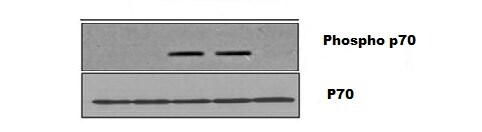
Application: Western BlotSample Tested: Cartilage tissueSpecies: MouseVerified Customer | Posted 08/14/2019Dilution 1:100 overnight incubation, we used the P70 as a baseline for Phospho-P70
There are no reviews that match your criteria.
Protocols
Find general support by application which include: protocols, troubleshooting, illustrated assays, videos and webinars.
- 7-Amino Actinomycin D (7-AAD) Cell Viability Flow Cytometry Protocol
- Antigen Retrieval Protocol (PIER)
- Antigen Retrieval for Frozen Sections Protocol
- Appropriate Fixation of IHC/ICC Samples
- Cellular Response to Hypoxia Protocols
- Chromogenic IHC Staining of Formalin-Fixed Paraffin-Embedded (FFPE) Tissue Protocol
- Chromogenic Immunohistochemistry Staining of Frozen Tissue
- ClariTSA™ Fluorophore Kits
- Detection & Visualization of Antibody Binding
- Extracellular Membrane Flow Cytometry Protocol
- Flow Cytometry Protocol for Cell Surface Markers
- Flow Cytometry Protocol for Staining Membrane Associated Proteins
- Flow Cytometry Staining Protocols
- Flow Cytometry Troubleshooting Guide
- Fluorescent IHC Staining of Frozen Tissue Protocol
- Graphic Protocol for Heat-induced Epitope Retrieval
- Graphic Protocol for the Preparation and Fluorescent IHC Staining of Frozen Tissue Sections
- Graphic Protocol for the Preparation and Fluorescent IHC Staining of Paraffin-embedded Tissue Sections
- Graphic Protocol for the Preparation of Gelatin-coated Slides for Histological Tissue Sections
- IHC Sample Preparation (Frozen sections vs Paraffin)
- Immunofluorescent IHC Staining of Formalin-Fixed Paraffin-Embedded (FFPE) Tissue Protocol
- Immunohistochemistry (IHC) and Immunocytochemistry (ICC) Protocols
- Immunohistochemistry Frozen Troubleshooting
- Immunohistochemistry Paraffin Troubleshooting
- Intracellular Flow Cytometry Protocol Using Alcohol (Methanol)
- Intracellular Flow Cytometry Protocol Using Detergents
- Intracellular Nuclear Staining Flow Cytometry Protocol Using Detergents
- Intracellular Staining Flow Cytometry Protocol Using Alcohol Permeabilization
- Intracellular Staining Flow Cytometry Protocol Using Detergents to Permeabilize Cells
- Preparing Samples for IHC/ICC Experiments
- Preventing Non-Specific Staining (Non-Specific Binding)
- Primary Antibody Selection & Optimization
- Propidium Iodide Cell Viability Flow Cytometry Protocol
- Protocol for Heat-Induced Epitope Retrieval (HIER)
- Protocol for Liperfluo
- Protocol for Making a 4% Formaldehyde Solution in PBS
- Protocol for VisUCyte™ HRP Polymer Detection Reagent
- Protocol for the Characterization of Human Th22 Cells
- Protocol for the Characterization of Human Th9 Cells
- Protocol for the Preparation & Fixation of Cells on Coverslips
- Protocol for the Preparation and Chromogenic IHC Staining of Frozen Tissue Sections
- Protocol for the Preparation and Chromogenic IHC Staining of Frozen Tissue Sections - Graphic
- Protocol for the Preparation and Chromogenic IHC Staining of Paraffin-embedded Tissue Sections
- Protocol for the Preparation and Chromogenic IHC Staining of Paraffin-embedded Tissue Sections - Graphic
- Protocol for the Preparation and Fluorescent IHC Staining of Frozen Tissue Sections
- Protocol for the Preparation and Fluorescent IHC Staining of Paraffin-embedded Tissue Sections
- Protocol for the Preparation of Gelatin-coated Slides for Histological Tissue Sections
- Protocol: Annexin V and PI Staining by Flow Cytometry
- Protocol: Annexin V and PI Staining for Apoptosis by Flow Cytometry
- R&D Systems Quality Control Western Blot Protocol
- TUNEL and Active Caspase-3 Detection by IHC/ICC Protocol
- The Importance of IHC/ICC Controls
- Troubleshooting Guide: Fluorokine Flow Cytometry Kits
- Troubleshooting Guide: Immunohistochemistry
- Troubleshooting Guide: Western Blot Figures
- Western Blot Conditions
- Western Blot Protocol
- Western Blot Protocol for Cell Lysates
- Western Blot Troubleshooting
- Western Blot Troubleshooting Guide
- View all Protocols, Troubleshooting, Illustrated assays and Webinars
Loading...
Associated Pathways


IL-7 Signaling Pathways
 IL-9 Signaling Pathways
 IL-15 Signaling Pathways
 IL-21 Signaling Pathways
 MAPK Signaling Pathway: Mitogen Stimulation Pathway
 mTOR Signaling Pathway
 TGF-beta Signaling Pathways
