Human PD-1 Antibody (2335A)
R&D Systems | Catalog # MAB10863

Key Product Details
Validated by
Species Reactivity
Applications
Label
Antibody Source
Product Specifications
Immunogen
Met1-Gln167
Accession # Q15116
Specificity
Clonality
Host
Isotype
Scientific Data Images for Human PD-1 Antibody (2335A)
Detection of Human PD‑1 by Western Blot.
Western blot shows lysates of HEK293T human embryonic kidney cell line either mock transfected or transfected with human PD-1. PVDF membrane was probed with 0.5 µg/mL of Rabbit Anti-Human PD-1 Monoclonal Antibody (Catalog # MAB10863) followed by HRP-conjugated Anti-Rabbit IgG Secondary Antibody (Catalog # HAF008). Specific bands were detected for PD-1 at approximately 40 kDa and 80 kDa (as indicated). This experiment was conducted under reducing conditions and using Immunoblot Buffer Group 1.
Detection of PD‑1 in Human PBMCs by Flow Cytometry.
Human peripheral blood mononuclear cells (PBMCs) either (A) treated with 5 ug/mL PHA for 5 days or (B) untreated, were stained with Rabbit Anti-Human PD-1 Monoclonal Antibody (Catalog # MAB10863) followed by Allophycocyanin-conjugated Anti-Rabbit IgG Secondary Antibody (Catalog # F0111) and Mouse Anti-Human CD3 epsilon PE-conjugated Monoclonal Antibody (Catalog # FAB100P). Quadrant markers were set based on control antibody staining (Catalog # MAB1050). View our protocol for Staining Membrane-associated Proteins.
Human PD‑1 ELISA Standard Curve.
Recombinant Human PD-1 protein was serially diluted 2-fold and captured by Rabbit Anti-Human PD-1 Monoclonal Antibody (Catalog # MAB10863) coated on a Clear Polystyrene Microplate (Catalog # DY990). Mouse Anti-Human PD-1 Monoclonal Antibody(Catalog # MAB10864) was biotinylated and incubated with the protein captured on the plate. Detection of the standard curve was achieved by incubating Streptavidin-HRP (Catalog # DY998) followed by Substrate Solution (Catalog # DY999) and stopping the enzymatic reaction with Stop Solution (Catalog # DY994).Applications for Human PD-1 Antibody (2335A)
CyTOF-ready
ELISA
This antibody functions as an ELISA capture antibody when paired with Mouse Anti-Human PD‑1 Monoclonal Antibody(Catalog # MAB10864).
This product is intended for assay development on various assay platforms requiring antibody pairs. We recommend the Human PD-1 DuoSet ELISA Kit (Catalog # DY1086) for convenient development of a sandwich ELISA.
Flow Cytometry
Sample: Human PBMC treated with PHA
Western Blot
Sample: HEK293T human embryonic kidney cell line transfected with human PD-1
Reviewed Applications
Read 1 review rated 4 using MAB10863 in the following applications:
Flow Cytometry Panel Builder
Bio-Techne Knows Flow Cytometry
Save time and reduce costly mistakes by quickly finding compatible reagents using the Panel Builder Tool.
Advanced Features
- Spectra Viewer - Custom analysis of spectra from multiple fluorochromes
- Spillover Popups - Visualize the spectra of individual fluorochromes
- Antigen Density Selector - Match fluorochrome brightness with antigen density

Formulation, Preparation, and Storage
Purification
Reconstitution
Reconstitute at 0.5 mg/mL in sterile PBS. For liquid material, refer to CoA for concentration.
Formulation
Shipping
Stability & Storage
- 12 months from date of receipt, -20 to -70 °C as supplied.
- 1 month, 2 to 8 °C under sterile conditions after reconstitution.
- 6 months, -20 to -70 °C under sterile conditions after reconstitution.
Calculators
Background: PD-1
References
- Ishida, Y. et al. (1992) EMBO J. 11:3887.
- Sharpe, A.H. and G. J. Freeman (2002) Nat. Rev. Immunol. 2:116.
- Coyle, A. and J. Gutierrez-Ramos (2001) Nat. Immunol. 2:203.
- Nishimura, H. and T. Honjo (2001) Trends Immunol. 22:265.
- Watanabe, N et al. (2003) Nat. Immunol. 4:670.
- Zhang, X. et al. (2004) Immunity 20:337.
- Lázár-Molnár, E. et al. (2008) Proc. Natl. Acad. Sci. USA 105:10483.
- Nishimura, H et al. (1996) Int. Immunol. 8:773.
- Keir, M.E. et al. (2008) Annu. Rev. Immunol. 26:677.
- Butte, M.J. et al. (2007) Immunity 27:111.
- Okazaki, T. et al. (2013) Nat. Immunol. 14:1212.
- Iwai, Y. et al. (2002) Proc. Natl. Acad. Sci. USA 99: 12293.
- Nogrady, B. (2014) Nature 513:S10.
- Swaika, A. et al. (2015) Mol. Immunol. 67: 4
Long Name
Alternate Names
Entrez Gene IDs
Gene Symbol
UniProt
Additional PD-1 Products
Product Documents for Human PD-1 Antibody (2335A)
Certificate of Analysis
To download a Certificate of Analysis, please enter a lot or batch number in the search box below.
Note: Certificate of Analysis not available for kit components.
Product Specific Notices for Human PD-1 Antibody (2335A)
For research use only
Citations for Human PD-1 Antibody (2335A)
 Powered by Bioz
Customer Reviews for Human PD-1 Antibody (2335A) (1)
Have you used Human PD-1 Antibody (2335A)?
Submit a review and receive an Amazon gift card!
$25/€18/£15/$25CAN/¥2500 Yen for a review with an image
$10/€7/£6/$10CAN/¥1110 Yen for a review without an image
Submit a review
Customer Images
-
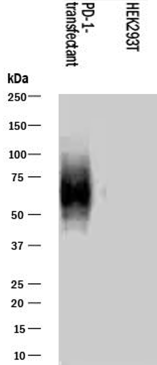
Application: Western BlotSample Tested: 293T human embryonic kidney cell lineSpecies: HumanVerified Customer | Posted 08/28/2023A Western blot analysis was performed using lysates from the HEK293T human embryonic kidney cell line that had been transfected with human PD-1. A PVDF membrane was utilized and probed with the antibody at a concentration of 0.5 µg/mL. The bands corresponding to PD-1 were observed at around 70 kDa. The experimental conditions involved conducting the assay under reducing conditions.

There are no reviews that match your criteria.
Protocols
Find general support by application which include: protocols, troubleshooting, illustrated assays, videos and webinars.
- 7-Amino Actinomycin D (7-AAD) Cell Viability Flow Cytometry Protocol
- Cellular Response to Hypoxia Protocols
- ELISA Sample Preparation & Collection Guide
- ELISA Troubleshooting Guide
- Extracellular Membrane Flow Cytometry Protocol
- Flow Cytometry Protocol for Cell Surface Markers
- Flow Cytometry Protocol for Staining Membrane Associated Proteins
- Flow Cytometry Staining Protocols
- Flow Cytometry Troubleshooting Guide
- How to Run an R&D Systems DuoSet ELISA
- How to Run an R&D Systems Quantikine ELISA
- How to Run an R&D Systems Quantikine™ QuicKit™ ELISA
- Intracellular Flow Cytometry Protocol Using Alcohol (Methanol)
- Intracellular Flow Cytometry Protocol Using Detergents
- Intracellular Nuclear Staining Flow Cytometry Protocol Using Detergents
- Intracellular Staining Flow Cytometry Protocol Using Alcohol Permeabilization
- Intracellular Staining Flow Cytometry Protocol Using Detergents to Permeabilize Cells
- Propidium Iodide Cell Viability Flow Cytometry Protocol
- Protocol for Liperfluo
- Protocol for the Characterization of Human Th22 Cells
- Protocol for the Characterization of Human Th9 Cells
- Protocol: Annexin V and PI Staining by Flow Cytometry
- Protocol: Annexin V and PI Staining for Apoptosis by Flow Cytometry
- Quantikine HS ELISA Kit Assay Principle, Alkaline Phosphatase
- Quantikine HS ELISA Kit Principle, Streptavidin-HRP Polymer
- R&D Systems Quality Control Western Blot Protocol
- Sandwich ELISA (Colorimetric) – Biotin/Streptavidin Detection Protocol
- Sandwich ELISA (Colorimetric) – Direct Detection Protocol
- Troubleshooting Guide: ELISA
- Troubleshooting Guide: Fluorokine Flow Cytometry Kits
- Troubleshooting Guide: Western Blot Figures
- Western Blot Conditions
- Western Blot Protocol
- Western Blot Protocol for Cell Lysates
- Western Blot Troubleshooting
- Western Blot Troubleshooting Guide
- View all Protocols, Troubleshooting, Illustrated assays and Webinars