
Key Product Details
Species Reactivity
Validated:
Human, Primate
Cited:
Human, Mouse
Applications
Validated:
Immunohistochemistry, Western Blot, ELISA Capture (Matched Antibody Pair), Immunoprecipitation
Cited:
Immunohistochemistry, Immunohistochemistry-Paraffin, Immunohistochemistry-Frozen, Western Blot, ELISA Development
Label
Unconjugated
Antibody Source
Polyclonal Goat IgG
Loading...
Product Specifications
Immunogen
Mouse myeloma cell line NS0-derived recombinant human MMP‑3
Tyr18-Cys477 (Lys45Glu)
Accession # P08254
Tyr18-Cys477 (Lys45Glu)
Accession # P08254
Specificity
Detects human and primate MMP-3 in ELISAs and Western blots. In Direct ELISA's, less than 10% cross-reactivity with recombinant human (rh) MMP‑10 is observed and less than 1% cross-reactivity with rhMMP-1, -2, -7, -8, -9, -12, -13, and recombinant mouse MMP‑9 is observed.
Clonality
Polyclonal
Host
Goat
Isotype
IgG
Scientific Data Images for MMP-3 Antibody
Detection of Human MMP‑3 by Western Blot.
Western blot shows Recombinant Human MMP-3 Western Blot Standard Protein (WBC015) and lysate of U-118-MG human glioblastoma/astrocytoma cell line. PVDF membrane was probed with 2 µg/mL of Goat Anti-Human/Primate MMP-3 Antigen Affinity-purified Polyclonal Antibody (Catalog # AF513) followed by HRP-conjugated Anti-Goat IgG Secondary Antibody (HAF017). A specific band was detected for MMP-3 at approximately 57-60 kDa (as indicated). This experiment was conducted under reducing conditions and using Immunoblot Buffer Group 1.
MMP‑3 in Human Bladder Cancer Tissue.
MMP-3 was detected in immersion fixed paraffin-embedded sections of human bladder cancer tissue using Goat Anti-Human/ Primate MMP-3 Antigen Affinity-purified Polyclonal Antibody (Catalog # AF513) at 10 µg/mL overnight at 4 °C. Tissue was stained using the Anti-Goat HRP-DAB Cell & Tissue Staining Kit (brown; CTS008) and counterstained with hematoxylin (blue). Specific staining was localized to epithelial cells. View our protocol for Chromogenic IHC Staining of Paraffin-embedded Tissue Sections.
MMP‑3 in Human Lung Cancer.
MMP-3 was detected in immersion fixed paraffin-embedded sections of human lung cancer using Goat Anti-Human/Primate MMP-3 Antigen Affinity-purified Polyclonal Antibody (Catalog # AF513) at 15 µg/mL overnight at 4 °C. Tissue was stained using the Anti-Goat HRP-DAB Cell & Tissue Staining Kit (brown; CTS008) and counterstained with hematoxylin (blue). Specific staining was localized to cytoplasm. View our protocol for Chromogenic IHC Staining of Paraffin-embedded Tissue Sections.Applications for MMP-3 Antibody
Application
Recommended Usage
Immunohistochemistry
5-15 µg/mL
Sample: Immersion fixed paraffin-embedded sections of human lung cancer, human colon cancer tissue, and human bladder cancer tissue
Sample: Immersion fixed paraffin-embedded sections of human lung cancer, human colon cancer tissue, and human bladder cancer tissue
Immunoprecipitation
25 µg/mL
Sample: Conditioned cell culture medium spiked with Recombinant Human MMP‑3 (Catalog # 513-MP), see our available Western blot detection antibodies
Sample: Conditioned cell culture medium spiked with Recombinant Human MMP‑3 (Catalog # 513-MP), see our available Western blot detection antibodies
Western Blot
2 µg/mL
Sample: Recombinant Human MMP-3 Western Blot Standard Proteins (Catalog # WBC015) and lysates of U‑118‑MG human glioblastoma/astrocytoma cell line
Sample: Recombinant Human MMP-3 Western Blot Standard Proteins (Catalog # WBC015) and lysates of U‑118‑MG human glioblastoma/astrocytoma cell line
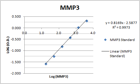
Human/Primate MMP-3 Sandwich Immunoassay
Please Note: Optimal dilutions of this antibody should be experimentally determined.
Reviewed Applications
Read 1 review rated 4 using AF513 in the following applications:
Formulation, Preparation, and Storage
Purification
Antigen Affinity-purified
Reconstitution
Reconstitute at 0.2 mg/mL in sterile PBS. For liquid material, refer to CoA for concentration.
Loading...
Formulation
Lyophilized from a 0.2 μm filtered solution in PBS with Trehalose. See Certificate of Analysis for details.
*Small pack size (-SP) is supplied either lyophilized or as a 0.2 µm filtered solution in PBS.
*Small pack size (-SP) is supplied either lyophilized or as a 0.2 µm filtered solution in PBS.
Shipping
Lyophilized product is shipped at ambient temperature. Liquid small pack size (-SP) is shipped with polar packs. Upon receipt, store immediately at the temperature recommended below.
Stability & Storage
Use a manual defrost freezer and avoid repeated freeze-thaw cycles.
- 12 months from date of receipt, -20 to -70 °C as supplied.
- 1 month, 2 to 8 °C under sterile conditions after reconstitution.
- 6 months, -20 to -70 °C under sterile conditions after reconstitution.
Calculators
Background: MMP-3
Long Name
Matrix Metalloproteinase 3
Alternate Names
MMP3, Stromelysin 1
Gene Symbol
MMP3
UniProt
Additional MMP-3 Products
Product Documents for MMP-3 Antibody
Certificate of Analysis
To download a Certificate of Analysis, please enter a lot or batch number in the search box below.
Note: Certificate of Analysis not available for kit components.
Product Specific Notices for MMP-3 Antibody
For research use only
Related Research Areas
Citations for MMP-3 Antibody
 Powered by Bioz
Customer Reviews for MMP-3 Antibody (1)
4 out of 5
1 Customer Rating
Have you used MMP-3 Antibody?
Submit a review and receive an Amazon gift card!
$25/€18/£15/$25CAN/¥2500 Yen for a review with an image
$10/€7/£6/$10CAN/¥1110 Yen for a review without an image
Submit a review
Customer Images
Showing
1
-
1 of
1 review
Showing All
Filter By:
-
Application: ELISASample Tested: Serum and PlasmaSpecies: HumanVerified Customer | Posted 11/09/2017An ELISA was built using this antibody as both the capture and detection molecule. MMP-3 in human serum samples was measured using this assay.

There are no reviews that match your criteria.
Protocols
Find general support by application which include: protocols, troubleshooting, illustrated assays, videos and webinars.
- Antigen Retrieval Protocol (PIER)
- Antigen Retrieval for Frozen Sections Protocol
- Appropriate Fixation of IHC/ICC Samples
- Cellular Response to Hypoxia Protocols
- Chromogenic IHC Staining of Formalin-Fixed Paraffin-Embedded (FFPE) Tissue Protocol
- Chromogenic Immunohistochemistry Staining of Frozen Tissue
- ClariTSA™ Fluorophore Kits
- Detection & Visualization of Antibody Binding
- Fluorescent IHC Staining of Frozen Tissue Protocol
- Graphic Protocol for Heat-induced Epitope Retrieval
- Graphic Protocol for the Preparation and Fluorescent IHC Staining of Frozen Tissue Sections
- Graphic Protocol for the Preparation and Fluorescent IHC Staining of Paraffin-embedded Tissue Sections
- Graphic Protocol for the Preparation of Gelatin-coated Slides for Histological Tissue Sections
- IHC Sample Preparation (Frozen sections vs Paraffin)
- Immunofluorescent IHC Staining of Formalin-Fixed Paraffin-Embedded (FFPE) Tissue Protocol
- Immunohistochemistry (IHC) and Immunocytochemistry (ICC) Protocols
- Immunohistochemistry Frozen Troubleshooting
- Immunohistochemistry Paraffin Troubleshooting
- Immunoprecipitation Protocol
- Preparing Samples for IHC/ICC Experiments
- Preventing Non-Specific Staining (Non-Specific Binding)
- Primary Antibody Selection & Optimization
- Protocol for Heat-Induced Epitope Retrieval (HIER)
- Protocol for Making a 4% Formaldehyde Solution in PBS
- Protocol for VisUCyte™ HRP Polymer Detection Reagent
- Protocol for the Preparation & Fixation of Cells on Coverslips
- Protocol for the Preparation and Chromogenic IHC Staining of Frozen Tissue Sections
- Protocol for the Preparation and Chromogenic IHC Staining of Frozen Tissue Sections - Graphic
- Protocol for the Preparation and Chromogenic IHC Staining of Paraffin-embedded Tissue Sections
- Protocol for the Preparation and Chromogenic IHC Staining of Paraffin-embedded Tissue Sections - Graphic
- Protocol for the Preparation and Fluorescent IHC Staining of Frozen Tissue Sections
- Protocol for the Preparation and Fluorescent IHC Staining of Paraffin-embedded Tissue Sections
- Protocol for the Preparation of Gelatin-coated Slides for Histological Tissue Sections
- R&D Systems Quality Control Western Blot Protocol
- TUNEL and Active Caspase-3 Detection by IHC/ICC Protocol
- The Importance of IHC/ICC Controls
- Troubleshooting Guide: Immunohistochemistry
- Troubleshooting Guide: Western Blot Figures
- Western Blot Conditions
- Western Blot Protocol
- Western Blot Protocol for Cell Lysates
- Western Blot Troubleshooting
- Western Blot Troubleshooting Guide
- View all Protocols, Troubleshooting, Illustrated assays and Webinars
Loading...