Pro Matrix Metalloproteinase 9 (Pro-MMP-9) is an inactive proenzyme secreted by multiple cell types. The N-terminal pro region is proteolytically removed, resulting in active MMP-9, which can degrade collagens and elastin as well as several non-ECM molecules.

Key Product Details
Species Reactivity
Validated:
Human, Primate
Cited:
Human, Primate - Macaca mulatta (Rhesus Macaque)
Applications
Validated:
Immunohistochemistry, Western Blot, ELISA Capture (Matched Antibody Pair), Immunocytochemistry, Immunoprecipitation
Cited:
Immunohistochemistry, Immunohistochemistry-Paraffin, Western Blot, Flow Cytometry, Immunocytochemistry, ELISA Capture, ELISA Development (Capture)
Label
Unconjugated
Antibody Source
Monoclonal Mouse IgG1 Clone # 36020
Loading...
Product Specifications
Immunogen
Chinese hamster ovary cell line CHO-derived recombinant human MMP-9
Specificity
Detects human MMP-9 in Western blots. Does not react with the C-terminal truncated form (65 kDa) of recombinant human (rh) MMP-9. In Western blots, approximately 5% cross-reactivity with rhMMP-2 and no cross-reactivity with rhMMP-1 or rhMMP-3 is observed.
Clonality
Monoclonal
Host
Mouse
Isotype
IgG1
Scientific Data Images for MMP-9 Antibody (36020)
MMP‑9 in SW1353 Human Cell Line.
MMP-9 was detected in immersion fixed SW1353 human chondrosarcoma cell line using Mouse Anti-Human/Primate MMP-9 Monoclonal Antibody (Catalog # MAB936) at 3 µg/mL for 3 hours at room temperature. Cells were stained using the NorthernLights™ 557-conjugated Anti-Mouse IgG Secondary Antibody (red; Catalog # NL007) and counterstained with DAPI (blue). Specific staining was localized to cytoplasm. View our protocol for Fluorescent ICC Staining of Cells on Coverslips.
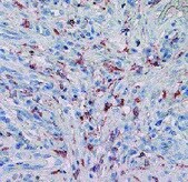
MMP‑9 in Human Lung.
MMP-9 was detected in immersion fixed paraffin-embedded sections of human lung using Mouse Anti-Human/Primate MMP-9 Monoclonal Antibody (Catalog # MAB936) at 15 µg/mL for 1 hour at room temperature followed by incubation with the Anti-Mouse IgG VisUCyte™ HRP Polymer Antibody (Catalog # VC001). Tissue was stained using DAB (brown) and counterstained with hematoxylin (blue). Specific staining was localized to cytoplasm in macrophages. View our protocol for IHC Staining with VisUCyte HRP Polymer Detection Reagents.
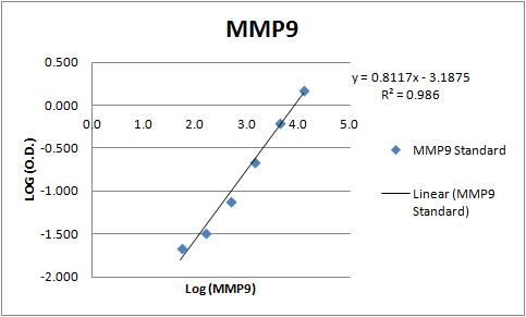
Human MMP-9 ELISA Standard Curve
Recombinant Human MMP‑9 (Catalog # 911-MP) was serially diluted and captured by Mouse Anti-Human/Primate MMP‑9 Monoclonal Antibody (Catalog # MAB936) coated on a Clear Polystyrene Microplate (Catalog # DY990). Goat Anti-Human MMP‑9 Antigen Affinity-purified Polyclonal Antibody (Catalog # AF911) was biotinylated and incubated with the protein captured on the plate. Detection of the standard curve was achieved by incubating Streptavidin-HRP (Catalog # DY998)Applications for MMP-9 Antibody (36020)
Application
Recommended Usage
Immunocytochemistry
3-25 µg/mL
Sample: Immersion fixed SW1353 human chondrosarcoma cell line
Sample: Immersion fixed SW1353 human chondrosarcoma cell line
Immunohistochemistry
8-25 µg/mL
Sample:
Sample:
Immersion fixed paraffin-embedded sections of human lung
Immunoprecipitation
25 µg/mL
Sample: Conditioned cell culture medium spiked with Recombinant Human MMP‑9 (Catalog # 911-MP), see our available Western blot detection antibodies
Sample: Conditioned cell culture medium spiked with Recombinant Human MMP‑9 (Catalog # 911-MP), see our available Western blot detection antibodies
Western Blot
1 µg/mL
Sample: Recombinant Human MMP‑9 Western Blot Standard (Catalog # WBC018)
Sample: Recombinant Human MMP‑9 Western Blot Standard (Catalog # WBC018)
Human/Primate MMP-9 Sandwich Immunoassay
Please Note: Optimal dilutions of this antibody should be experimentally determined.
Reviewed Applications
Read 3 reviews rated 4.3 using MAB936 in the following applications:
Formulation, Preparation, and Storage
Purification
Protein A or G purified from hybridoma culture supernatant
Reconstitution
Reconstitute at 0.5 mg/mL in sterile PBS. For liquid material, refer to CoA for concentration.
Loading...
Formulation
Lyophilized from a 0.2 μm filtered solution in PBS with Trehalose. *Small pack size (SP) is supplied either lyophilized or as a 0.2 µm filtered solution in PBS.
Shipping
Lyophilized product is shipped at ambient temperature. Liquid small pack size (-SP) is shipped with polar packs. Upon receipt, store immediately at the temperature recommended below.
Stability & Storage
Use a manual defrost freezer and avoid repeated freeze-thaw cycles.
- 12 months from date of receipt, -20 to -70 °C as supplied.
- 1 month, 2 to 8 °C under sterile conditions after reconstitution.
- 6 months, -20 to -70 °C under sterile conditions after reconstitution.
Calculators
Background: MMP-9
Long Name
Matrix Metalloproteinase 9
Alternate Names
CLG4B, Gelatinase B, GELB, MANDP2, MMP9
Gene Symbol
MMP9
Additional MMP-9 Products
Product Documents for MMP-9 Antibody (36020)
Certificate of Analysis
To download a Certificate of Analysis, please enter a lot or batch number in the search box below.
Note: Certificate of Analysis not available for kit components.
Product Specific Notices for MMP-9 Antibody (36020)
For research use only
Citations for MMP-9 Antibody (36020)
 Powered by Bioz
Customer Reviews for MMP-9 Antibody (36020) (3)
4.3 out of 5
3 Customer Ratings
Have you used MMP-9 Antibody (36020)?
Submit a review and receive an Amazon gift card!
$25/€18/£15/$25CAN/¥2500 Yen for a review with an image
$10/€7/£6/$10CAN/¥1110 Yen for a review without an image
Submit a review
Customer Images
Showing
1
-
3 of
3 reviews
Showing All
Filter By:
-
Application: ImmunohistochemistrySample Tested: Squamous cell carcinomaSpecies: HumanVerified Customer | Posted 10/04/2021

-
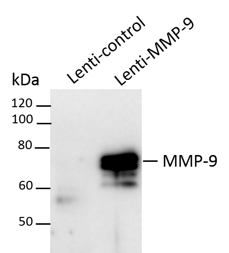
Application: Western BlotSample Tested: 293T human embryonic kidney cell lineSpecies: HumanVerified Customer | Posted 07/23/2018293T cells were infected by lentivirus overexpression of control or MMP-9 for 72h. Total cell lysates were subjected to western blot. PVDF membrane were probed with 1 um/ml Human MMP-9 Antibody (MAB936). A specific band was detected for MMP-9 at approximately 74 kDa. This experiment was conducted under reducing conditions
-
Application: ELISASample Tested: Serum and PlasmaSpecies: HumanVerified Customer | Posted 11/10/2017A sandwich ELISA was built using this antibody as the capture and AF911 as the detection antibody. MMP-9 was measured in human serum and plasma samples.
There are no reviews that match your criteria.
Protocols
Find general support by application which include: protocols, troubleshooting, illustrated assays, videos and webinars.
- Antigen Retrieval Protocol (PIER)
- Antigen Retrieval for Frozen Sections Protocol
- Appropriate Fixation of IHC/ICC Samples
- Cellular Response to Hypoxia Protocols
- Chromogenic IHC Staining of Formalin-Fixed Paraffin-Embedded (FFPE) Tissue Protocol
- Chromogenic Immunohistochemistry Staining of Frozen Tissue
- ClariTSA™ Fluorophore Kits
- Detection & Visualization of Antibody Binding
- Fluorescent IHC Staining of Frozen Tissue Protocol
- Graphic Protocol for Heat-induced Epitope Retrieval
- Graphic Protocol for the Preparation and Fluorescent IHC Staining of Frozen Tissue Sections
- Graphic Protocol for the Preparation and Fluorescent IHC Staining of Paraffin-embedded Tissue Sections
- Graphic Protocol for the Preparation of Gelatin-coated Slides for Histological Tissue Sections
- ICC Cell Smear Protocol for Suspension Cells
- ICC Immunocytochemistry Protocol Videos
- ICC for Adherent Cells
- IHC Sample Preparation (Frozen sections vs Paraffin)
- Immunocytochemistry (ICC) Protocol
- Immunocytochemistry Troubleshooting
- Immunofluorescence of Organoids Embedded in Cultrex Basement Membrane Extract
- Immunofluorescent IHC Staining of Formalin-Fixed Paraffin-Embedded (FFPE) Tissue Protocol
- Immunohistochemistry (IHC) and Immunocytochemistry (ICC) Protocols
- Immunohistochemistry Frozen Troubleshooting
- Immunohistochemistry Paraffin Troubleshooting
- Immunoprecipitation Protocol
- Preparing Samples for IHC/ICC Experiments
- Preventing Non-Specific Staining (Non-Specific Binding)
- Primary Antibody Selection & Optimization
- Protocol for Heat-Induced Epitope Retrieval (HIER)
- Protocol for Making a 4% Formaldehyde Solution in PBS
- Protocol for VisUCyte™ HRP Polymer Detection Reagent
- Protocol for the Fluorescent ICC Staining of Cell Smears - Graphic
- Protocol for the Fluorescent ICC Staining of Cultured Cells on Coverslips - Graphic
- Protocol for the Preparation & Fixation of Cells on Coverslips
- Protocol for the Preparation and Chromogenic IHC Staining of Frozen Tissue Sections
- Protocol for the Preparation and Chromogenic IHC Staining of Frozen Tissue Sections - Graphic
- Protocol for the Preparation and Chromogenic IHC Staining of Paraffin-embedded Tissue Sections
- Protocol for the Preparation and Chromogenic IHC Staining of Paraffin-embedded Tissue Sections - Graphic
- Protocol for the Preparation and Fluorescent ICC Staining of Cells on Coverslips
- Protocol for the Preparation and Fluorescent ICC Staining of Non-adherent Cells
- Protocol for the Preparation and Fluorescent ICC Staining of Stem Cells on Coverslips
- Protocol for the Preparation and Fluorescent IHC Staining of Frozen Tissue Sections
- Protocol for the Preparation and Fluorescent IHC Staining of Paraffin-embedded Tissue Sections
- Protocol for the Preparation of Gelatin-coated Slides for Histological Tissue Sections
- Protocol for the Preparation of a Cell Smear for Non-adherent Cell ICC - Graphic
- R&D Systems Quality Control Western Blot Protocol
- TUNEL and Active Caspase-3 Detection by IHC/ICC Protocol
- The Importance of IHC/ICC Controls
- Troubleshooting Guide: Immunohistochemistry
- Troubleshooting Guide: Western Blot Figures
- Western Blot Conditions
- Western Blot Protocol
- Western Blot Protocol for Cell Lysates
- Western Blot Troubleshooting
- Western Blot Troubleshooting Guide
- View all Protocols, Troubleshooting, Illustrated assays and Webinars


