Human PSGL-1 (P-Selectin Glycoprotein Ligand-1; also CD162), is a 120 kDa mucin-type glycoprotein that plays a key role in leukocyte adhesion (1-3). It is synthesized as a 412 amino acid (aa) preproprecursor that contains a 17 aa signal sequence, a 24 aa propeptide, a 279 aa extracellular domain (ECD), a 21 aa transmembrane segment and a 71 aa cytoplasmic region (4, 5). Following cleavage of the pre- and prosegments, it is expressed as a 240 kDa disulfide-linked homodimer. The extreme N-terminus (aa 1-16 of the mature molecule) contains one threonine (aa 16) and three tyrosines (aa 5, 7, and 10) that are involved in ligand binding. The Thr residue allows for O-linked glycosylation in the form of a core-2 structure (GalNAc-Gal) linked in a beta 1,6 bond to a sialylated Lewis X motif (GlcNAc linked to both Fuc and Gal with a terminal sialic acid residue) (1, 2, 5, 6, 7). The three tyrosine residues allow for sulfation (8, 9). When binding to P-selectin, Tyr sulfation and glycosylation are essential. Tyr7 provides the most efficient sulfate moiety, while Fuc and sialic acid are essentially mandatory (7). When binding to E-selectin, only carbohydrate is needed, while both carbohydrate and Tyr10 are used for L-selectin binding (6, 8). There are 16 decameric aa repeats in the ECD of the longform of PSGL-1. This form is referred to as the A allele, and represents 65 - 80% of the population. Alleles B and C show deletions of decameric repeats #2 (aa 132-141) plus #9 and 10 (aa 222-241), respectively. Shorter forms may show weaker binding to P-selectin (9, 10). Soluble forms of PSGL-1 are also known. Neutrophil elastase will cleave somewhere within repeats #5-9, while cathepsin G cleaves after Tyr7 (11). The loss of Tyr5 and 7 should impact binding affinity. PSGL-1 is found on virtually all leukocytes and macrophages/DC’s (1). Although there is similarity in the organization of the ECD between species, there is little aa identity. Human PSGL-1 ECD shares 51%, 52% and 43% aa sequence identity with equine, canine and mouse ECD, respectively.
Human PSGL-1/CD162 Antibody (688101)
R&D Systems | Catalog # MAB9961

Key Product Details
Species Reactivity
Validated:
Human
Cited:
Human
Applications
Validated:
Immunohistochemistry, Western Blot, Flow Cytometry, Immunocytochemistry, CyTOF-ready
Cited:
Flow Cytometry
Label
Unconjugated
Antibody Source
Monoclonal Mouse IgG2A Clone # 688101
Loading...
Product Specifications
Immunogen
Chinese hamster ovary cell line CHO-derived recombinant human PSGL-1/CD162
Gln42-Gly295
Accession # NP_002997
Gln42-Gly295
Accession # NP_002997
Specificity
Detects human PSGL-1/CD162 in direct ELISAs and Western blots.
Clonality
Monoclonal
Host
Mouse
Isotype
IgG2A
Scientific Data Images for Human PSGL-1/CD162 Antibody (688101)
Detection of Human PSGL‑1/CD162 by Western Blot.
Western blot shows lysates of Jurkat human acute T cell leukemia cell line. PVDF membrane was probed with 2 µg/mL of Mouse Anti-Human PSGL-1/CD162 Monoclonal Antibody (Catalog # MAB9961) followed by HRP-conjugated Anti-Mouse IgG Secondary Antibody (Catalog # HAF018). Specific bands were detected for PSGL1/ CD162 homodimer at approximately 250 kDa and PSGL1/ CD162 monomer at approximately 110 kDa (as indicated). This experiment was conducted under reducing conditions and using Immunoblot Buffer Group 1.
Detection of PSGL-1 in Human Peripheral Blood Monocytes by Flow Cytometry.
Human peripheral blood monocytes were stained with Mouse Anti-Human PSGL-1/CD162 Monoclonal Antibody (Catalog # MAB9961, filled histogram) or isotype control antibody (Catalog # MAB003, open histogram), followed by Allophycocyanin-conjugated Anti-Mouse IgG Secondary Antibody (Catalog # F0101B).
PSGL‑1/CD162 in Human PBMCs.
PSGL-1/CD162 was detected in immersion fixed human peripheral blood mononuclear cells (PBMCs) using Mouse Anti-Human PSGL-1/CD162 Monoclonal Antibody (Catalog # MAB9961) at 8 µg/mL for 3 hours at room temperature. Cells were stained using the NorthernLights™ 557-conjugated Anti-Mouse IgG Secondary Antibody (red; Catalog # NL007) and counterstained with DAPI (blue). Specific staining was localized to cytoplasm. View our protocol for Fluorescent ICC Staining of Non-adherent Cells.
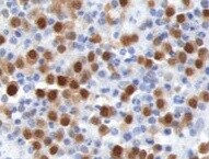
PSGL‑1/CD162 in Human Tonsil.
PSGL-1/CD162 was detected in immersion fixed paraffin-embedded sections of human tonsil using Mouse Anti-Human PSGL-1/CD162 Monoclonal Antibody (Catalog # MAB9961) at 5 µg/mL for 1 hour at room temperature followed by incubation with the Anti-Mouse IgG VisUCyte™ HRP Polymer Antibody (Catalog # VC001). Tissue was stained using DAB (brown) and counterstained with hematoxylin (blue). Specific staining was localized to cytoplasm. View our protocol for IHC Staining with VisUCyte HRP Polymer Detection Reagents.Applications for Human PSGL-1/CD162 Antibody (688101)
Application
Recommended Usage
CyTOF-ready
Ready to be labeled using established conjugation methods. No BSA or other carrier proteins that could interfere with conjugation.
Flow Cytometry
2.5 µg/106 cells
Sample: Human peripheral blood monocytes
Sample: Human peripheral blood monocytes
Immunocytochemistry
8-25 µg/mL
Sample: Immersion fixed human peripheral blood mononuclear cells (PBMCs)
Sample: Immersion fixed human peripheral blood mononuclear cells (PBMCs)
Immunohistochemistry
5-25 µg/mL
Sample: Immersion fixed paraffin-embedded sections of human tonsil
Sample: Immersion fixed paraffin-embedded sections of human tonsil
Western Blot
2 µg/mL
Sample: Jurkat human acute T cell leukemia cell line
Sample: Jurkat human acute T cell leukemia cell line
Reviewed Applications
Read 1 review rated 5 using MAB9961 in the following applications:
Flow Cytometry Panel Builder
Bio-Techne Knows Flow Cytometry
Save time and reduce costly mistakes by quickly finding compatible reagents using the Panel Builder Tool.
Advanced Features
- Spectra Viewer - Custom analysis of spectra from multiple fluorochromes
- Spillover Popups - Visualize the spectra of individual fluorochromes
- Antigen Density Selector - Match fluorochrome brightness with antigen density

Formulation, Preparation, and Storage
Purification
Protein A or G purified from hybridoma culture supernatant
Reconstitution
Sterile PBS to a final concentration of 0.5 mg/mL. For liquid material, refer to CoA for concentration.
Loading...
Formulation
Lyophilized from a 0.2 μm filtered solution in PBS with Trehalose. *Small pack size (SP) is supplied either lyophilized or as a 0.2 µm filtered solution in PBS.
Shipping
Lyophilized product is shipped at ambient temperature. Liquid small pack size (-SP) is shipped with polar packs. Upon receipt, store immediately at the temperature recommended below.
Stability & Storage
Use a manual defrost freezer and avoid repeated freeze-thaw cycles.
- 12 months from date of receipt, -20 to -70 °C as supplied.
- 1 month, 2 to 8 °C under sterile conditions after reconstitution.
- 6 months, -20 to -70 °C under sterile conditions after reconstitution.
Calculators
Background: PSGL-1/CD162
References
- Yang, J. et al. (1999) Thromb. Haemost. 81:1.
- Cummings, R.D. (1999) Braz. J. Med. Biol. Res. 32:519.
- McEver, R.P. and R.D. Cummings (1997) J. Clin. Invest. 100:485.
- Sako, D. et al. (1993) Cell 75:1179.
- Veldman, G.M. et al. (1995) J. Biol. Chem. 270:16470.
- Bernimoulin, M.P. et al. (2003) J. Biol. Chem. 278:37.
- Leppanen, A. et al. (2000) J. Biol. Chem. 275:39569.
- Sako, D. et al. (1995) Cell 83:323.
- Afshar-Kharghan, V. et al. (2001) Blood 97:3306.
- Lozano, M.L. et al. (2001) Br. J. Haematol. 115:969.
- Gardiner, E.E. et al. (2001) Blood 98:1440.
Long Name
P-Selectin Glycoprotein Ligand 1
Alternate Names
CD162, PSGL1, SELPLG
Gene Symbol
SELPLG
UniProt
Additional PSGL-1/CD162 Products
Product Documents for Human PSGL-1/CD162 Antibody (688101)
Certificate of Analysis
To download a Certificate of Analysis, please enter a lot or batch number in the search box below.
Note: Certificate of Analysis not available for kit components.
Product Specific Notices for Human PSGL-1/CD162 Antibody (688101)
For research use only
Citations for Human PSGL-1/CD162 Antibody (688101)
 Powered by Bioz
Customer Reviews for Human PSGL-1/CD162 Antibody (688101) (1)
5 out of 5
1 Customer Rating
Have you used Human PSGL-1/CD162 Antibody (688101)?
Submit a review and receive an Amazon gift card!
$25/€18/£15/$25CAN/¥2500 Yen for a review with an image
$10/€7/£6/$10CAN/¥1110 Yen for a review without an image
Submit a review
Customer Images
Showing
1
-
1 of
1 review
Showing All
Filter By:
-
Application: ImmunohistochemistrySample Tested: MyelomaSpecies: HumanVerified Customer | Posted 12/15/2021

There are no reviews that match your criteria.
Protocols
Find general support by application which include: protocols, troubleshooting, illustrated assays, videos and webinars.
- 7-Amino Actinomycin D (7-AAD) Cell Viability Flow Cytometry Protocol
- Antigen Retrieval Protocol (PIER)
- Antigen Retrieval for Frozen Sections Protocol
- Appropriate Fixation of IHC/ICC Samples
- Cellular Response to Hypoxia Protocols
- Chromogenic IHC Staining of Formalin-Fixed Paraffin-Embedded (FFPE) Tissue Protocol
- Chromogenic Immunohistochemistry Staining of Frozen Tissue
- ClariTSA™ Fluorophore Kits
- Detection & Visualization of Antibody Binding
- Extracellular Membrane Flow Cytometry Protocol
- Flow Cytometry Protocol for Cell Surface Markers
- Flow Cytometry Protocol for Staining Membrane Associated Proteins
- Flow Cytometry Staining Protocols
- Flow Cytometry Troubleshooting Guide
- Fluorescent IHC Staining of Frozen Tissue Protocol
- Graphic Protocol for Heat-induced Epitope Retrieval
- Graphic Protocol for the Preparation and Fluorescent IHC Staining of Frozen Tissue Sections
- Graphic Protocol for the Preparation and Fluorescent IHC Staining of Paraffin-embedded Tissue Sections
- Graphic Protocol for the Preparation of Gelatin-coated Slides for Histological Tissue Sections
- ICC Cell Smear Protocol for Suspension Cells
- ICC Immunocytochemistry Protocol Videos
- ICC for Adherent Cells
- IHC Sample Preparation (Frozen sections vs Paraffin)
- Immunocytochemistry (ICC) Protocol
- Immunocytochemistry Troubleshooting
- Immunofluorescence of Organoids Embedded in Cultrex Basement Membrane Extract
- Immunofluorescent IHC Staining of Formalin-Fixed Paraffin-Embedded (FFPE) Tissue Protocol
- Immunohistochemistry (IHC) and Immunocytochemistry (ICC) Protocols
- Immunohistochemistry Frozen Troubleshooting
- Immunohistochemistry Paraffin Troubleshooting
- Intracellular Flow Cytometry Protocol Using Alcohol (Methanol)
- Intracellular Flow Cytometry Protocol Using Detergents
- Intracellular Nuclear Staining Flow Cytometry Protocol Using Detergents
- Intracellular Staining Flow Cytometry Protocol Using Alcohol Permeabilization
- Intracellular Staining Flow Cytometry Protocol Using Detergents to Permeabilize Cells
- Preparing Samples for IHC/ICC Experiments
- Preventing Non-Specific Staining (Non-Specific Binding)
- Primary Antibody Selection & Optimization
- Propidium Iodide Cell Viability Flow Cytometry Protocol
- Protocol for Heat-Induced Epitope Retrieval (HIER)
- Protocol for Liperfluo
- Protocol for Making a 4% Formaldehyde Solution in PBS
- Protocol for VisUCyte™ HRP Polymer Detection Reagent
- Protocol for the Characterization of Human Th22 Cells
- Protocol for the Characterization of Human Th9 Cells
- Protocol for the Fluorescent ICC Staining of Cell Smears - Graphic
- Protocol for the Fluorescent ICC Staining of Cultured Cells on Coverslips - Graphic
- Protocol for the Preparation & Fixation of Cells on Coverslips
- Protocol for the Preparation and Chromogenic IHC Staining of Frozen Tissue Sections
- Protocol for the Preparation and Chromogenic IHC Staining of Frozen Tissue Sections - Graphic
- Protocol for the Preparation and Chromogenic IHC Staining of Paraffin-embedded Tissue Sections
- Protocol for the Preparation and Chromogenic IHC Staining of Paraffin-embedded Tissue Sections - Graphic
- Protocol for the Preparation and Fluorescent ICC Staining of Cells on Coverslips
- Protocol for the Preparation and Fluorescent ICC Staining of Non-adherent Cells
- Protocol for the Preparation and Fluorescent ICC Staining of Stem Cells on Coverslips
- Protocol for the Preparation and Fluorescent IHC Staining of Frozen Tissue Sections
- Protocol for the Preparation and Fluorescent IHC Staining of Paraffin-embedded Tissue Sections
- Protocol for the Preparation of Gelatin-coated Slides for Histological Tissue Sections
- Protocol for the Preparation of a Cell Smear for Non-adherent Cell ICC - Graphic
- Protocol: Annexin V and PI Staining by Flow Cytometry
- Protocol: Annexin V and PI Staining for Apoptosis by Flow Cytometry
- R&D Systems Quality Control Western Blot Protocol
- TUNEL and Active Caspase-3 Detection by IHC/ICC Protocol
- The Importance of IHC/ICC Controls
- Troubleshooting Guide: Fluorokine Flow Cytometry Kits
- Troubleshooting Guide: Immunohistochemistry
- Troubleshooting Guide: Western Blot Figures
- Western Blot Conditions
- Western Blot Protocol
- Western Blot Protocol for Cell Lysates
- Western Blot Troubleshooting
- Western Blot Troubleshooting Guide
- View all Protocols, Troubleshooting, Illustrated assays and Webinars
Loading...