Sphingomyelin phosphodiesterase, also known as acid sphingomyelinase and encoded by the SMPD1 gene, is a lysosomal phosphodiesterase which belongs to the acid sphingomyelinase family (1). SMPD1 catalyzes the hydrolysis of sphingomyelin to ceramide and phosphorylcholine. Ceramide, a bioactive lipid, has emerged as an important signaling molecule involved in a variety of cellular processes such as cell differentiation, apoptosis, and proliferation (2). Activation of SMPD1 occurs by the removal, chemical modification or dimerization of its C-terminal cysteine residue (3). Deficiencies of SMPD1 result in a lysosomal storage disorder referred to as Niemann-Pick disease (4). rhSMPD1 was expressed without the last three C-terminal residues, and is therefore constitutively active.

Key Product Details
Validated by
Knockout/Knockdown
Species Reactivity
Validated:
Human
Cited:
Human, Mouse
Applications
Validated:
Western Blot, Neutralization, Immunoprecipitation
Cited:
Western Blot, Immunocytochemistry
Label
Unconjugated
Antibody Source
Monoclonal Mouse IgG2A Clone # 563418
Loading...
Product Specifications
Immunogen
Chinese hamster ovary cell line CHO-derived recombinant human SMPD1 isoform 1
His62-Pro628
Accession # NP_000534
His62-Pro628
Accession # NP_000534
Specificity
Detects human SMPD1 in direct ELISAs. In direct ELISAs, no cross-reactivity with recombinant human SMPD3 and recombinant mouse SMPD1 is observed.
Clonality
Monoclonal
Host
Mouse
Isotype
IgG2A
Scientific Data Images for Human SMPD1 Antibody (563418)
Western Blot Shows SMPD1 Specificity Using Knockdown Cell Line.
Western blot shows culture media of U‑87 MG human glioblastoma/astrocytoma parental cell line and SMPD1 knockdown U-87 MG cell line (KD). Nitrocellulose membrane was probed with Mouse Anti-Human SMPD1 Monoclonal Antibody (Catalog # MAB5348) followed by HRP-conjugated secondary antibody. A specific band was detected for SMPD1 at approximately 69.9 kDa (as indicated) in the parental U-87 MG cell line, but is significantly reduced in the knockdown U-87 MG cell line. Primary antibody concentration used: 2.5 µg/mL. The Ponceau stained transfer of the blot is shown. This experiment was conducted under reducing conditions. Image, protocol, and testing courtesy of YCharOS Inc. See ycharos.com for additional details.
Detection of SMPD1 by Immunoprecipitation.
U‑87 MG human glioblastoma/astrocytoma cell line culture medium were prepared and immunoprecipitation was performed using 2.0 μg of Mouse Anti-Human SMPD1 Monoclonal Antibody (Catalog # MAB5348) pre-coupled to Dynabeads Protein G. Immunoprecipitated SMPD1 was detected in Western Blot with MAB5348 used at 1/500. The Ponceau stained transfer of the blot is shown. SM=4% starting material; UB=4% unbound fraction; IP=immunoprecipitate; HC=antibody heavy chain. Image, protocol and testing courtesy of YCharOS Inc. (ycharos.com).Applications for Human SMPD1 Antibody (563418)
Application
Recommended Usage
Immunoprecipitation
25 µg/mL
Sample: Conditioned cell culture medium spiked with Recombinant Human SMPD1
(Catalog # 5348-PD), see our available Western blot detection antibodies.
Sample: Conditioned cell culture medium spiked with Recombinant Human SMPD1
(Catalog # 5348-PD), see our available Western blot detection antibodies.
Western Blot
A specific band was detected for SMPD1 at approximately 69.9 kDa (as indicated) in the parental U-87 MG cell line, but is significantly reduced in the knockdown U-87 MG cell line.
Neutralization
Measured by its ability to neutralize Recombinant Human SMPD1 (0.5 µg/mL, Catalog # 5348-PD) cleavage of the substrate 2-N-Hexadecanoylamino- 4-nitrophenylphosphorylcholine (HNPPC, 250 µM). The Neutralization Dose (ND50) is typically 3.0 µg/mL
Reviewed Applications
Read 1 review rated 5 using MAB5348 in the following applications:
Formulation, Preparation, and Storage
Purification
Protein A or G purified from hybridoma culture supernatant
Reconstitution
Sterile PBS to a final concentration of 0.5 mg/mL. For liquid material, refer to CoA for concentration.
Loading...
Formulation
Lyophilized from a 0.2 μm filtered solution in PBS with Trehalose. See Certificate of Analysis for details.
*Small pack size (-SP) is supplied either lyophilized or as a 0.2 µm filtered solution in PBS.
*Small pack size (-SP) is supplied either lyophilized or as a 0.2 µm filtered solution in PBS.
Shipping
Lyophilized product is shipped at ambient temperature. Liquid small pack size (-SP) is shipped with polar packs. Upon receipt, store immediately at the temperature recommended below.
Stability & Storage
Use a manual defrost freezer and avoid repeated freeze-thaw cycles.
- 12 months from date of receipt, -20 to -70 °C as supplied.
- 1 month, 2 to 8 °C under sterile conditions after reconstitution.
- 6 months, -20 to -70 °C under sterile conditions after reconstitution.
Calculators
Background: SMPD1
References
- Schuchman, E.H. et al. (1991) J. Biol. Chem. 266:8531.
- Melendez, A.J. et al. (2008) Biochim. Biophys. Acta 1784:66.
- Qiu, H. et al. (2003) J. Biol. Chem. 278:32744.
- Smith, E.L. and Schuchman, E.H. (2008) FASEB J. 22:3419.
Long Name
Sphingomyelin Phosphodiesterase 1, Acid Lysosomal
Alternate Names
aSMase, NPD
Gene Symbol
SMPD1
UniProt
Additional SMPD1 Products
Product Documents for Human SMPD1 Antibody (563418)
Certificate of Analysis
To download a Certificate of Analysis, please enter a lot or batch number in the search box below.
Note: Certificate of Analysis not available for kit components.
Product Specific Notices for Human SMPD1 Antibody (563418)
For research use only
Related Research Areas
Citations for Human SMPD1 Antibody (563418)
 Powered by Bioz
Customer Reviews for Human SMPD1 Antibody (563418) (1)
5 out of 5
1 Customer Rating
Have you used Human SMPD1 Antibody (563418)?
Submit a review and receive an Amazon gift card!
$25/€18/£15/$25CAN/¥2500 Yen for a review with an image
$10/€7/£6/$10CAN/¥1110 Yen for a review without an image
Submit a review
Customer Images
Showing
1
-
1 of
1 review
Showing All
Filter By:
-
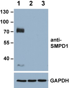
Application: Western BlotSample Tested: 143B osteosarcoma cellsSpecies: HumanVerified Customer | Posted 02/03/201730 micrograms of proteins per lane. Nitrocellulose membrane. 5% milk/PBS-Tween was used for blocking. Antibody dilution: 1:1000 in 1% milk/PBS-Tween. Lane 1=wild-type cells, lane 2 and 3 show two different knock-out clones obtained by CRISPR/Cas9 genome editing. A loading control (GAPDH) is also shown.

There are no reviews that match your criteria.
Protocols
Find general support by application which include: protocols, troubleshooting, illustrated assays, videos and webinars.
- Cellular Response to Hypoxia Protocols
- Immunoprecipitation Protocol
- R&D Systems Quality Control Western Blot Protocol
- Troubleshooting Guide: Western Blot Figures
- Western Blot Conditions
- Western Blot Protocol
- Western Blot Protocol for Cell Lysates
- Western Blot Troubleshooting
- Western Blot Troubleshooting Guide
- View all Protocols, Troubleshooting, Illustrated assays and Webinars
Loading...