Sialic acid molecules attached to glycoproteins or glycosphingolipids play important roles in various biological processes such as immune recognition, pathogen infection, and cell adhesion (1). Sialyltransferases are key enzymes that regulate the cellular levels of sialic acid-containing molecules. Beta-galactosamide alpha‑2,6‑sialyltransferase 1 encoded by the ST6GAL1 gene is a type-II membrane protein localized in the trans-Golgi network and catalyzes 2,6-sialylation of Gal beta 1,4‑GlcNAc structures on N-glycans (2). The enzyme is involved in the generation of the cell-surface carbohydrate determinants and differentiation antigens HB‑6, CD75, and CD76 (3). ST6GAL1 is highly expressed in the liver and also expressed in most other tissues to some extent (4). ST6GAL1 deficiency causes abnormalities in B-cell immunoreactivity (5). The expression and activity of ST6GAL1 are associated with tumor metastasis in breast (6) and colon (7) cancers. The majority of ST6GAL1 in the liver is cleaved and secreted into the serum (8) and may be used as a biomarker for hepatitis diseases (9).
Human ST6 Gal Sialyltransferase 1/
ST6GAL1 Antibody
R&D Systems | Catalog # AF5924

Key Product Details
Validated by
Knockout/Knockdown, Biological Validation
Species Reactivity
Validated:
Human
Cited:
Human, Mouse, Hamster, Transgenic Mouse
Applications
Validated:
Immunohistochemistry, Western Blot, Immunocytochemistry, Simple Western
Cited:
Immunohistochemistry, Immunohistochemistry-Paraffin, Immunohistochemistry-Frozen, Western Blot, Flow Cytometry, Immunocytochemistry
Label
Unconjugated
Antibody Source
Polyclonal Goat IgG
Loading...
Product Specifications
Immunogen
Mouse myeloma cell line NS0-derived recombinant human ST6 Gal Sialyltransferase 1/ST6GAL1
Lys27-Cys406
Accession # P15907
Lys27-Cys406
Accession # P15907
Specificity
Detects human ST6 Gal Sialyltransferase 1/ST6GAL1 in direct ELISAs and Western blots.
Clonality
Polyclonal
Host
Goat
Isotype
IgG
Scientific Data Images for Human ST6 Gal Sialyltransferase 1/
ST6GAL1 Antibody
Detection of Human ST6 Gal Sialyltransferase 1/ST6GAL1 by Western Blot.
Western blot shows lysates of Daudi human Burkitt's lymphoma cell line. PVDF Membrane was probed with 1 µg/mL of Goat Anti-Human ST6 Gal Sialyltransferase 1/ST6GAL1 Antigen Affinity-purified Polyclonal Antibody (Catalog # AF5924) followed by HRP-conjugated Anti-Goat IgG Secondary Antibody (HAF019). A specific band was detected for ST6GAL1 at approximately 56 kDa (as indicated). This experiment was conducted under reducing conditions and using Immunoblot Buffer Group 8.
ST6 Gal Sialyltransferase 1/ST6GAL1 in Human Liver.
ST6 Gal Sialyltransferase 1/ST6GAL1 was detected in immersion fixed paraffin-embedded sections of human liver using Goat Anti-Human ST6 Gal Sialyltransferase 1/ST6GAL1 Antigen Affinity-purified Polyclonal Antibody (Catalog # AF5924) at 0.3 µg/mL for 1 hour at room temperature followed by incubation with the Anti-Goat IgG VisUCyte™ HRP Polymer Antibody (VC004). Before incubation with the primary antibody, tissue was subjected to heat-induced epitope retrieval using Antigen Retrieval Reagent-Basic (CTS013). Tissue was stained using DAB (brown) and counterstained with hematoxylin (blue). Specific staining was localized to cytoplasm in hepatocytes. Staining was performed using our protocol for IHC Staining with VisUCyte HRP Polymer Detection Reagents.
ST6 Gal Sialyltransferase 1/ST6GAL1 in Human Prostate.
ST6 Gal Sialyltransferase 1/ST6GAL1 was detected in immersion fixed paraffin-embedded sections of human prostate using Goat Anti-Human ST6 Gal Sialyltransferase 1/ST6GAL1 Antigen Affinity-purified Polyclonal Antibody (Catalog # AF5924) at 1 µg/mL for 1 hour at room temperature followed by incubation with the Anti-Goat IgG VisUCyte™ HRP Polymer Antibody (VC004). Before incubation with the primary antibody, tissue was subjected to heat-induced epitope retrieval using Antigen Retrieval Reagent-Basic (CTS013). Tissue was stained using DAB (brown) and counterstained with hematoxylin (blue). Specific staining was localized to cytoplasm in glandular epithelial cells. Staining was performed using our protocol for IHC Staining with VisUCyte HRP Polymer Detection Reagents.
Detection of Human ST6 Gal Sialyltransferase 1/ST6GAL1 by Simple WesternTM.
Simple Western lane view shows lysates of Daudi human Burkitt's lymphoma cell line, loaded at 0.2 mg/mL. A specific band was detected for ST6 Gal Sialyltransferase 1/ST6GAL1 at approximately 64 kDa (as indicated) using 10 µg/mL of Goat Anti-Human ST6 Gal Sialyltransferase 1/ST6GAL1 Antigen Affinity-purified Polyclonal Antibody (Catalog # AF5924) followed by 1:50 dilution of HRP-conjugated Anti-Goat IgG Secondary Antibody (HAF109). This experiment was conducted under reducing conditions and using the 12-230 kDa separation system.
Detection of Human ST6 Gal Sialyltransferase 1/ST6GAL1/CD75 by Western Blot
ST6Gal-I expression in OV4 ovarian cancer cells. OV4 cells that have no endogenous ST6Gal-I were stably transduced with either empty vector or ST6Gal-I-expressing lentivirus. ST6Gal-I expression was confirmed by immunocytochemistry (A) and immunoblotting (B). ST6Gal-I localization to the Golgi is shown by co-localization with the Golgi marker GM-130 in the ST6Gal-I forced expression line (C) Par = parental; EV = empty vector; ST6 = cells with forced ST6Gal-I expression. Image collected and cropped by CiteAb from the following publication (https://ovarianresearch.biomedcentral.com/articles/10.1186/1757-2215-6-…), licensed under a CC-BY license. Not internally tested by R&D Systems. & RT‑4 human urinary bladder transitional cell papilloma cell line (Negative).")
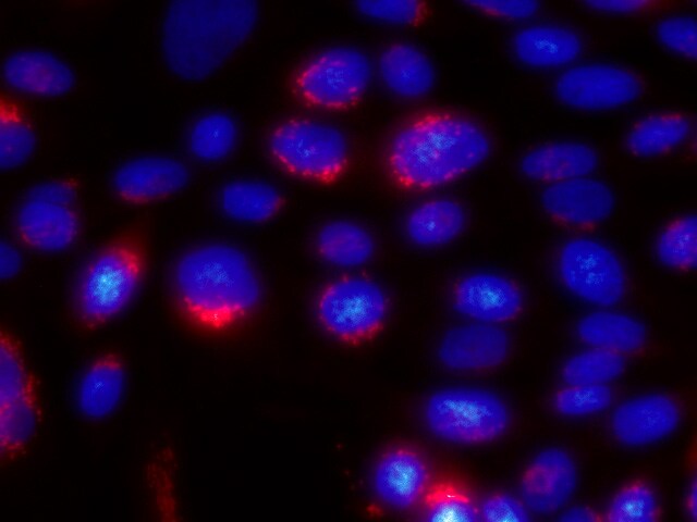
Detection of ST6 Gal Sialyltransferase 1/ST6GAL1 in U266 human myeloma cell line (Positive) & RT‑4 human urinary bladder transitional cell papilloma cell line (Negative).
ST6 Gal Sialyltransferase 1/ST6GAL1 was detected in immersion fixed U266 human myeloma cell line (Positive) & RT‑4 human urinary bladder transitional cell papilloma cell line (Negative) using Goat Anti-Human ST6 Gal Sialyltransferase 1/ST6GAL1 Antigen Affinity-purified Polyclonal Antibody (Catalog # AF5924) at 5 µg/mL for 3 hours at room temperature. Cells were stained using the NorthernLights™ 557-conjugated Anti-Goat IgG Secondary Antibody (red; Catalog # NL001) and counterstained with DAPI (blue). Specific staining was localized to Golgi. View our protocol for Fluorescent ICC Staining of Non-adherent Cells.
Detection of Human ST6 Gal Sialyltransferase 1/ST6GAL1/CD75 by Immunocytochemistry/Immunofluorescence
ST6Gal-I expression in OV4 ovarian cancer cells. OV4 cells that have no endogenous ST6Gal-I were stably transduced with either empty vector or ST6Gal-I-expressing lentivirus. ST6Gal-I expression was confirmed by immunocytochemistry (A) and immunoblotting (B). ST6Gal-I localization to the Golgi is shown by co-localization with the Golgi marker GM-130 in the ST6Gal-I forced expression line (C) Par = parental; EV = empty vector; ST6 = cells with forced ST6Gal-I expression. Image collected and cropped by CiteAb from the following publication (https://ovarianresearch.biomedcentral.com/articles/10.1186/1757-2215-6-…), licensed under a CC-BY license. Not internally tested by R&D Systems.
Detection of Human ST6 Gal Sialyltransferase 1/ST6GAL1/CD75 by Western Blot
Cells that are resistant to cisplatin have upregulated endogenous ST6Gal-I. (A) Pa-1 cells with ST6Gal-I knockdown (sh.ST6) were exposed to cisplatin for 3 weeks, and the remaining viable population (sh.ST6 cis-res) was expanded and immunoblotted for ST6Gal-I. (B) Pa-1 cells with ST6Gal-I knockdown do not upregulate ST6Gal-I expression following a 24-hr treatment with cisplatin (sh.ST6 + cis). (C) Parental A2780 ovarian cancer cells (Par) and a cisplatin-resistant derivative population (Cis-res) were immunoblotted for endogenous ST6Gal-I. Image collected and cropped by CiteAb from the following publication (https://ovarianresearch.biomedcentral.com/articles/10.1186/1757-2215-6-…), licensed under a CC-BY license. Not internally tested by R&D Systems.
Detection of Human ST6 Gal Sialyltransferase 1/ST6GAL1/CD75 by Immunocytochemistry/Immunofluorescence
ST6Gal-I expression in OV4 ovarian cancer cells. OV4 cells that have no endogenous ST6Gal-I were stably transduced with either empty vector or ST6Gal-I-expressing lentivirus. ST6Gal-I expression was confirmed by immunocytochemistry (A) and immunoblotting (B). ST6Gal-I localization to the Golgi is shown by co-localization with the Golgi marker GM-130 in the ST6Gal-I forced expression line (C) Par = parental; EV = empty vector; ST6 = cells with forced ST6Gal-I expression. Image collected and cropped by CiteAb from the following publication (https://ovarianresearch.biomedcentral.com/articles/10.1186/1757-2215-6-…), licensed under a CC-BY license. Not internally tested by R&D Systems.
Detection of Human ST6 Gal Sialyltransferase 1/ST6GAL1/CD75 by Immunocytochemistry/Immunofluorescence
ST6Gal-I knockdown in Pa-1 ovarian cancer cells. Pa-1 cells that have high endogenous ST6Gal-I were stably transduced with either empty vector lentivirus, or virus expressing shRNA for ST6Gal-I. ST6Gal-I knockdown was confirmed by immunocytochemistry (A) and immunoblotting (B). ST6Gal-I localization to the Golgi was confirmed by co-localization with the Golgi marker GM-130 in the empty vector transduced line (C) Par = parental; EV = empty vector; sh.ST6 = cells stably expressing shRNA for ST6Gal-I. Image collected and cropped by CiteAb from the following publication (https://ovarianresearch.biomedcentral.com/articles/10.1186/1757-2215-6-…), licensed under a CC-BY license. Not internally tested by R&D Systems.
Detection of Human ST6 Gal Sialyltransferase 1/ST6GAL1/CD75 by Western Blot
ST6Gal-I knockdown in Pa-1 ovarian cancer cells. Pa-1 cells that have high endogenous ST6Gal-I were stably transduced with either empty vector lentivirus, or virus expressing shRNA for ST6Gal-I. ST6Gal-I knockdown was confirmed by immunocytochemistry (A) and immunoblotting (B). ST6Gal-I localization to the Golgi was confirmed by co-localization with the Golgi marker GM-130 in the empty vector transduced line (C) Par = parental; EV = empty vector; sh.ST6 = cells stably expressing shRNA for ST6Gal-I. Image collected and cropped by CiteAb from the following publication (https://ovarianresearch.biomedcentral.com/articles/10.1186/1757-2215-6-…), licensed under a CC-BY license. Not internally tested by R&D Systems.
Detection of Human Human ST6 Gal Sialyltransferase 1/ ST6GAL1 Antibody by Western Blot
Cells that are resistant to cisplatin have upregulated endogenous ST6Gal-I. (A) Pa-1 cells with ST6Gal-I knockdown (sh.ST6) were exposed to cisplatin for 3 weeks, and the remaining viable population (sh.ST6 cis-res) was expanded and immunoblotted for ST6Gal-I. (B) Pa-1 cells with ST6Gal-I knockdown do not upregulate ST6Gal-I expression following a 24-hr treatment with cisplatin (sh.ST6 + cis). (C) Parental A2780 ovarian cancer cells (Par) and a cisplatin-resistant derivative population (Cis-res) were immunoblotted for endogenous ST6Gal-I. Image collected and cropped by CiteAb from the following publication (https://pubmed.ncbi.nlm.nih.gov/23578204), licensed under a CC-BY license. Not internally tested by R&D Systems.Applications for Human ST6 Gal Sialyltransferase 1/ST6GAL1 Antibody
Application
Recommended Usage
Immunocytochemistry
5-15 µg/mL
Sample: Immersion fixed U266 human myeloma cell line (Positive) & RT‑4 human urinary bladder transitional cell papilloma cell line (Negative)
Sample: Immersion fixed U266 human myeloma cell line (Positive) & RT‑4 human urinary bladder transitional cell papilloma cell line (Negative)
Immunohistochemistry
0.3-15 µg/mL
Sample: Immersion fixed paraffin-embedded sections of human liver and immersion fixed paraffin-embedded sections of human prostate
Sample: Immersion fixed paraffin-embedded sections of human liver and immersion fixed paraffin-embedded sections of human prostate
Simple Western
10 µg/mL
Sample: Daudi human Burkitt's lymphoma cell line
Sample: Daudi human Burkitt's lymphoma cell line
Western Blot
1 µg/mL
Sample: Daudi human Burkitt's lymphoma cell line
Sample: Daudi human Burkitt's lymphoma cell line
Reviewed Applications
Read 1 review rated 5 using AF5924 in the following applications:
Formulation, Preparation, and Storage
Purification
Antigen Affinity-purified
Reconstitution
Reconstitute at 0.2 mg/mL in sterile PBS. For liquid material, refer to CoA for concentration.
Loading...
Formulation
Lyophilized from a 0.2 μm filtered solution in PBS with Trehalose. *Small pack size (SP) is supplied either lyophilized or as a 0.2 µm filtered solution in PBS.
Shipping
Lyophilized product is shipped at ambient temperature. Liquid small pack size (-SP) is shipped with polar packs. Upon receipt, store immediately at the temperature recommended below.
Stability & Storage
Use a manual defrost freezer and avoid repeated freeze-thaw cycles.
- 12 months from date of receipt, -20 to -70 °C as supplied.
- 1 month, 2 to 8 °C under sterile conditions after reconstitution.
- 6 months, -20 to -70 °C under sterile conditions after reconstitution.
Calculators
Background: ST6 Gal Sialyltransferase 1/ST6GAL1
References
- Varki, A. et al. (1999) Essentials of Glycobiology, Cold Spring Harbor Laboratory Press, pp195.
- Weinstein, J. et al. (1987) J. Biol. Chem. 262:17735.
- Bast, B.J. et al. (1992) J. Cell Biol. 116:423.
- Kitagawa, H. and J.C. Paulson (1994) J. Biol. Chem. 269:17872.
- Hennet, T. et al. (1998) Proc. Natl. Acad. Sci. USA 95:4504.
- Recchi, M.A. et al. (1998) Cancer Res. 58:4066.
- Dall’Olio, F. et al. (2001) Eur. J. Biochem. 268:5876.
- Weinstein, J. et al. (1987) J. Biol. Chem. 262:17735.
- Kitazume, S. et al. (2009) Glycobiology 19:479.
Long Name
ST6 beta-Galactoside alpha-2,6-Sialyltransferase 1
Alternate Names
Alpha 2,6-ST, B-cell antigen CD75, CD75, SIAT1, ST6N
Gene Symbol
ST6GAL1
UniProt
Additional ST6 Gal Sialyltransferase 1/ST6GAL1 Products
- All Products for ST6 Gal Sialyltransferase 1/ST6GAL1
- ST6 Gal Sialyltransferase 1/ST6GAL1 cDNA Clones
- ST6 Gal Sialyltransferase 1/ST6GAL1 ELISA Kits
- ST6 Gal Sialyltransferase 1/ST6GAL1 Lysates
- ST6 Gal Sialyltransferase 1/ST6GAL1 Primary Antibodies
- ST6 Gal Sialyltransferase 1/ST6GAL1 Proteins and Enzymes
Product Documents for Human ST6 Gal Sialyltransferase 1/ST6GAL1 Antibody
Certificate of Analysis
To download a Certificate of Analysis, please enter a lot or batch number in the search box below.
Note: Certificate of Analysis not available for kit components.
Product Specific Notices for Human ST6 Gal Sialyltransferase 1/ST6GAL1 Antibody
For research use only
Related Research Areas
Citations for Human ST6 Gal Sialyltransferase 1/
ST6GAL1 Antibody
 Powered by Bioz
Customer Reviews for Human ST6 Gal Sialyltransferase 1/ST6GAL1 Antibody (1)
5 out of 5
1 Customer Rating
Have you used Human ST6 Gal Sialyltransferase 1/ST6GAL1 Antibody?
Submit a review and receive an Amazon gift card!
$25/€18/£15/$25CAN/¥2500 Yen for a review with an image
$10/€7/£6/$10CAN/¥1110 Yen for a review without an image
Submit a review
Customer Images
Showing
1
-
1 of
1 review
Showing All
Filter By:
-
Application: Immunocytochemistry/ImmunofluorescenceSample Tested: ST6GAL1 transfected CHO cellsSpecies: MouseVerified Customer | Posted 03/21/2018Used this Ab for both Western and immunofluorescence on mouse ST6GAL1 transfected CHO cells. Fixed cells with 4% Paraformaldehyde and permeabilized with 0.5% saponin.

There are no reviews that match your criteria.
Protocols
Find general support by application which include: protocols, troubleshooting, illustrated assays, videos and webinars.
- Antigen Retrieval Protocol (PIER)
- Antigen Retrieval for Frozen Sections Protocol
- Appropriate Fixation of IHC/ICC Samples
- Cellular Response to Hypoxia Protocols
- Chromogenic IHC Staining of Formalin-Fixed Paraffin-Embedded (FFPE) Tissue Protocol
- Chromogenic Immunohistochemistry Staining of Frozen Tissue
- ClariTSA™ Fluorophore Kits
- Detection & Visualization of Antibody Binding
- Fluorescent IHC Staining of Frozen Tissue Protocol
- Graphic Protocol for Heat-induced Epitope Retrieval
- Graphic Protocol for the Preparation and Fluorescent IHC Staining of Frozen Tissue Sections
- Graphic Protocol for the Preparation and Fluorescent IHC Staining of Paraffin-embedded Tissue Sections
- Graphic Protocol for the Preparation of Gelatin-coated Slides for Histological Tissue Sections
- ICC Cell Smear Protocol for Suspension Cells
- ICC Immunocytochemistry Protocol Videos
- ICC for Adherent Cells
- IHC Sample Preparation (Frozen sections vs Paraffin)
- Immunocytochemistry (ICC) Protocol
- Immunocytochemistry Troubleshooting
- Immunofluorescence of Organoids Embedded in Cultrex Basement Membrane Extract
- Immunofluorescent IHC Staining of Formalin-Fixed Paraffin-Embedded (FFPE) Tissue Protocol
- Immunohistochemistry (IHC) and Immunocytochemistry (ICC) Protocols
- Immunohistochemistry Frozen Troubleshooting
- Immunohistochemistry Paraffin Troubleshooting
- Preparing Samples for IHC/ICC Experiments
- Preventing Non-Specific Staining (Non-Specific Binding)
- Primary Antibody Selection & Optimization
- Protocol for Heat-Induced Epitope Retrieval (HIER)
- Protocol for Making a 4% Formaldehyde Solution in PBS
- Protocol for VisUCyte™ HRP Polymer Detection Reagent
- Protocol for the Fluorescent ICC Staining of Cell Smears - Graphic
- Protocol for the Fluorescent ICC Staining of Cultured Cells on Coverslips - Graphic
- Protocol for the Preparation & Fixation of Cells on Coverslips
- Protocol for the Preparation and Chromogenic IHC Staining of Frozen Tissue Sections
- Protocol for the Preparation and Chromogenic IHC Staining of Frozen Tissue Sections - Graphic
- Protocol for the Preparation and Chromogenic IHC Staining of Paraffin-embedded Tissue Sections
- Protocol for the Preparation and Chromogenic IHC Staining of Paraffin-embedded Tissue Sections - Graphic
- Protocol for the Preparation and Fluorescent ICC Staining of Cells on Coverslips
- Protocol for the Preparation and Fluorescent ICC Staining of Non-adherent Cells
- Protocol for the Preparation and Fluorescent ICC Staining of Stem Cells on Coverslips
- Protocol for the Preparation and Fluorescent IHC Staining of Frozen Tissue Sections
- Protocol for the Preparation and Fluorescent IHC Staining of Paraffin-embedded Tissue Sections
- Protocol for the Preparation of Gelatin-coated Slides for Histological Tissue Sections
- Protocol for the Preparation of a Cell Smear for Non-adherent Cell ICC - Graphic
- R&D Systems Quality Control Western Blot Protocol
- TUNEL and Active Caspase-3 Detection by IHC/ICC Protocol
- The Importance of IHC/ICC Controls
- Troubleshooting Guide: Immunohistochemistry
- Troubleshooting Guide: Western Blot Figures
- Western Blot Conditions
- Western Blot Protocol
- Western Blot Protocol for Cell Lysates
- Western Blot Troubleshooting
- Western Blot Troubleshooting Guide
- View all Protocols, Troubleshooting, Illustrated assays and Webinars
Loading...