STX1A (Syntaxin [Greek for 'organizing']1A; also HPC-1) is a 34-36 kDa member of the syntaxin family of proteins. It is a t-SNARE that is widely expressed in neurons, and is involved in the exocytosis of neurotransmitters at the presynaptic membrane. STX1A is transported intracellularly by microtubule-associated syntabulin, and its availability appears to be regulated through binding to LGI3. When released from LGI3, STX1A interacts with SNAP25 and VAMP2 to form the SNARE complex involved in exocytotic vesicle release. Human STX1A is a type IV single-pass transmembrane protein (very long cytoplasmic N-terminus) that is 288 amino acids (aa) in length. It contains a 265 aa N-terminal cytoplasmic domain that contains one coiled‑coil region (aa 68-109), a t-SNARE domain with a coiled‑coil region (aa 192-254), and a C-terminal transmembrane sequence (aa 266-286). There are three potential isoform variants. One shows a 34 aa substitution for aa 227‑288, while another termed STX1C is likely soluble, and contains a 25 aa substitution for the same aa sequence above encompassing aa 227‑288. A third variant shows a five aa substitution for aa 1-10. Over aa 1-165, human STX1A shares 99% aa sequence identity with mouse STX1A.

Key Product Details
Species Reactivity
Human
Applications
Immunohistochemistry, Western Blot
Label
Unconjugated
Antibody Source
Monoclonal Mouse IgG2A Clone # 761811
Loading...
Product Specifications
Immunogen
E. coli-derived recombinant human Syntaxin 1A
Met1-Leu165
Accession # Q16623
Met1-Leu165
Accession # Q16623
Specificity
Detects human Syntaxin 1A in direct ELISAs and Western blots. In direct ELISAs, approximately 50% cross-reactivity
with recombinant human (rh) Epimorphin/Syntaxin 2 is observed, 25%
cross-reactivity with rhSyntaxin 1B2 and rhSyntaxin 3 is observed, and no
cross-reactivity with rhSyntaxin 1B1 is observed.
Clonality
Monoclonal
Host
Mouse
Isotype
IgG2A
Scientific Data Images for Human Syntaxin 1A Antibody (761811)
Detection of Human and Rat Syntaxin 1A by Western Blot.
Western blot shows lysates of human brain tissue and rat brain tissue. PVDF membrane was probed with 0.1 µg/mL of Mouse Anti-Human Syntaxin 1A Monoclonal Antibody (Catalog # MAB7237) followed by HRP-conjugated Anti-Mouse IgG Secondary Antibody (Catalog # HAF018). A specific band was detected for Syntaxin 1A at approximately 35 kDa (as indicated). This experiment was conducted under reducing conditions and using Immunoblot Buffer Group 1.
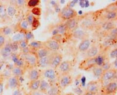
Syntaxin 1A in Human Brain.
Syntaxin 1A was detected in immersion fixed paraffin-embedded sections of human brain using Mouse Anti-Human Syntaxin 1A Monoclonal Antibody (Catalog # MAB7237) at 15 µg/mL overnight at 4 °C. Before incubation with the primary antibody, tissue was subjected to heat-induced epitope retrieval using Antigen Retrieval Reagent-Basic (Catalog # CTS013). Tissue was stained using the Anti-Mouse HRP-DAB Cell & Tissue Staining Kit (brown; Catalog # CTS002) and counterstained with hematoxylin (blue). Specific staining was localized to synaptic puncta. View our protocol for Chromogenic IHC Staining of Paraffin-embedded Tissue Sections.Applications for Human Syntaxin 1A Antibody (761811)
Application
Recommended Usage
Immunohistochemistry
8-25 µg/mL
Sample: Immersion fixed paraffin-embedded sections of human brain
Sample: Immersion fixed paraffin-embedded sections of human brain
Western Blot
0.1 µg/mL
Sample: Human brain tissue and rat brain tissue
Sample: Human brain tissue and rat brain tissue
Reviewed Applications
Read 1 review rated 5 using MAB7237 in the following applications:
Formulation, Preparation, and Storage
Purification
Protein A or G purified from hybridoma culture supernatant
Reconstitution
Sterile PBS to a final concentration of 0.5 mg/mL. For liquid material, refer to CoA for concentration.
Loading...
Formulation
Lyophilized from a 0.2 μm filtered solution in PBS with Trehalose. *Small pack size (SP) is supplied either lyophilized or as a 0.2 µm filtered solution in PBS.
Shipping
Lyophilized product is shipped at ambient temperature. Liquid small pack size (-SP) is shipped with polar packs. Upon receipt, store immediately at the temperature recommended below.
Stability & Storage
Use a manual defrost freezer and avoid repeated freeze-thaw cycles.
- 12 months from date of receipt, -20 to -70 °C as supplied.
- 1 month, 2 to 8 °C under sterile conditions after reconstitution.
- 6 months, -20 to -70 °C under sterile conditions after reconstitution.
Calculators
Background: Syntaxin 1A
Alternate Names
Brain Syntaxin, HPC-1, p35-1, STX1A, SYN1A
Gene Symbol
STX1A
UniProt
Additional Syntaxin 1A Products
Product Documents for Human Syntaxin 1A Antibody (761811)
Certificate of Analysis
To download a Certificate of Analysis, please enter a lot or batch number in the search box below.
Note: Certificate of Analysis not available for kit components.
Product Specific Notices for Human Syntaxin 1A Antibody (761811)
For research use only
Related Research Areas
Customer Reviews for Human Syntaxin 1A Antibody (761811) (1)
5 out of 5
1 Customer Rating
Have you used Human Syntaxin 1A Antibody (761811)?
Submit a review and receive an Amazon gift card!
$25/€18/£15/$25CAN/¥2500 Yen for a review with an image
$10/€7/£6/$10CAN/¥1110 Yen for a review without an image
Submit a review
Customer Images
Showing
1
-
1 of
1 review
Showing All
Filter By:
-
Application: ImmunohistochemistrySample Tested: Brain tissueSpecies: HumanVerified Customer | Posted 03/03/2022

There are no reviews that match your criteria.
Protocols
Find general support by application which include: protocols, troubleshooting, illustrated assays, videos and webinars.
- Antigen Retrieval Protocol (PIER)
- Antigen Retrieval for Frozen Sections Protocol
- Appropriate Fixation of IHC/ICC Samples
- Cellular Response to Hypoxia Protocols
- Chromogenic IHC Staining of Formalin-Fixed Paraffin-Embedded (FFPE) Tissue Protocol
- Chromogenic Immunohistochemistry Staining of Frozen Tissue
- ClariTSA™ Fluorophore Kits
- Detection & Visualization of Antibody Binding
- Fluorescent IHC Staining of Frozen Tissue Protocol
- Graphic Protocol for Heat-induced Epitope Retrieval
- Graphic Protocol for the Preparation and Fluorescent IHC Staining of Frozen Tissue Sections
- Graphic Protocol for the Preparation and Fluorescent IHC Staining of Paraffin-embedded Tissue Sections
- Graphic Protocol for the Preparation of Gelatin-coated Slides for Histological Tissue Sections
- IHC Sample Preparation (Frozen sections vs Paraffin)
- Immunofluorescent IHC Staining of Formalin-Fixed Paraffin-Embedded (FFPE) Tissue Protocol
- Immunohistochemistry (IHC) and Immunocytochemistry (ICC) Protocols
- Immunohistochemistry Frozen Troubleshooting
- Immunohistochemistry Paraffin Troubleshooting
- Preparing Samples for IHC/ICC Experiments
- Preventing Non-Specific Staining (Non-Specific Binding)
- Primary Antibody Selection & Optimization
- Protocol for Heat-Induced Epitope Retrieval (HIER)
- Protocol for Making a 4% Formaldehyde Solution in PBS
- Protocol for VisUCyte™ HRP Polymer Detection Reagent
- Protocol for the Preparation & Fixation of Cells on Coverslips
- Protocol for the Preparation and Chromogenic IHC Staining of Frozen Tissue Sections
- Protocol for the Preparation and Chromogenic IHC Staining of Frozen Tissue Sections - Graphic
- Protocol for the Preparation and Chromogenic IHC Staining of Paraffin-embedded Tissue Sections
- Protocol for the Preparation and Chromogenic IHC Staining of Paraffin-embedded Tissue Sections - Graphic
- Protocol for the Preparation and Fluorescent IHC Staining of Frozen Tissue Sections
- Protocol for the Preparation and Fluorescent IHC Staining of Paraffin-embedded Tissue Sections
- Protocol for the Preparation of Gelatin-coated Slides for Histological Tissue Sections
- R&D Systems Quality Control Western Blot Protocol
- TUNEL and Active Caspase-3 Detection by IHC/ICC Protocol
- The Importance of IHC/ICC Controls
- Troubleshooting Guide: Immunohistochemistry
- Troubleshooting Guide: Western Blot Figures
- Western Blot Conditions
- Western Blot Protocol
- Western Blot Protocol for Cell Lysates
- Western Blot Troubleshooting
- Western Blot Troubleshooting Guide
- View all Protocols, Troubleshooting, Illustrated assays and Webinars
Loading...