Vascular Endothelial Growth Factor Receptor 1 (VEGFR1) is a receptor tyrosine kinase that is expressed primarily on endothelial cells and plays a role in vasculogenesis and angiogenesis. A soluble variant of VEGFR1 was also reported to bind VEGF and PlGF with high affinity and function as a potent VEGF antagonist.

Key Product Details
Species Reactivity
Validated:
Human
Cited:
Human, Bovine, Chinese Hamster
Applications
Validated:
Immunohistochemistry, Western Blot, Blockade of Receptor-ligand Interaction, Flow Cytometry, CyTOF-ready
Cited:
Immunohistochemistry, Immunohistochemistry-Paraffin, Immunohistochemistry-Frozen, Western Blot, Neutralization, Flow Cytometry, Immunocytochemistry, Blocking, Functional Assay, Proximity Ligation Assay (PLA)
Label
Unconjugated
Antibody Source
Polyclonal Goat IgG
Loading...
Product Specifications
Immunogen
S. frugiperda insect ovarian cell line Sf 21-derived recombinant human VEGFR1/Flt-1
Ser27-His687
Accession # NP_001153392
Ser27-His687
Accession # NP_001153392
Specificity
Detects human VEGFR1/Flt-1 in direct ELISAs and Western blots.
Clonality
Polyclonal
Host
Goat
Isotype
IgG
Endotoxin Level
<0.10 EU per 1 μg of the antibody by the LAL method.
Scientific Data Images for Human VEGFR1/Flt-1 Antibody
VEGFR1/Flt‑1 in Human Breast Cancer Tissue.
VEGFR1/Flt‑1 was detected in immersion fixed paraffin-embedded sections of human breast cancer tissue using 15 µg/mL Human VEGFR1/Flt‑1 Antigen Affinity-purified Polyclonal Antibody (Catalog # AF321) overnight at 4 °C. Tissue was stained (red) and counterstained with hematoxylin (blue). View our protocol for Chromogenic IHC Staining of Paraffin-embedded Tissue Sections.
VEGFR1/Flt‑1 in Human Ovarian Cancer Tissue.
VEGFR1/Flt-1 was detected in immersion fixed paraffin-embedded sections of human ovarian cancer tissue using Human VEGFR1/Flt-1 Antigen Affinity-purified Polyclonal Antibody (Catalog # AF321) at 3 µg/mL overnight at 4 °C. Tissue was stained using the Anti-Goat HRP-DAB Cell & Tissue Staining Kit (brown; CTS008) and counterstained with hematoxylin (blue). View our protocol for Chromogenic IHC Staining of Paraffin-embedded Tissue Sections.
Detection of Human VEGFR1/Flt-1 by Western Blot
Exogenous VEGF165 suppresses RASSF1A expression in normal epithelial and endothelial cells. Metastatic colon cancer cells (mCRC, (A) T84 and (B) Colo 205) were stimulated with VEGF165 as indicated before and cell metabolism was performed by MTT assay for 6 h (black dash), 24 h (green) and 48 h (pink). Immortalized benign protate ((C) RWPE1), human bronchial epithelial cells ((D) HBEpc), immortalized human breast epithelial cells ((E) MCF-10A), endothelial cells (F, EC) were stimulated with or without VEGF165 (12.5 and 25 ng/mL) for 24 h. Cell lysates were monitored by Western blot for p-VEGFR1, p-VEGFR2, RASSF1A, and GAPDH as indicated. Immunoblot was quantified by scanning densitometry and normalized against GAPDH expression for RWPE1 (G), HBEpc (H), MCF 10A (I) and EC (lower panel of (F)). Results are from three independent experiments and statistical significance was determined using one way-ANOVA followed Bonferroni test. (* p < 0.05, ** p < 0.01, *** p < 0.001). Image collected and cropped by CiteAb from the following open publication (https://pubmed.ncbi.nlm.nih.gov/30744076), licensed under a CC-BY license. Not internally tested by R&D Systems.
Detection of VEGFR1/Flt-1 in HUVEC cells by Flow Cytometry
HUVEC cells were stained with Goat Anti-Human VEGFR1/Flt-1 Antigen Affinity-purified Polyclonal Antibody (Catalog # AF321, filled histogram) or isotype control antibody (Catalog # AB-108-C, open histogram) followed by Allophycocyanin-conjugated Anti-Goat IgG Secondary Antibody (Catalog # F0108). View our protocol for Staining Membrane-associated Proteins.
Detection of Human VEGFR1/Flt-1 by Western Blot
VEGFR1 isoforms in human degenerated and bovine healthy discs. A representative immunoblotting assay showed the expression of the full-length membrane form (~ 200 kDa), the soluble form (~ 130 kDa) and a cytoplasmic fragment (~ 60 kDa) in human degenerated (a) and healthy bovine (c) disc cells. beta -Actin was used as control. Western blot quantification of membrane and soluble VEGFR1 was performed on human (b; n = 3) and bovine (d; n = 2) disc cells. Results represent an average of area (square pixels) ± SD normalized on beta Actin. White bars represent AF, grey bars NP. No statistical significance with non-parametric Mann–Whitney–Wilcoxon U test for independent variables Image collected and cropped by CiteAb from the following open publication (https://pubmed.ncbi.nlm.nih.gov/29784013), licensed under a CC-BY license. Not internally tested by R&D Systems.
Detection of VEGFR1/Flt-1 by Western Blot
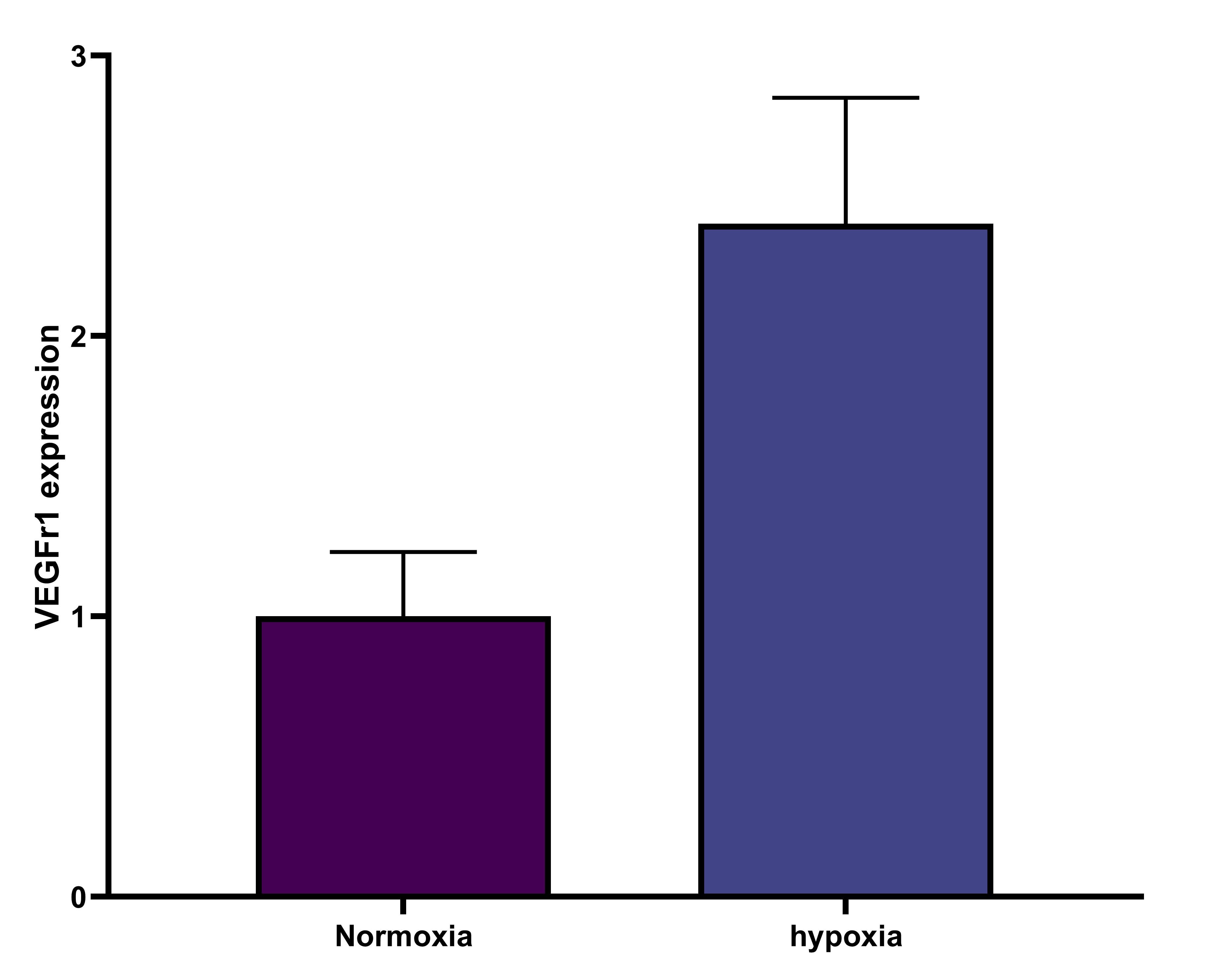
Decreased levels of VEGFR-2 in hypoxia and in early first trimester placentae.A: Representative VEGFR-2 and sFlt-1 immunoblots of placental villous explants cultured at 3% and 20% O2. Densitometry analysis comparing normalized VEGFR-2 immunoreactivity between explants cultured at 3% (n = 7) and 20% O2 (n = 7). VEGFR-2 transcript levels were assessed by qPCR analysis comparing 3% (n = 4) and 20% O2 (n = 4) samples. B: Representative VEGFR-2 immunoblots of early (EFT) and late (LFT) first trimester placentae. Densitometry analysis comparing normalized VEGFR-2 immunoreactivity between samples were not significantly different. Transcript levels were assessed by qPCR analysis comparing VEGFR-2 levels in EFT and LFT placentae. (EFT, n = 12; LFT, n = 14; *P<0.05). All values are represented as the means±SEM. Image collected and cropped by CiteAb from the following open publication (https://pubmed.ncbi.nlm.nih.gov/24260556), licensed under a CC-BY license. Not internally tested by R&D Systems.
Detection of VEGFR1/Flt-1 by Western Blot
sFlt-1 directly attenuates VEGFR-2 expression and downstream signalling in placental villous explants.A: Representative VEGFR-2, sFlt-1, pAkt and pERK immunoblots of first trimester placental villous explants treated with sFlt-1 or a blocking antibody to sFlt-1. Densitometry analyses comparing normalized VEGFR-2, sFlt-1, pAkt and pERK in treated samples (Con, control; sFlt-1, sFlt-1 protein; alpha -Flt, Flt-1 neutralizing antibody; n = 3, *P<0.05). All values are represented as the means±SEM. B: Representative VEGFR-2 immunoblot of first trimester placental villous explants treated with VEGF. Densitometry analyses comparing normalized VEGFR-2 between control and VEGF-treated samples (Con, control; VEGF, VEGF-treated; n = 3, *P<0.05). All values are represented as the means±SEM. Image collected and cropped by CiteAb from the following open publication (https://pubmed.ncbi.nlm.nih.gov/24260556), licensed under a CC-BY license. Not internally tested by R&D Systems.
Detection of VEGFR1/Flt-1 by Western Blot
An inverse correlation between VEGFR-2 and sFlt-1 levels in preeclamptic placentae.A: Representative VEGFR-2 and sFlt-1 immunoblots of PE and PTC placentae. Densitometry analysis comparing normalized VEGFR-2 immunoreactivity between PE (n = 13) and PTC (n = 8) samples. VEGFR-2 transcript levels were assessed by qPCR analysis comparing PE (n = 12) and PTC (n = 7) samples. B: Representative VEGFR-2 and sFlt-1 immunoblots of placental tissues from PE and control (Con) dichorionic twins. (PE, preeclampsia; PTC, age-matched preterm control; *P<0.05). All values are represented as the means±SEM. Image collected and cropped by CiteAb from the following open publication (https://pubmed.ncbi.nlm.nih.gov/24260556), licensed under a CC-BY license. Not internally tested by R&D Systems.
Detection of Human VEGFR1/Flt-1 by Western Blot
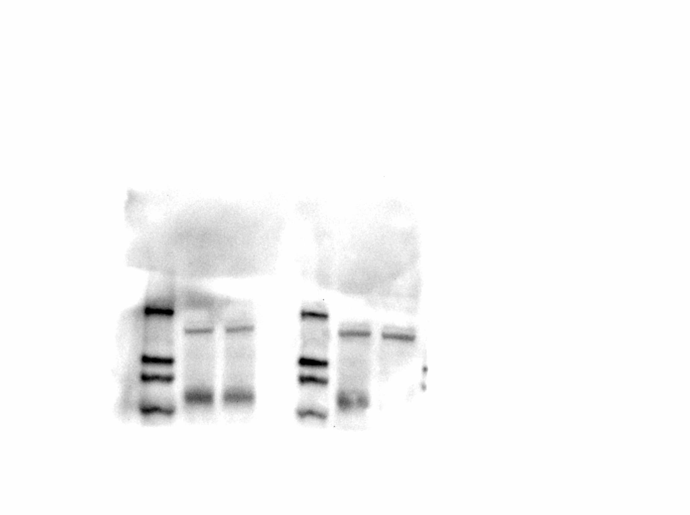
Effect of the metalloprotease inhibitors, GM6001 and TAPI-1 on Flt1 N-terminal cleavage. Panel A: HUVECs were incubated with GM6001 (10 µg/ml) and PMA (30 nM) for the indicated times. GM6001 significantly reduces the PMA-induced soluble Flt1 levels measured by ELISA in conditioned media (CM). **p<0.001 and *p<0.05, n = 3. Panel B and C: HEK293 cells transiently expressing HA and Flag-tagged Flt1 were treated with metalloproteases inhibitors, GM6001 (10 µg/ml) and TAPI-1 (20 µM) and conditioned media was immunoblotted with HA, the epitope tag at the N-terminal end of Flt1 or with AF321, an antibody that recognizes the N-terninus of Flt1 and/or sFlt1. Both GM6001 and TAPI-1 significantly reduces the abundance of the cleaved N-terminal fragment. Representative immunoblot in B and pooled data quantified by densitometry shown in C. **p<0.001 by Kruskal Wallis ANOVAR, Mean ± SE, n = 3–7. Image collected and cropped by CiteAb from the following open publication (https://pubmed.ncbi.nlm.nih.gov/25387128), licensed under a CC0-1.0 license. Not internally tested by R&D Systems.Applications for Human VEGFR1/Flt-1 Antibody
Application
Recommended Usage
Blockade of Receptor-ligand Interaction
In a functional ELISA, 1-4 µg/mL of this antibody will block 50% of the binding of 2 ng/mL of Recombinant Human PlGF (Catalog # 264-PG) to immobilized Recombinant Human VEGFR1/Flt-1 Fc Chimera (Catalog # 321-FL) coated at 1 µg/mL (100 µL/well). At 30 μg/mL, this antibody will block >90% of the binding.
CyTOF-ready
Ready to be labeled using established conjugation methods. No BSA or other carrier proteins that could interfere with conjugation.
Flow Cytometry
0.25 µg/106 cells
Sample: HUVEC human umbilical vein endothelial cells
Sample: HUVEC human umbilical vein endothelial cells
Immunohistochemistry
5-15 µg/mL
Sample: Immersion fixed paraffin-embedded sections of human breast cancer and ovarian cancer tissues
Sample: Immersion fixed paraffin-embedded sections of human breast cancer and ovarian cancer tissues
Western Blot
0.1 µg/mL
Sample: Recombinant Human VEGFR1/Flt‑1 Fc Chimera (Catalog # 321-FL)
Sample: Recombinant Human VEGFR1/Flt‑1 Fc Chimera (Catalog # 321-FL)
Reviewed Applications
Read 7 reviews rated 4.6 using AF321 in the following applications:
Flow Cytometry Panel Builder
Bio-Techne Knows Flow Cytometry
Save time and reduce costly mistakes by quickly finding compatible reagents using the Panel Builder Tool.
Advanced Features
- Spectra Viewer - Custom analysis of spectra from multiple fluorochromes
- Spillover Popups - Visualize the spectra of individual fluorochromes
- Antigen Density Selector - Match fluorochrome brightness with antigen density

Formulation, Preparation, and Storage
Purification
Antigen Affinity-purified
Reconstitution
Reconstitute at 0.2 mg/mL in sterile PBS. For liquid material, refer to CoA for concentration.
Loading...
Formulation
Lyophilized from a 0.2 μm filtered solution in PBS with Trehalose. See Certificate of Analysis for details.
*Small pack size (-SP) is supplied either lyophilized or as a 0.2 µm filtered solution in PBS.
*Small pack size (-SP) is supplied either lyophilized or as a 0.2 µm filtered solution in PBS.
Shipping
Lyophilized product is shipped at ambient temperature. Liquid small pack size (-SP) is shipped with polar packs. Upon receipt, store immediately at the temperature recommended below.
Stability & Storage
Use a manual defrost freezer and avoid repeated freeze-thaw cycles.
- 12 months from date of receipt, -20 to -70 °C as supplied.
- 1 month, 2 to 8 °C under sterile conditions after reconstitution.
- 6 months, -20 to -70 °C under sterile conditions after reconstitution.
Calculators
Background: VEGFR1/Flt-1
Long Name
Vascular Endothelial Growth Factor Receptor 1
Alternate Names
Flt-1, FLT1, FRT, VEGF R1, VEGFR-1
Gene Symbol
FLT1
UniProt
Additional VEGFR1/Flt-1 Products
Product Documents for Human VEGFR1/Flt-1 Antibody
Certificate of Analysis
To download a Certificate of Analysis, please enter a lot or batch number in the search box below.
Note: Certificate of Analysis not available for kit components.
Product Specific Notices for Human VEGFR1/Flt-1 Antibody
For research use only
Citations for Human VEGFR1/Flt-1 Antibody
 Powered by Bioz
Customer Reviews for Human VEGFR1/Flt-1 Antibody (7)
4.6 out of 5
7 Customer Ratings
Have you used Human VEGFR1/Flt-1 Antibody?
Submit a review and receive an Amazon gift card!
$25/€18/£15/$25CAN/¥2500 Yen for a review with an image
$10/€7/£6/$10CAN/¥1110 Yen for a review without an image
Submit a review
Customer Images
Showing
1
-
5 of
7 reviews
Showing All
Filter By:
-
Application: ImmunohistochemistrySample Tested: Embryonic retinaSpecies: MouseVerified Customer | Posted 05/25/2024

-
Application: Western BlotSample Tested: HUVEC human umbilical vein endothelial cellsSpecies: HumanVerified Customer | Posted 03/24/2021
-
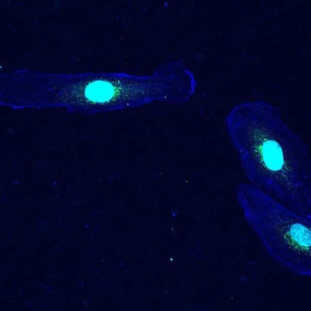
Application: Immunocytochemistry/ImmunofluorescenceSample Tested: HUVEC human umbilical vein endothelial cellsSpecies: HumanVerified Customer | Posted 03/24/2021
-
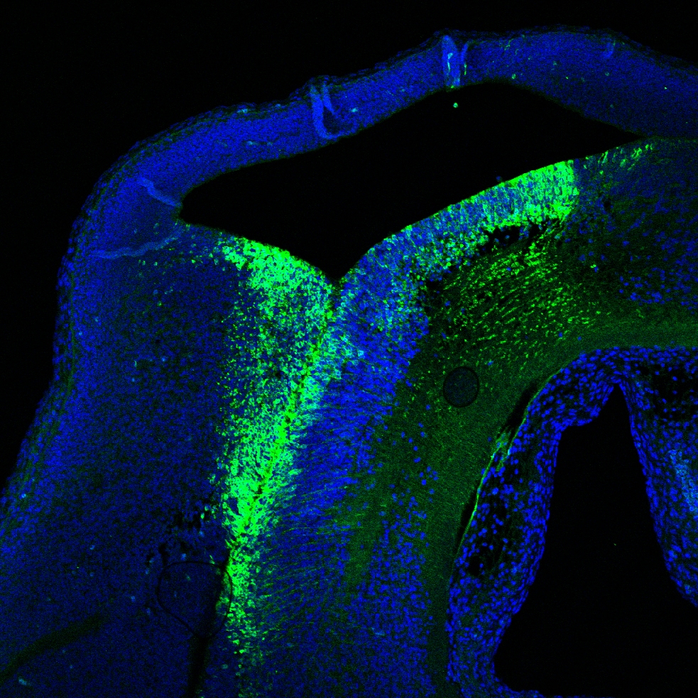
Application: Immunocytochemistry/ImmunofluorescenceSample Tested: Embryonic brainSpecies: MouseVerified Customer | Posted 08/08/2018transgenic mouse line -conditional induction of human sFlt1
-
Application: Immunocytochemistry/ImmunofluorescenceSample Tested: CT26 mouse colorectal cancer cell line and Human liver cancer cell lineSpecies: HumanVerified Customer | Posted 07/08/2016
-
Application: Flow CytometrySample Tested: See PMID 19757485Species: HumanVerified Customer | Posted 01/07/2015
-
Application: Western BlotSample Tested: See PMID 21699503Species: HumanVerified Customer | Posted 01/07/2015
There are no reviews that match your criteria.
Protocols
Find general support by application which include: protocols, troubleshooting, illustrated assays, videos and webinars.
- 7-Amino Actinomycin D (7-AAD) Cell Viability Flow Cytometry Protocol
- Antigen Retrieval Protocol (PIER)
- Antigen Retrieval for Frozen Sections Protocol
- Appropriate Fixation of IHC/ICC Samples
- Cellular Response to Hypoxia Protocols
- Chromogenic IHC Staining of Formalin-Fixed Paraffin-Embedded (FFPE) Tissue Protocol
- Chromogenic Immunohistochemistry Staining of Frozen Tissue
- ClariTSA™ Fluorophore Kits
- Detection & Visualization of Antibody Binding
- Extracellular Membrane Flow Cytometry Protocol
- Flow Cytometry Protocol for Cell Surface Markers
- Flow Cytometry Protocol for Staining Membrane Associated Proteins
- Flow Cytometry Staining Protocols
- Flow Cytometry Troubleshooting Guide
- Fluorescent IHC Staining of Frozen Tissue Protocol
- Graphic Protocol for Heat-induced Epitope Retrieval
- Graphic Protocol for the Preparation and Fluorescent IHC Staining of Frozen Tissue Sections
- Graphic Protocol for the Preparation and Fluorescent IHC Staining of Paraffin-embedded Tissue Sections
- Graphic Protocol for the Preparation of Gelatin-coated Slides for Histological Tissue Sections
- IHC Sample Preparation (Frozen sections vs Paraffin)
- Immunofluorescent IHC Staining of Formalin-Fixed Paraffin-Embedded (FFPE) Tissue Protocol
- Immunohistochemistry (IHC) and Immunocytochemistry (ICC) Protocols
- Immunohistochemistry Frozen Troubleshooting
- Immunohistochemistry Paraffin Troubleshooting
- Intracellular Flow Cytometry Protocol Using Alcohol (Methanol)
- Intracellular Flow Cytometry Protocol Using Detergents
- Intracellular Nuclear Staining Flow Cytometry Protocol Using Detergents
- Intracellular Staining Flow Cytometry Protocol Using Alcohol Permeabilization
- Intracellular Staining Flow Cytometry Protocol Using Detergents to Permeabilize Cells
- Preparing Samples for IHC/ICC Experiments
- Preventing Non-Specific Staining (Non-Specific Binding)
- Primary Antibody Selection & Optimization
- Propidium Iodide Cell Viability Flow Cytometry Protocol
- Protocol for Heat-Induced Epitope Retrieval (HIER)
- Protocol for Liperfluo
- Protocol for Making a 4% Formaldehyde Solution in PBS
- Protocol for VisUCyte™ HRP Polymer Detection Reagent
- Protocol for the Characterization of Human Th22 Cells
- Protocol for the Characterization of Human Th9 Cells
- Protocol for the Preparation & Fixation of Cells on Coverslips
- Protocol for the Preparation and Chromogenic IHC Staining of Frozen Tissue Sections
- Protocol for the Preparation and Chromogenic IHC Staining of Frozen Tissue Sections - Graphic
- Protocol for the Preparation and Chromogenic IHC Staining of Paraffin-embedded Tissue Sections
- Protocol for the Preparation and Chromogenic IHC Staining of Paraffin-embedded Tissue Sections - Graphic
- Protocol for the Preparation and Fluorescent IHC Staining of Frozen Tissue Sections
- Protocol for the Preparation and Fluorescent IHC Staining of Paraffin-embedded Tissue Sections
- Protocol for the Preparation of Gelatin-coated Slides for Histological Tissue Sections
- Protocol: Annexin V and PI Staining by Flow Cytometry
- Protocol: Annexin V and PI Staining for Apoptosis by Flow Cytometry
- R&D Systems Quality Control Western Blot Protocol
- TUNEL and Active Caspase-3 Detection by IHC/ICC Protocol
- The Importance of IHC/ICC Controls
- Troubleshooting Guide: Fluorokine Flow Cytometry Kits
- Troubleshooting Guide: Immunohistochemistry
- Troubleshooting Guide: Western Blot Figures
- Western Blot Conditions
- Western Blot Protocol
- Western Blot Protocol for Cell Lysates
- Western Blot Troubleshooting
- Western Blot Troubleshooting Guide
- View all Protocols, Troubleshooting, Illustrated assays and Webinars
Loading...
Associated Pathways


