Versican (versatile proteoglycan; also known as PG-M) is a 500-600 kDa, secreted member of the Aggrecan/Versican proteoglycan family. It is a variably glycanated, hyaluronan-binding, chondroitin sulfate-containing molecule that likely serves an anti-adhesion function and regulates the hydration properties of pericellular matrices. Human Versican is 3396 amino acids (aa) in length. It contains an N-terminal hyaluronan-binding region (G1; aa 21-346), two large, alternately spliced GAG attachment domains (GAG alpha ; aa 348-1335; GAG beta aa 1336-3088) and a C-terminal selectin-like region (G3; aa 3089-3396). Over aa 1344-1554, human Versican shares 80% and 60% aa sequence identity with canine and mouse Versican, respectively.
Human Versican Isoform V0 Antibody
R&D Systems | Catalog # AF3054

Key Product Details
Species Reactivity
Validated:
Human
Cited:
Human, Mouse
Applications
Validated:
Immunohistochemistry, Western Blot, Immunocytochemistry
Cited:
Immunohistochemistry, Western Blot, Immunocytochemistry
Label
Unconjugated
Antibody Source
Polyclonal Goat IgG
Loading...
Product Specifications
Immunogen
E. coli-derived recombinant human Versican isoform V0
Gly1344-Asp1554
Accession # P13611
Gly1344-Asp1554
Accession # P13611
Specificity
Detects human Versican isoform V0 in direct ELISAs and Western blots.
Clonality
Polyclonal
Host
Goat
Isotype
IgG
Scientific Data Images for Human Versican Isoform V0 Antibody
Versican in HeLa Human Cell Line.
Versican was detected in immersion fixed HeLa human cervical epithelial carcinoma cell line using 10 µg/mL Goat Anti-Human Versican Isoform V0 Antigen Affinity-purified Polyclonal Antibody (Catalog # AF3054) for 3 hours at room temperature. Cells were stained with the NorthernLights™ 557-conjugated Anti-Goat IgG Secondary Antibody (red; Catalog # NL001) and counterstained with DAPI (blue). View our protocol for Fluorescent ICC Staining of Cells on Coverslips.
Detection of Human Human Versican Isoform V0 Antibody by Immunocytochemistry/ Immunofluorescence
Analysis of the expression of specialized markers in the dermal spheroids: CD44 (green), alpha SMA (red); VERSICAN (green), Ki67 (red); VIMENTIN (green), FIBRONECTIN (red). Fluorescence microscopy, the scale length in all pictures is 100 µm. Image collected and cropped by CiteAb from the following publication (https://pubmed.ncbi.nlm.nih.gov/36078136), licensed under a CC-BY license. Not internally tested by R&D Systems.
Versican in U‑251 MG Human Cell Line.
Versican was detected in immersion fixed U‑251 MG human glioblastoma cell line (positive staining) and Daudi human Burkitt's lymphoma cell line (negative staining) using Goat Anti-Human Versican Isoform V0 Antigen Affinity-purified Polyclonal Antibody (Catalog # AF3054) at 5 µg/mL for 3 hours at room temperature. Cells were stained using the NorthernLights™ 557-conjugated Anti-Goat IgG Secondary Antibody (red; NL001) and counterstained with DAPI (blue). Specific staining was localized to cytoplasm. Staining was performed using our protocol for Fluorescent ICC Staining of Non-adherent Cells.Applications for Human Versican Isoform V0 Antibody
Application
Recommended Usage
Immunocytochemistry
5-15 µg/mL
Sample: Immersion fixed HeLa human cervical epithelial carcinoma cell line and U‑251 MG human glioblastoma cell line
Sample: Immersion fixed HeLa human cervical epithelial carcinoma cell line and U‑251 MG human glioblastoma cell line
Immunohistochemistry
5-15 µg/mL
Sample: Immersion fixed frozen sections of mouse embryo (E13.5)
Sample: Immersion fixed frozen sections of mouse embryo (E13.5)
Western Blot
0.1 µg/mL
Sample: Recombinant Human Versican Isoform V0
Sample: Recombinant Human Versican Isoform V0
Reviewed Applications
Read 1 review rated 4 using AF3054 in the following applications:
Formulation, Preparation, and Storage
Purification
Antigen Affinity-purified
Reconstitution
Reconstitute at 0.2 mg/mL in sterile PBS. For liquid material, refer to CoA for concentration.
Loading...
Formulation
Lyophilized from a 0.2 μm filtered solution in PBS with Trehalose. *Small pack size (SP) is supplied either lyophilized or as a 0.2 µm filtered solution in PBS.
Shipping
Lyophilized product is shipped at ambient temperature. Liquid small pack size (-SP) is shipped with polar packs. Upon receipt, store immediately at the temperature recommended below.
Stability & Storage
Use a manual defrost freezer and avoid repeated freeze-thaw cycles.
- 12 months from date of receipt, -20 to -70 °C as supplied.
- 1 month, 2 to 8 °C under sterile conditions after reconstitution.
- 6 months, -20 to -70 °C under sterile conditions after reconstitution.
Calculators
Background: Versican
Alternate Names
CSPG2, GHAP, PG-M, VCAN
Gene Symbol
VCAN
UniProt
Additional Versican Products
Product Documents for Human Versican Isoform V0 Antibody
Certificate of Analysis
To download a Certificate of Analysis, please enter a lot or batch number in the search box below.
Note: Certificate of Analysis not available for kit components.
Product Specific Notices for Human Versican Isoform V0 Antibody
For research use only
Related Research Areas
Citations for Human Versican Isoform V0 Antibody
 Powered by Bioz
Customer Reviews for Human Versican Isoform V0 Antibody (1)
4 out of 5
1 Customer Rating
Have you used Human Versican Isoform V0 Antibody?
Submit a review and receive an Amazon gift card!
$25/€18/£15/$25CAN/¥2500 Yen for a review with an image
$10/€7/£6/$10CAN/¥1110 Yen for a review without an image
Submit a review
Customer Images
Showing
1
-
1 of
1 review
Showing All
Filter By:
-
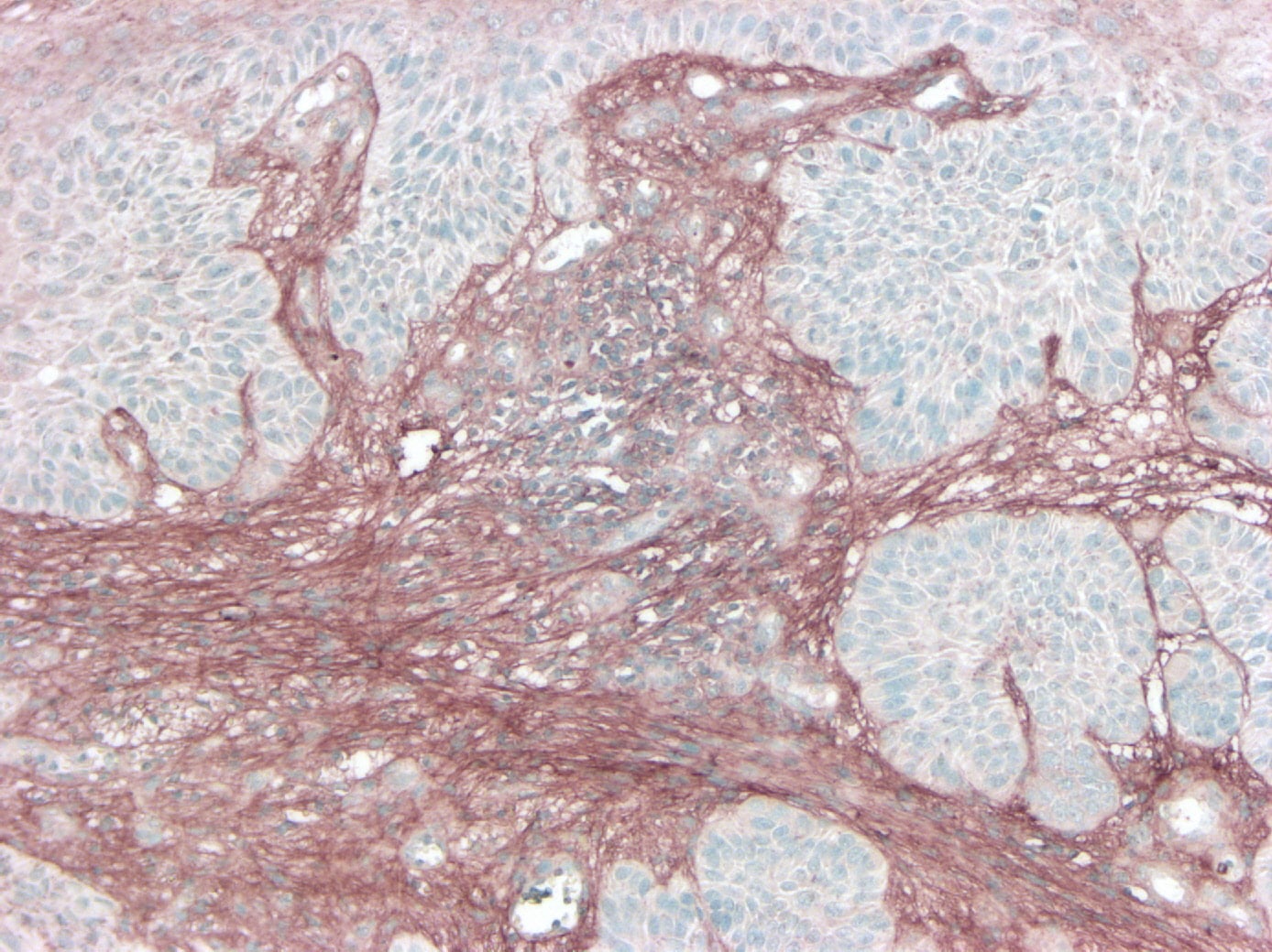
Application: ImmunohistochemistrySample Tested: Skin cancer tissueSpecies: HumanVerified Customer | Posted 11/11/2024FFPE section of cSCC tumour tissue stained with VCAN (AF3054) in brown. HIER of pH 6 was used. Primary antibody dilution of 1:200 was used and secondary anti-Goat HRP was used for detection. Blue staining represt nuclei.

There are no reviews that match your criteria.
Protocols
Find general support by application which include: protocols, troubleshooting, illustrated assays, videos and webinars.
- Antigen Retrieval Protocol (PIER)
- Antigen Retrieval for Frozen Sections Protocol
- Appropriate Fixation of IHC/ICC Samples
- Cellular Response to Hypoxia Protocols
- Chromogenic IHC Staining of Formalin-Fixed Paraffin-Embedded (FFPE) Tissue Protocol
- Chromogenic Immunohistochemistry Staining of Frozen Tissue
- ClariTSA™ Fluorophore Kits
- Detection & Visualization of Antibody Binding
- Fluorescent IHC Staining of Frozen Tissue Protocol
- Graphic Protocol for Heat-induced Epitope Retrieval
- Graphic Protocol for the Preparation and Fluorescent IHC Staining of Frozen Tissue Sections
- Graphic Protocol for the Preparation and Fluorescent IHC Staining of Paraffin-embedded Tissue Sections
- Graphic Protocol for the Preparation of Gelatin-coated Slides for Histological Tissue Sections
- ICC Cell Smear Protocol for Suspension Cells
- ICC Immunocytochemistry Protocol Videos
- ICC for Adherent Cells
- IHC Sample Preparation (Frozen sections vs Paraffin)
- Immunocytochemistry (ICC) Protocol
- Immunocytochemistry Troubleshooting
- Immunofluorescence of Organoids Embedded in Cultrex Basement Membrane Extract
- Immunofluorescent IHC Staining of Formalin-Fixed Paraffin-Embedded (FFPE) Tissue Protocol
- Immunohistochemistry (IHC) and Immunocytochemistry (ICC) Protocols
- Immunohistochemistry Frozen Troubleshooting
- Immunohistochemistry Paraffin Troubleshooting
- Preparing Samples for IHC/ICC Experiments
- Preventing Non-Specific Staining (Non-Specific Binding)
- Primary Antibody Selection & Optimization
- Protocol for Heat-Induced Epitope Retrieval (HIER)
- Protocol for Making a 4% Formaldehyde Solution in PBS
- Protocol for VisUCyte™ HRP Polymer Detection Reagent
- Protocol for the Fluorescent ICC Staining of Cell Smears - Graphic
- Protocol for the Fluorescent ICC Staining of Cultured Cells on Coverslips - Graphic
- Protocol for the Preparation & Fixation of Cells on Coverslips
- Protocol for the Preparation and Chromogenic IHC Staining of Frozen Tissue Sections
- Protocol for the Preparation and Chromogenic IHC Staining of Frozen Tissue Sections - Graphic
- Protocol for the Preparation and Chromogenic IHC Staining of Paraffin-embedded Tissue Sections
- Protocol for the Preparation and Chromogenic IHC Staining of Paraffin-embedded Tissue Sections - Graphic
- Protocol for the Preparation and Fluorescent ICC Staining of Cells on Coverslips
- Protocol for the Preparation and Fluorescent ICC Staining of Non-adherent Cells
- Protocol for the Preparation and Fluorescent ICC Staining of Stem Cells on Coverslips
- Protocol for the Preparation and Fluorescent IHC Staining of Frozen Tissue Sections
- Protocol for the Preparation and Fluorescent IHC Staining of Paraffin-embedded Tissue Sections
- Protocol for the Preparation of Gelatin-coated Slides for Histological Tissue Sections
- Protocol for the Preparation of a Cell Smear for Non-adherent Cell ICC - Graphic
- R&D Systems Quality Control Western Blot Protocol
- TUNEL and Active Caspase-3 Detection by IHC/ICC Protocol
- The Importance of IHC/ICC Controls
- Troubleshooting Guide: Immunohistochemistry
- Troubleshooting Guide: Western Blot Figures
- Western Blot Conditions
- Western Blot Protocol
- Western Blot Protocol for Cell Lysates
- Western Blot Troubleshooting
- Western Blot Troubleshooting Guide
- View all Protocols, Troubleshooting, Illustrated assays and Webinars
Loading...
Associated Pathways
