Vitronectin is a 71 kDa secreted glycoprotein produced by the liver and tumor cells. In blood, Vitronectin is called serum spreading factor. In the extracellular matrix, its function is determined by binding partners such as PAI-1, complement factors, integrins (notably alpha v beta 3) and thrombin. The 459 aa mature human Vitronectin shows 74% amino acid identity with mouse Vitronectin and contains somatomedin B-like and hemopexin-like domains, an RGD motif, a basic heparin-binding domain and sulfated tyrosines. Unbound Vitronectin is a monomer that may be cleaved to form a dimer of 65 kDa and 10 kDa components.
Human Vitronectin Antibody (342603)
R&D Systems | Catalog # MAB2349

Key Product Details
Species Reactivity
Validated:
Human
Cited:
Human
Applications
Validated:
Immunohistochemistry, Western Blot, Simple Western
Cited:
Western Blot
Label
Unconjugated
Antibody Source
Monoclonal Mouse IgG2B Clone # 342603
Loading...
Product Specifications
Immunogen
Human plasma-derived Vitronectin
Specificity
Detects human Vitronectin in direct ELISAs and Western blots. In direct ELISAs, less than 20% cross-reactivity with recombinant bovine vitronectin or recombinant mouse vitronectin is observed.
Clonality
Monoclonal
Host
Mouse
Isotype
IgG2B
Scientific Data Images for Human Vitronectin Antibody (342603)
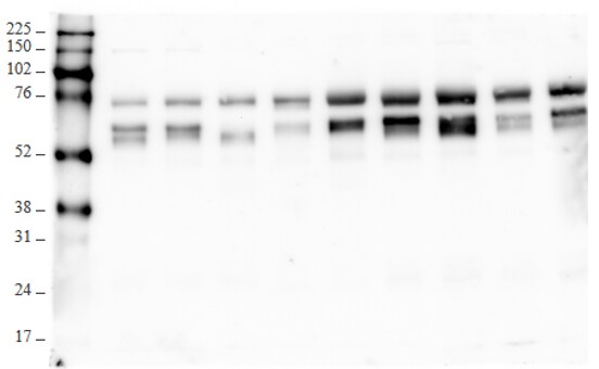
Detection of Human Vitronectin by Western Blot.
Western blot shows human serum. PVDF membrane was probed with 0.1 µg/mL of Mouse Anti-Human Vitronectin Monoclonal Antibody (Catalog # MAB2349) followed by HRP-conjugated Anti-Mouse IgG Secondary Antibody (Catalog # HAF007). A specific band was detected for Vitronectin at approximately 65 kDa (as indicated). This experiment was conducted under reducing conditions and using Immunoblot Buffer Group 1.
Vitronectin in Human Bladder.
Vitronectin was detected in immersion fixed paraffin-embedded sections of human bladder using Mouse Anti-Human Vitronectin Monoclonal Antibody (Catalog # MAB2349) at 8 µg/mL overnight at 4 °C. Tissue was stained using the Anti-Mouse HRP-DAB Cell & Tissue Staining Kit (brown; Catalog # CTS002) and counterstained with hematoxylin (blue). Specific labeling was localized to endothelial cells. View our protocol for Chromogenic IHC Staining of Paraffin-embedded Tissue Sections.
Detection of Human Vitronectin by Simple WesternTM.
Simple Western lane view shows human serum, loaded at 0.2 mg/mL. A specific band was detected for Vitronectin at approximately 85 kDa (as indicated) using 5 µg/mL of Mouse Anti-Human Vitronectin Monoclonal Antibody (Catalog # MAB2349). This experiment was conducted under reducing conditions and using the 12-230 kDa separation system.
Detection of Human Vitronectin by Western Blot
Vitronectin expression is modulated by PI3K/AKT axis.A. Displays the pseudo blot extracted from Simple Western experiments. There are four cell-lines- MDA-MB-231, MCF7, MDA-MB-468, HCC1599, were used to evaluate the signaling cascade relationship. Different protein expression levels were normalized with an averaged house-keeping GAPDH and beta -actin expression B. Compares vitronectin concentration levels in four BC cell lines. C. PI3K concentration levels in BC same cell-lines. D. compares AKT concentration and, E. P-AKT concentration levels in the same four BC cell lines. Image collected and cropped by CiteAb from the following open publication (https://pubmed.ncbi.nlm.nih.gov/33211735), licensed under a CC-BY license. Not internally tested by R&D Systems.Applications for Human Vitronectin Antibody (342603)
Application
Recommended Usage
Immunohistochemistry
8-25 µg/mL
Sample: Immersion fixed paraffin-embedded sections of human bladder and breast
Sample: Immersion fixed paraffin-embedded sections of human bladder and breast
Simple Western
5 µg/mL
Sample: Human serum
Sample: Human serum
Western Blot
0.1 µg/mL
Sample: Human serum
Sample: Human serum
Reviewed Applications
Read 2 reviews rated 4.5 using MAB2349 in the following applications:
Formulation, Preparation, and Storage
Purification
Protein A or G purified from hybridoma culture supernatant
Reconstitution
Reconstitute at 0.5 mg/mL in sterile PBS. For liquid material, refer to CoA for concentration.
Loading...
Formulation
Lyophilized from a 0.2 μm filtered solution in PBS with Trehalose. *Small pack size (SP) is supplied either lyophilized or as a 0.2 µm filtered solution in PBS.
Shipping
Lyophilized product is shipped at ambient temperature. Liquid small pack size (-SP) is shipped with polar packs. Upon receipt, store immediately at the temperature recommended below.
Stability & Storage
Use a manual defrost freezer and avoid repeated freeze-thaw cycles.
- 12 months from date of receipt, -20 to -70 °C as supplied.
- 1 month, 2 to 8 °C under sterile conditions after reconstitution.
- 6 months, -20 to -70 °C under sterile conditions after reconstitution.
Calculators
Background: Vitronectin
Alternate Names
Complement S-protein, Serum Spreading Factor, Somatomedin B, VTN
Gene Symbol
VTN
Additional Vitronectin Products
Product Documents for Human Vitronectin Antibody (342603)
Certificate of Analysis
To download a Certificate of Analysis, please enter a lot or batch number in the search box below.
Note: Certificate of Analysis not available for kit components.
Product Specific Notices for Human Vitronectin Antibody (342603)
For research use only
Related Research Areas
Citations for Human Vitronectin Antibody (342603)
 Powered by Bioz
Customer Reviews for Human Vitronectin Antibody (342603) (2)
4.5 out of 5
2 Customer Ratings
Have you used Human Vitronectin Antibody (342603)?
Submit a review and receive an Amazon gift card!
$25/€18/£15/$25CAN/¥2500 Yen for a review with an image
$10/€7/£6/$10CAN/¥1110 Yen for a review without an image
Submit a review
Customer Images
Showing
1
-
2 of
2 reviews
Showing All
Filter By:
-
Application: ELISASample Tested: Recombinant proteinSpecies: HumanVerified Customer | Posted 09/30/2019Paired with AF2349

-
Application: Western BlotSample Tested: HC11 cellsSpecies: HumanVerified Customer | Posted 02/22/2016
There are no reviews that match your criteria.
Protocols
Find general support by application which include: protocols, troubleshooting, illustrated assays, videos and webinars.
- Antigen Retrieval Protocol (PIER)
- Antigen Retrieval for Frozen Sections Protocol
- Appropriate Fixation of IHC/ICC Samples
- Cellular Response to Hypoxia Protocols
- Chromogenic IHC Staining of Formalin-Fixed Paraffin-Embedded (FFPE) Tissue Protocol
- Chromogenic Immunohistochemistry Staining of Frozen Tissue
- ClariTSA™ Fluorophore Kits
- Detection & Visualization of Antibody Binding
- Fluorescent IHC Staining of Frozen Tissue Protocol
- Graphic Protocol for Heat-induced Epitope Retrieval
- Graphic Protocol for the Preparation and Fluorescent IHC Staining of Frozen Tissue Sections
- Graphic Protocol for the Preparation and Fluorescent IHC Staining of Paraffin-embedded Tissue Sections
- Graphic Protocol for the Preparation of Gelatin-coated Slides for Histological Tissue Sections
- IHC Sample Preparation (Frozen sections vs Paraffin)
- Immunofluorescent IHC Staining of Formalin-Fixed Paraffin-Embedded (FFPE) Tissue Protocol
- Immunohistochemistry (IHC) and Immunocytochemistry (ICC) Protocols
- Immunohistochemistry Frozen Troubleshooting
- Immunohistochemistry Paraffin Troubleshooting
- Preparing Samples for IHC/ICC Experiments
- Preventing Non-Specific Staining (Non-Specific Binding)
- Primary Antibody Selection & Optimization
- Protocol for Heat-Induced Epitope Retrieval (HIER)
- Protocol for Making a 4% Formaldehyde Solution in PBS
- Protocol for VisUCyte™ HRP Polymer Detection Reagent
- Protocol for the Preparation & Fixation of Cells on Coverslips
- Protocol for the Preparation and Chromogenic IHC Staining of Frozen Tissue Sections
- Protocol for the Preparation and Chromogenic IHC Staining of Frozen Tissue Sections - Graphic
- Protocol for the Preparation and Chromogenic IHC Staining of Paraffin-embedded Tissue Sections
- Protocol for the Preparation and Chromogenic IHC Staining of Paraffin-embedded Tissue Sections - Graphic
- Protocol for the Preparation and Fluorescent IHC Staining of Frozen Tissue Sections
- Protocol for the Preparation and Fluorescent IHC Staining of Paraffin-embedded Tissue Sections
- Protocol for the Preparation of Gelatin-coated Slides for Histological Tissue Sections
- R&D Systems Quality Control Western Blot Protocol
- TUNEL and Active Caspase-3 Detection by IHC/ICC Protocol
- The Importance of IHC/ICC Controls
- Troubleshooting Guide: Immunohistochemistry
- Troubleshooting Guide: Western Blot Figures
- Western Blot Conditions
- Western Blot Protocol
- Western Blot Protocol for Cell Lysates
- Western Blot Troubleshooting
- Western Blot Troubleshooting Guide
- View all Protocols, Troubleshooting, Illustrated assays and Webinars
Loading...