Key Product Details

Species Reactivity

Validated:

Mouse

Cited:

Mouse, Rat

Applications

Validated:

Immunohistochemistry, Western Blot, ELISA Capture (Matched Antibody Pair), Intracellular Staining by Flow Cytometry, Simple Western, CyTOF-ready

Cited:

Immunohistochemistry, Immunohistochemistry-Paraffin, Immunohistochemistry-Frozen, Western Blot, Neutralization, Flow Cytometry, Immunocytochemistry, Immunoprecipitation, ELISA Capture

Label

Unconjugated

Antibody Source

Polyclonal Goat IgG
Loading...

Product Specifications

Immunogen

E. coli-derived recombinant mouse Galectin-1
Ala2-Glu135
Accession # P16045

Specificity

Detects mouse Galectin-1 in ELISAs and Western blots. In sandwich ELISAs, less than 5% cross-reactivity with recombinant human (rh) Galectin‑1 and less than 0.5% cross-reactivity with recombinant mouse (rm) Galectin-3, rhGalectin-4, rmGalectin-7, and rhGalectin‑8 is observed.

Clonality

Polyclonal

Host

Goat

Isotype

IgG

Scientific Data Images for Mouse Galectin‑1 Antibody

Galectin‑1 antibody in Mouse Thymus by Immunohistochemistry (IHC-Fr).

Galectin‑1 in Mouse Thymus.

Galectin‑1 was detected in perfusion fixed frozen sections of mouse thymus using Goat Anti-Mouse Galectin‑1 Antigen Affinity-purified Polyclonal Antibody (Catalog # AF1245) at 5 µg/mL overnight at 4 °C. Tissue was stained using the Anti-Goat HRP-DAB Cell & Tissue Staining Kit (brown; Catalog # CTS008) and counterstained with hematoxylin (blue). Specific labeling was localized to the cytoplasm of lymphocytes. View our protocol for Chromogenic IHC Staining of Frozen Tissue Sections.

Detection of Galectin-1 antibody in Mouse Splenocytes antibody by Flow Cytometry.

Detection of Galectin‑1 in Mouse Splenocytes by Flow Cytometry.

Mouse splenocytes were stained with Rat Anti-Mouse CD3 APC-conjugated Monoclonal Antibody (Catalog # FAB4841A) and either (A) Normal Goat IgG Control (Catalog # AB-108-C) or (B) Goat Anti-Mouse Galectin-1 Antigen Affinity-purified Polyclonal Antibody (Catalog # AF1245) followed by Phycoerythrin-conjugated Anti-Goat IgG Secondary Antibody (Catalog # F0107). To facilitate intracellular staining, cells were fixed with paraformaldehyde and permeabilized with saponin.

Detection of Mouse Galectin‑1 by Simple WesternTM.

Simple Western lane view shows lysates of L6 rat myoblast cell line, C2C12 mouse myoblast cell line, THP‑1 human acute monocytic leukemia cell line and Neuro‑2A mouse neuroblastoma cell line, loaded at 0.2 mg/mL. A specific band was detected for Galectin‑1 at approximately 18 kDa (as indicated) using 5 µg/mL of Goat Anti-Mouse Galectin‑1 Antigen Affinity-purified Polyclonal Antibody (Catalog # AF1245). This experiment was conducted under reducing conditions and using the 12-230kDa separation system.

Detection of Mouse Galectin‑1 by Western Blot.

Western blot shows lysates of THP‑1 human acute monocytic leukemia cell line, L6 rat myoblast cell line, C2C12 mouse myoblast cell line, Neuro‑2A mouse neuroblastoma cell line and Mouse Thymus. PVDF membrane was probed with 0.1 µg/mL of Goat Anti-Mouse Galectin‑1 Antigen Affinity-purified Polyclonal Antibody (Catalog # AF1245) followed by HRP-conjugated Anti-Goat IgG Secondary Antibody (Catalog # HAF017). A specific band was detected for Galectin‑1 at approximately 14 kDa (as indicated). This experiment was conducted under reducing conditions and using Western Blot Buffer Group 1.

Applications for Mouse Galectin‑1 Antibody

Application
Recommended Usage

CyTOF-ready

Ready to be labeled using established conjugation methods. No BSA or other carrier proteins that could interfere with conjugation.

Immunohistochemistry

5-15 µg/mL
Sample: Perfusion fixed frozen sections of mouse thymus

Intracellular Staining by Flow Cytometry

2.5 µg/106 cells
Sample: Mouse splenocytes

Simple Western

5 µg/mL
Sample: L6 rat myoblast cell line, C2C12 mouse myoblast cell line, THP‑1 human acute monocytic leukemia cell line and Neuro‑2A mouse neuroblastoma cell line

Western Blot

0.1 µg/mL
Sample: THP‑1 human acute monocytic leukemia cell line, L6 rat myoblast cell line, C2C12 mouse myoblast cell line, Neuro‑2A mouse neuroblastoma cell line and Mouse Thymus

Mouse Galectin-1 Sandwich Immunoassay

ELISA Capture (Matched Antibody Pair)
Recommended Concentration: 0.2-0.8 µg/mL
Use in combination with these reagents:
  • Detection Reagent: Mouse Galectin‑1 Biotinylated Antibody (Catalog # BAF1245)
  • Standard: Recombinant Mouse Galectin-1 Protein (Catalog # 1245-GA)
Please Note: Optimal dilutions of this antibody should be experimentally determined.

Reviewed Applications

Read 5 reviews rated 5 using AF1245 in the following applications:

Flow Cytometry Panel Builder

Bio-Techne Knows Flow Cytometry

Save time and reduce costly mistakes by quickly finding compatible reagents using the Panel Builder Tool.

Advanced Features

  • Spectra Viewer - Custom analysis of spectra from multiple fluorochromes
  • Spillover Popups - Visualize the spectra of individual fluorochromes
  • Antigen Density Selector - Match fluorochrome brightness with antigen density
Build Your Panel Now

Formulation, Preparation, and Storage

Purification

Antigen Affinity-purified

Reconstitution

Reconstitute at 0.2 mg/mL in sterile PBS. For liquid material, refer to CoA for concentration.


Loading...

Formulation

Lyophilized from a 0.2 μm filtered solution in PBS with Trehalose. *Small pack size (SP) is supplied either lyophilized or as a 0.2 µm filtered solution in PBS.

Shipping

Lyophilized product is shipped at ambient temperature. Liquid small pack size (-SP) is shipped with polar packs. Upon receipt, store immediately at the temperature recommended below.

Stability & Storage

Use a manual defrost freezer and avoid repeated freeze-thaw cycles.
  • 12 months from date of receipt, -20 to -70 °C as supplied.
  • 1 month, 2 to 8 °C under sterile conditions after reconstitution.
  • 6 months, -20 to -70 °C under sterile conditions after reconstitution.

Calculators

The reconstitution calculator allows you to quickly calculate the volume of a reagent to reconstitute your vial. Simply enter the mass of reagent and the target concentration and the calculator will determine the rest.

=
÷

Background: Galectin-1

The galectins constitute a large family of carbohydrate-binding proteins with specificity for N-acetyl-lactosamine-containing glycoproteins. At least 14 mammalian galectins, which share structural similarities in their carbohydrate recognition domains (CRD), have been identified to date. The galectins have been classified into the prototype galectins (-1, -2, -5, -7, -10, -11, -13, -14), which contain one CRD and exist either as a monomer or a noncovalent homodimer; the chimera galectins (Galectin-3) containing one CRD linked to a nonlectin domain; and the tandem-repeat galectins (-4, -6, -8, -9, -12) consisting of two CRDs joined by a linker peptide. Galectins lack a classical signal peptide and can be localized to the cytosolic compartments where they have intracellular functions. However, via one or more as yet unidentified non-classical secretory pathways, galectins can also be secreted to function extracellularly. Individual members of the galectin family have different tissue distribution profiles and exhibit subtle differences in their carbohydrate-binding specificities. Each family member may preferentially bind to a unique subset of cell-surface glycoproteins (1-4).

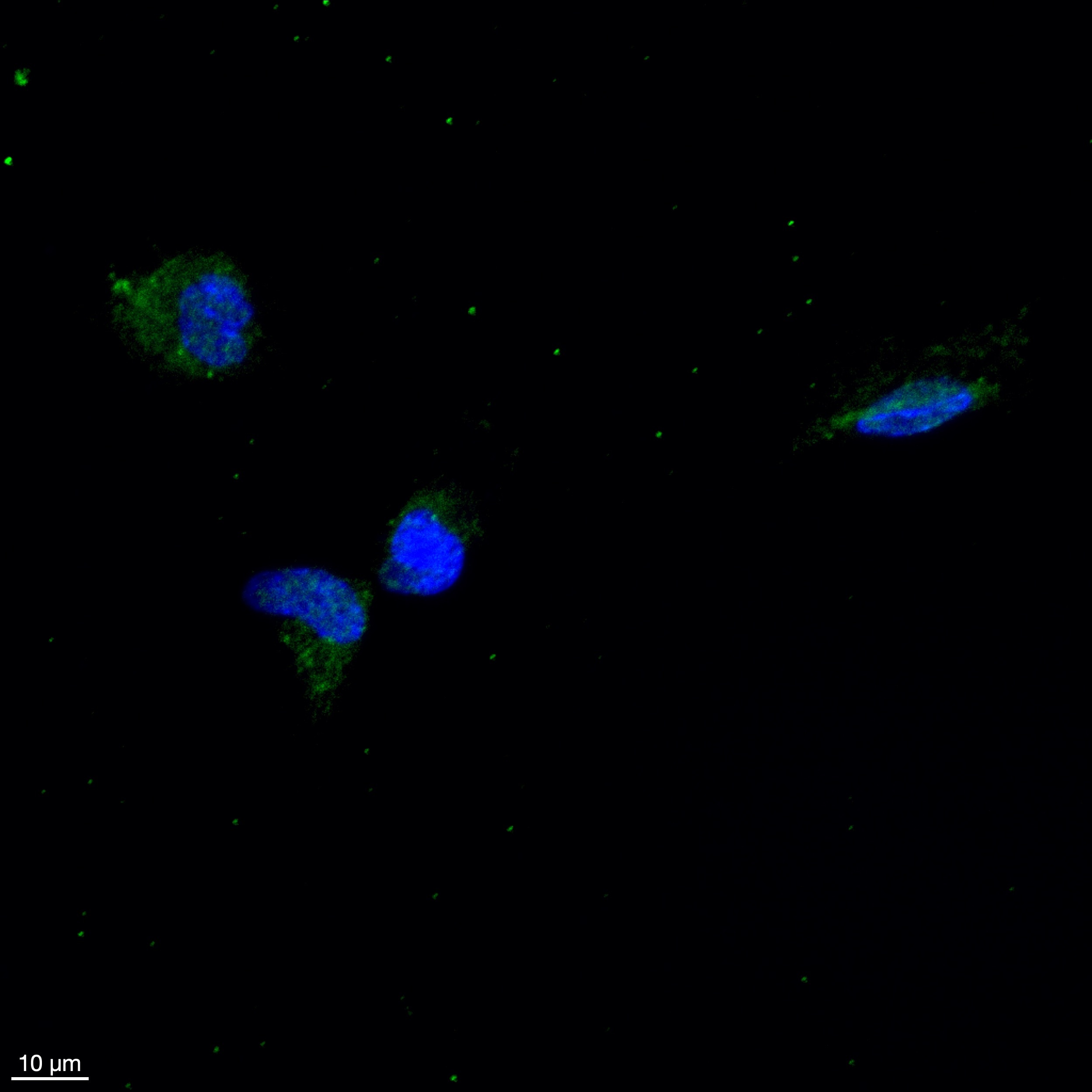
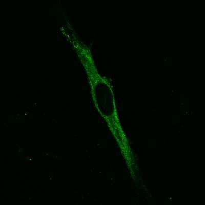
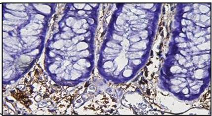
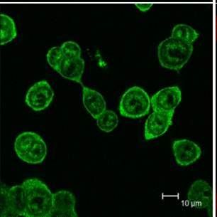
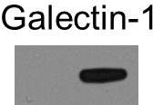
Mouse Galectin-1, also known as beta-galactoside-binding lectin L-14-I, lactose-binding lectin 1, S-Lac lectin 1, galaptin and 14 kDa lectin, is a monomeric or homodimeric prototype galectin that is expressed in a variety of cells and tissues including muscle, heart, lymph nodes, spleen, thymus, macrophages, B cells, T cells, dendritic cells, and tumor cells. It preferentially binds laminin, fibronectin, 90K/Mac-2BP, CD45, CD43, CD7, CD2, CD3, and ganglioside GM1. Galectin-1 modulates cell growth, proliferation and differentiation, either positively or negatively, depending on the cell type and activation status. It controls cell survival by inducing apoptosis of activated T cells and immature thymocytes. It modulates cytokine secretion by inducing Th2 type cytokines and inhibiting pro-inflammatory cytokine production. Galectin-1 can also modulate cell-cell as well as cell-matrix interactions and depending on the cell type and developmental stage, promote cell attachment or detachment. Galectin-1 has immunosuppressive and anti-inflammatory properties and has been shown to suppress acute and chronic inflammation and autoimmunity. Mouse and human Galectin-1 share about 88% amino acid sequence similarity (1-5).

References

  1. Rabinovich, A. et al. (2002) Trends in Immunol. 23:313.
  2. Rabinovich, A. et al. (2002) J. Leukocyte Biology 71:741.
  3. Hughes, R.C. (2001) Biochimie 83:667.
  4. R&D Systems’ Cytokine Bulletin, Summer (2002).
  5. Goldring, K. et al. (2002) J. Cell Science 115:355.

Alternate Names

BHL, Galaptin, Galectin1, GBP, L-14, LGALS1

Entrez Gene IDs

3956 (Human); 16852 (Mouse)

Gene Symbol

LGALS1

UniProt

Additional Galectin-1 Products

Product Documents for Mouse Galectin‑1 Antibody

Certificate of Analysis

To download a Certificate of Analysis, please enter a lot or batch number in the search box below.

Note: Certificate of Analysis not available for kit components.

Product Specific Notices for Mouse Galectin‑1 Antibody

For research use only

Citations for Mouse Galectin‑1 Antibody

Customer Reviews for Mouse Galectin‑1 Antibody (5)

5 out of 5
5 Customer Ratings
5 Stars
100%
4 Stars
0%
3 Stars
0%
2 Stars
0%
1 Stars
0%

Have you used Mouse Galectin‑1 Antibody?

Submit a review and receive an Amazon gift card!

$25/€18/£15/$25CAN/¥2500 Yen for a review with an image

$10/€7/£6/$10CAN/¥1110 Yen for a review without an image

Submit a review
Amazon Gift Card

Customer Images


Showing  1 - 5 of 5 reviews Showing All
Filter By:
  • Mouse Galectin-1 Antibody
    Name: Anonymous
    Application: Western Blot
    Sample Tested: U-87 MG human glioblastoma/astrocytoma cell line
    Species: Mouse
    Verified Customer | Posted 08/17/2016
    1:2000 dilution in TBS-Tween 20 + 5% BSA
    Mouse Galectin‑1 Antibody AF1245
  • Mouse Galectin‑1 Antibody
    Name: Anonymous
    Application: Immunocytochemistry/Immunofluorescence
    Sample Tested: Human fibroblast
    Species: Human
    Verified Customer | Posted 10/26/2015
    These are HFF-2 cells that show the staining for this antibody. <br />Specificity: Specific<br />Sensitivity: Sensitive<br />Buffer: PBS`<br />Dilution: 1:500
    Mouse Galectin‑1 Antibody AF1245
  • Mouse Galectin‑1 Antibody
    Name: Anonymous
    Application: Immunohistochemistry
    Sample Tested: Colon tissue
    Species: Mouse
    Verified Customer | Posted 10/26/2015
    The antibody specifically detects the stromal galectin-1 in normal tissues. <br />Specificity: Specific<br />Sensitivity: Sensitive<br />Buffer: PBS<br />Dilution: 1:1000
    Mouse Galectin‑1 Antibody AF1245
  • Mouse Galectin‑1 Antibody
    Name: Anonymous
    Application: Immunocytochemistry/Immunofluorescence
    Sample Tested: Cancer Cells
    Species: Human
    Verified Customer | Posted 10/26/2015
    Very specific in detection of galectin-1 in cancer cells. <br />Specificity: Specific<br />Sensitivity: Sensitive<br />Buffer: PBS<br />Dilution: 1:1000
    Mouse Galectin‑1 Antibody AF1245
  • Mouse Galectin‑1 Antibody
    Name: Anonymous
    Application: Western Blot
    Sample Tested: Cell Lysates
    Species: Human
    Verified Customer | Posted 10/26/2015
    The antibody is very specific at 1:1000 and also at 1:2500 dilutions. <br />Specificity: Specific<br />Sensitivity: Sensitive<br />Buffer: BSA<br />Dilution: 1:1000
    Mouse Galectin‑1 Antibody AF1245

There are no reviews that match your criteria.

Showing  1 - 5 of 5 reviews Showing All

Protocols

Find general support by application which include: protocols, troubleshooting, illustrated assays, videos and webinars.

FAQs

No product specific FAQs exist for this product.

View all FAQs for Antibodies