Interleukin 17 (also known as CTLA-8) is a T cell-expressed pleiotropic cytokine that exhibits a high degree of homology to a protein encoded by the ORF13 gene of herpesvirus Saimiri. cDNA clones encoding IL-17 have been isolated from activated rat, mouse, and human T cells. Mouse IL-17 cDNA encodes a 158 amino acid (aa) residue precursor protein with a 21 amino acid residue signal peptide that is cleaved to yield the 137 aa residue mature IL-17. Both recombinant and natural IL-17 have been shown to exist as disulfide linked homodimers. At the amino acid level, mIL-17 shows 57% and 87% sequence identity with herpesvirus and rat IL-17, respectively. An IL-17 specific mouse cell surface receptor (IL-17 R) has been cloned. While the expression of IL-17 mRNA is restricted to activated alpha beta TCR+CD4-CD8-T cells, the expression of mIL-17 R mRNA has been detected in virtually all cells and tissues tested. IL-17 exhibits multiple biological activities on a variety of cells including: the induction of IL-6 and IL-8 production in fibroblasts; the enhancement of surface expression of ICAM-1 in fibroblasts; activation of NF-kappa B and costimulation of T cell proliferation.

Key Product Details
Validated by
Biological Validation
Species Reactivity
Validated:
Mouse
Cited:
Mouse
Applications
Validated:
Western Blot, Neutralization, Intracellular Staining by Flow Cytometry, CyTOF-ready
Cited:
Immunohistochemistry, Immunohistochemistry-Frozen, Western Blot, Neutralization
Label
Unconjugated
Antibody Source
Polyclonal Goat IgG
Loading...
Product Specifications
Immunogen
E. coli-derived recombinant mouse IL‑17
Thr22-Ala158
Accession # Q62386
Thr22-Ala158
Accession # Q62386
Specificity
Detects mouse and rat IL-17 in direct ELISAs and Western blots. In direct ELISAs, approximately 10% cross-reactivity with recombinant human IL-17A is observed.
Clonality
Polyclonal
Host
Goat
Isotype
IgG
Endotoxin Level
<0.10 EU per 1 μg of the antibody by the LAL method.
Scientific Data Images for Mouse IL-17/IL-17A Antibody
Detection of IL-17 in EL-4 Mouse Cell Line by Flow Cytometry.
EL-4 mouse lymphoblast cell line was treated for 16 hours with 50 ng/mL PMA then stained with Goat Anti-Mouse IL-17 Antigen Affinity-purified Polyclonal Antibody (Catalog # AF-421-NA, filled histogram) or isotype control antibody (Catalog # AB-108-C, open histogram), followed by Phycoerythrin-conjugated Anti-Goat IgG Secondary Antibody (Catalog # F0107). To facilitate intracellular staining, cells were fixed with paraformaldehyde and permeabilized with saponin.
IL-6 Secretion Induced by IL-17 and Neutralization by Mouse IL-17 Antibody.
Recombinant Mouse IL-17 (Catalog # 421-ML) stimulates IL-6 secretion in the NIH-3T3 mouse embryonic fibroblast cell line in a dose-dependent manner (orange line), as measured by the Mouse IL-6 Quantikine ELISA Kit (Catalog # M6000B). IL-6 secretion elicited by Recombinant Mouse IL-17 (10 ng/mL) is neutralized (green line) by increasing concentrations of Goat Anti-Mouse IL-17 Antigen Affinity-purified Polyclonal Anti-body (Catalog # AF-421-NA). The ND50 is typically 0.05-0.25 µg/mL.
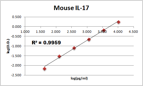
Mouse IL-17 / IL-17A ELISA Standard Curve
Recombinant Mouse IL‑17/IL‑17A (Catalog # 421-ML) was serially diluted and captured by Rat Anti-Mouse IL‑17/IL‑17A Monoclonal Antibody (Catalog # MAB721) coated on a Clear Polystyrene Microplate (Catalog # DY990). Goat Anti-Mouse IL‑17/IL‑17A Antigen Affinity-purified Polyclonal Antibody (Catalog # AF-421-NA) was biotinylated and incubated with the protein captured on the plate. Detection of the standard curve was achieved by incubating Streptavidin-HRP (Catalog # DY998)Applications for Mouse IL-17/IL-17A Antibody
Application
Recommended Usage
CyTOF-ready
Ready to be labeled using established conjugation methods. No BSA or other carrier proteins that could interfere with conjugation.
Intracellular Staining by Flow Cytometry
2.5 µg/106 cells
Sample: EL‑4 mouse lymphoblast cell line treated with LPS or PMA and Ca ionomycin, fixed with paraformaldehyde, and permeabilized with saponin
Sample: EL‑4 mouse lymphoblast cell line treated with LPS or PMA and Ca ionomycin, fixed with paraformaldehyde, and permeabilized with saponin
Western Blot
0.1 µg/mL
Sample: Recombinant Mouse IL‑17 (Catalog # 421-ML)
Sample: Recombinant Mouse IL‑17 (Catalog # 421-ML)
Neutralization
Measured by its ability to neutralize IL‑17-induced IL‑6 secretion in the NIH‑3T3 mouse embryonic fibroblast cell line. Yao, Z. et al. (1995) Immunity 3:811. The Neutralization Dose (ND50) is typically 0.05-0.25 µg/mL in the presence of 10 ng/mL Recombinant Mouse IL‑17.
Reviewed Applications
Read 1 review rated 5 using AF-421-NA in the following applications:
Flow Cytometry Panel Builder
Bio-Techne Knows Flow Cytometry
Save time and reduce costly mistakes by quickly finding compatible reagents using the Panel Builder Tool.
Advanced Features
- Spectra Viewer - Custom analysis of spectra from multiple fluorochromes
- Spillover Popups - Visualize the spectra of individual fluorochromes
- Antigen Density Selector - Match fluorochrome brightness with antigen density

Formulation, Preparation, and Storage
Purification
Antigen Affinity-purified
Reconstitution
Reconstitute at 0.2 mg/mL in sterile PBS. For liquid material, refer to CoA for concentration.
Loading...
Formulation
Lyophilized from a 0.2 μm filtered solution in PBS with Trehalose. *Small pack size (SP) is supplied either lyophilized or as a 0.2 µm filtered solution in PBS.
Shipping
Lyophilized product is shipped at ambient temperature. Liquid small pack size (-SP) is shipped with polar packs. Upon receipt, store immediately at the temperature recommended below.
Stability & Storage
Use a manual defrost freezer and avoid repeated freeze-thaw cycles.
- 12 months from date of receipt, -20 to -70 °C as supplied.
- 1 month, 2 to 8 °C under sterile conditions after reconstitution.
- 6 months, -20 to -70 °C under sterile conditions after reconstitution.
Calculators
Background: IL-17/IL-17A
References
- Kennedy, J. et al. (1996) J. Interferon Cytokine Res. 16:611.
- Yao, Z. et al. (1995) J. Immunol. 155:5483.
- Yao, Z. et al. (1995) Immunity 3:811.
- Rouvier, E. et al. (1993) J. Immunol. 150:5445.
Long Name
Interleukin 17
Alternate Names
CTLA-8, CTLA8, IL-17A, IL17, IL17A
Entrez Gene IDs
Gene Symbol
IL17A
UniProt
Additional IL-17/IL-17A Products
Product Documents for Mouse IL-17/IL-17A Antibody
Certificate of Analysis
To download a Certificate of Analysis, please enter a lot or batch number in the search box below.
Note: Certificate of Analysis not available for kit components.
Product Specific Notices for Mouse IL-17/IL-17A Antibody
For research use only
Citations for Mouse IL-17/IL-17A Antibody
 Powered by Bioz
Customer Reviews for Mouse IL-17/IL-17A Antibody (1)
5 out of 5
1 Customer Rating
Have you used Mouse IL-17/IL-17A Antibody?
Submit a review and receive an Amazon gift card!
$25/€18/£15/$25CAN/¥2500 Yen for a review with an image
$10/€7/£6/$10CAN/¥1110 Yen for a review without an image
Submit a review
Customer Images
Showing
1
-
1 of
1 review
Showing All
Filter By:
-
Application: ELISASample Tested: Serum and PlasmaSpecies: MouseVerified Customer | Posted 11/27/2017Antibody MAB421 was used as the capture with this antibody (AF421) as the detection in an ELISA.

There are no reviews that match your criteria.
Protocols
Find general support by application which include: protocols, troubleshooting, illustrated assays, videos and webinars.
- 7-Amino Actinomycin D (7-AAD) Cell Viability Flow Cytometry Protocol
- Cellular Response to Hypoxia Protocols
- Extracellular Membrane Flow Cytometry Protocol
- Flow Cytometry Protocol for Cell Surface Markers
- Flow Cytometry Protocol for Staining Membrane Associated Proteins
- Flow Cytometry Staining Protocols
- Flow Cytometry Troubleshooting Guide
- Intracellular Flow Cytometry Protocol Using Alcohol (Methanol)
- Intracellular Flow Cytometry Protocol Using Detergents
- Intracellular Nuclear Staining Flow Cytometry Protocol Using Detergents
- Intracellular Staining Flow Cytometry Protocol Using Alcohol Permeabilization
- Intracellular Staining Flow Cytometry Protocol Using Detergents to Permeabilize Cells
- Propidium Iodide Cell Viability Flow Cytometry Protocol
- Protocol for Liperfluo
- Protocol for the Characterization of Human Th22 Cells
- Protocol for the Characterization of Human Th9 Cells
- Protocol: Annexin V and PI Staining by Flow Cytometry
- Protocol: Annexin V and PI Staining for Apoptosis by Flow Cytometry
- R&D Systems Quality Control Western Blot Protocol
- Troubleshooting Guide: Fluorokine Flow Cytometry Kits
- Troubleshooting Guide: Western Blot Figures
- Western Blot Conditions
- Western Blot Protocol
- Western Blot Protocol for Cell Lysates
- Western Blot Troubleshooting
- Western Blot Troubleshooting Guide
- View all Protocols, Troubleshooting, Illustrated assays and Webinars
Loading...
Associated Pathways


