
Key Product Details
Validated by
Species Reactivity
Validated:
Cited:
Applications
Validated:
Cited:
Label
Antibody Source
Product Specifications
Immunogen
Ala11-Phe483
Accession # Q60793
Specificity
Clonality
Host
Isotype
Scientific Data Images for Mouse KLF4 Antibody
Detection of KLF4-regulated Genes by Chromatin Immunoprecipitation.
D3 mouse embryonic stem cell line was fixed using formaldehyde, resuspended in lysis buffer, and sonicated to shear chromatin. KLF4/DNA complexes were immunoprecipitated using 5 µg Goat Anti-Mouse KLF4 Antigen Affinity-purified Polyclonal Antibody (Catalog # AF3158) or control antibody (AB-108-C) for 15 minutes in an ultrasonic bath, followed by Biotinylated Anti-Goat IgG Secondary Antibody (BAF109). Immunocomplexes were captured using 50 µL of MagCellect Streptavidin Ferrofluid (MAG999) and DNA was purified using chelating resin solution. TheB2Rpromoter was detected by standard PCR.
KLF4 in D3 Mouse Cell Line.
KLF4 was detected in immersion fixed D3 mouse embryonic stem cell line using Mouse KLF4 Antigen Affinity-purified Polyclonal Antibody (Catalog # AF3158) at 10 µg/mL for 3 hours at room temperature. Cells were stained using the NorthernLights™ 557-conjugated Anti-Goat IgG Secondary Antibody (red; NL001) and counterstained with DAPI (blue). View our protocol for Fluorescent ICC Staining of Cells on Coverslips.
KLF4 in Mouse Colon Tissue.
KLF4 was detected in perfusion fixed frozen sections of mouse colon tissue using Goat Anti-Mouse KLF4 Antigen Affinity-purified Polyclonal Antibody (Catalog # AF3158) at 1 µg/mL overnight at 4 °C. Before incubation with the primary antibody, tissue was subjected to heat-induced epitope retrieval using Antigen Retrieval Reagent-Basic (CTS013). Tissue was stained using the Anti-Goat IgG VisUCyte™ HRP Polymer Antibody (brown; VC004) and counterstained with hematoxylin (blue). Specific staining was localized to cell nuclei. View our protocol for IHC Staining with VisUCyte HRP Polymer Detection Reagents.
Detection of Mouse KLF4 by Immunohistochemistry
Overexpression of beta -catenin maintains B6 ESC self-renewal in the presence of LIF.(A) Immunoblot analysis of phospho-beta -catenin, beta -catenin and beta -actin levels in B6 ES cells cultured in 1000 U/ml LIF condition supplemented without or with 3 µM CH for 4 days. (B) Immunofluorescence analysis of 3 µM CH-treated and non-treated B6 ESC clones stained for beta -catenin and Hoechst for nuclei in the presence of LIF for 4 days. Scale bars represent 100 µm. (C) Quantitative RT-PCR analysis of Axin2, T and Cdx1 transcript levels in 3 µM CH-treated and control B6 ES cells. Error bars represent the SD of three biological replicates. (D) beta -catenin S33A protein stably expressed in ES cells was detected by immunoblotting. (E) Phase contrast microscopy image and alkaline phosphatase staining of control and beta -catenin S33A-transfected B6 ES cells in the presence of 1000 U/ml LIF for 4 days. Scale bars represent 100 µm. (F) Immunofluorescence staining for OCT4, NANOG, and KLF4 in mock and beta -catenin S33A-transfected B6 ES cells in the presence of 1000 U/ml LIF for 4 days. Cell nuclei were stained with Hoechst. Scale bars represent 100 µm. (G) Quantitative RT-PCR analysis of Oct4, Nanog, Rex1, Klf4, Tbx3, and Fgf5 transcript levels in mock and beta -catenin S33A-transfected B6 ES cells in the presence of 1000 U/ml LIF for 4 days. Error bars represent the SD of three biological replicates. (H) beta -catenin S33A delta C, beta -catenin S33A and TCF4 delta N protein were detected by immunoblotting with anti-flag tag antibody from extracts of ES cells transfected with PiggyBac plasmids. Quantitative RT-PCR analysis of Axin2, T and Cdx1 transcript levels in beta -catenin S33A delta C, beta -catenin S33A, TCF4 delta N, and mock-transfected B6 ES cells. Error bars represent the SD of three biological replicates. (I) Alkaline phosphatase staining of mock-transfected, and beta -catenin delta C over-expressing B6 ES cells cultured in LIF for 5 passages, and mock-transfected and TCF4 delta N-transfected B6 ESCs cultured in CHIR99021 plus LIF for 5 passages. Scale
Detection of Mouse KLF4 by Immunocytochemistry/Immunofluorescence
LIF maintains B6 ES cell self-renewal in the presence of CHIR99021.(A) Phase contrast image of B6 ES cells in the presence of 1000 U/ml LIF supplemented without or with 3 µM CHIR99021 (CH) for 4 days. Scale bars represent 100 µm. (B) Alkaline phosphatase staining of B6 ES cells in the presence of 1000 U/ml LIF supplemented without or with 3 µM CH for 4 days. Scale bars represent 100 µm. (C) Immunofluorescence staining for OCT4, NANOG, and KLF4 in B6 ES cells treated with 1000 U/ml LIF and 3 µM CH for 4 days. Cell nuclei were stained using Hoechst. Scale bars represent 100 µm. (D) Relative quantification of Oct4, Nanog, Rex1, Klf4, Tbx3, and Fgf5 mRNA in B6 ES cells in 1000 U/ml LIF culture without or with 3 µM CH, by qRT-PCR. Error bars represent the SD of three biological replicates. (E) B6 ES cells,1×104/well, cultured in different concentrations of LIF, ranging from 0 to 10,000 U/ml, in the absence or presence of 3 µM CH for 5 days, were assessed for alkaline phosphatase activity. Scale bars represent 100 µm. Image collected and cropped by CiteAb from the following publication (https://dx.plos.org/10.1371/journal.pone.0035892), licensed under a CC-BY license. Not internally tested by R&D Systems.
Detection of Mouse KLF4 by Chromatin Immunoprecipitation
Overlapping SIM2 occupancy with master transcription factor binding sites.a. Frequency distribution of OCT4, SOX2 and NANOG DNA binding sites in a 40kb window centered to the newly identified SIM2 DNA binding sites. Plots show a significant enrichment for the OSN binding sites at the SIM2 peak localization in SIM2 A6 expressing cells. Pie charts show the proportion of SIM2 DNA binding sites overlapping with the OCT4, SOX2 or NANOG binding sites (in grey) (100bp window). p = Fisher’s exact test p-value; F score: measure of the significance of the association (1 = perfect match). b. Protein co-immunoprecipitation experiments of SIM2-FLAG with endogenous OCT4, SOX2, KLF4 (left panel) and NANOG (right panel). Cellular protein extracts from Sim2 expressing cells (A6) or EB3 cells were immunoprecipitated by using antibodies directed against each of the pluripotency factors (N-terminal and C-terminal part of NANOG) or IgG as a negative control for co-immunoprecipitation. Associated proteins were immunoblotted using an anti-FLAG antibody. Red star shows the SIM2-FLAG protein, blue star the signal given by the recognition of the IgG heavy chains. Ø: Beads only; kDa: kilodaltons; protein lysat: protein lysat was loaded as an input control for the immunoblot. Image collected and cropped by CiteAb from the following publication (https://pubmed.ncbi.nlm.nih.gov/25955728), licensed under a CC-BY license. Not internally tested by R&D Systems.
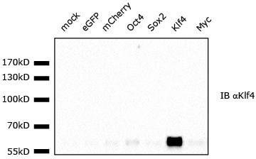
Detection of KLF4 by Western Blot
CMTR1 is required for histone and ribosomal protein gene expression during differentiation. (A) Cmtr1WT,Cmtr1KD and Cmtr1KD/HA-CMTR1 ESCs were cultured in 35S cysteine/methionine for 30 min and incorporation into cellular proteins was determined (N = 3). Average and standard deviation is presented. Student’s T test; ‘*’ indicates P-value < 0.05 relative to control. (B,C) Western blot analysis of ESC cultured in (B) LIF and (C) without LIF for 2 days following CMTR1 siRNA transfection; N = 3. (D) Cmtr1WT and Cmtr1KD ESC were cultured in and without LIF for the days indicated. Cell number was counted; N = 3. Average and standard deviation presented. Student’s T test; ‘*’ indicates P-value < 0.05 relative to control timepoint. (E) Cmtr1WT and Cmtr1KDESC were induced to differentiate (E) by LIF withdrawal, (F) towards a neural fate. Phase contrast images and representative western blot analysis. (G) The ESC lines indicated were cultured without LIF for 6 days, then stained with alkaline phosphatase. Resultant colonies were scored as alkaline phosphatase (AP) negative (-), positive (+) or mixed; N = 4. Average and standard deviation presented. Student’s T test performed on data from AP- colonies; ‘*’ indicates P-value < 0.05 relative to control timepoint. Image collected and cropped by CiteAb from the following open publication (https://pubmed.ncbi.nlm.nih.gov/35212377), licensed under a CC-BY license. Not internally tested by R&D Systems.Applications for Mouse KLF4 Antibody
Chromatin Immunoprecipitation (ChIP)
Sample: D3 mouse embryonic stem cell line chromatin, B2R promoter detected by standard PCR.
Immunocytochemistry
Sample: Immersion fixed D3 mouse embryonic stem cell line
Immunohistochemistry
Sample: Perfusion fixed frozen sections of mouse colon tissue
Western Blot
Sample: Recombinant Mouse KLF4
Reviewed Applications
Read 4 reviews rated 4.5 using AF3158 in the following applications:
Formulation, Preparation, and Storage
Purification
Reconstitution
Reconstitute at 0.2 mg/mL in sterile PBS. For liquid material, refer to CoA for concentration.
Formulation
Shipping
Stability & Storage
- 12 months from date of receipt, -20 to -70 °C as supplied.
- 1 month, 2 to 8 °C under sterile conditions after reconstitution.
- 6 months, -20 to -70 °C under sterile conditions after reconstitution.
Calculators
Background: KLF4
Long Name
Alternate Names
Entrez Gene IDs
Gene Symbol
UniProt
Additional KLF4 Products
Product Documents for Mouse KLF4 Antibody
Certificate of Analysis
To download a Certificate of Analysis, please enter a lot or batch number in the search box below.
Note: Certificate of Analysis not available for kit components.
Product Specific Notices for Mouse KLF4 Antibody
For research use only
Citations for Mouse KLF4 Antibody
 Powered by Bioz
Customer Reviews for Mouse KLF4 Antibody (4)
Have you used Mouse KLF4 Antibody?
Submit a review and receive an Amazon gift card!
$25/€18/£15/$25CAN/¥2500 Yen for a review with an image
$10/€7/£6/$10CAN/¥1110 Yen for a review without an image
Submit a review
Customer Images
-
Application: Western BlotSample Tested: Mouse intestinal tissue, Human colorectal cancer tissue, HCT116 , Mouse embryonic fibroblasts and Caco-2 human colorectal adenocarcinoma cell lineSpecies: Mouse and HumanVerified Customer | Posted 02/22/2016
-
Application: Western BlotSample Tested: Mouse embryonic fibroblastsSpecies: MouseVerified Customer | Posted 10/26/2015MEFs expressing indicated constructs were lysed and total lysates run on 10% gels.<br /> Blots were blocked in 5% milk in TBST, incubated with antibody in 1% milk in TBST over night at 4C, washed 3x in TBST for 15min, incubated with HRP-conjugated secondary antibody in 1% milk in TBST for 1h, washed again 3x in TBST for 15min and then developed.<br /> Bands in other than Klf4 lane show endogenous Klf4 proteins. <br />Specificity: Specific<br />Sensitivity: Sensitive<br />Buffer: TBST + 1% milk<br />Dilution: 1:1000

-
Application: Western BlotSample Tested: See PMID 23395636Species: MouseVerified Customer | Posted 01/07/2015
-
Application: Western BlotSample Tested: See PMID 20550931Species: MouseVerified Customer | Posted 01/07/2015
There are no reviews that match your criteria.
Protocols
Find general support by application which include: protocols, troubleshooting, illustrated assays, videos and webinars.
- Antigen Retrieval Protocol (PIER)
- Antigen Retrieval for Frozen Sections Protocol
- Appropriate Fixation of IHC/ICC Samples
- Cellular Response to Hypoxia Protocols
- ChIP Protocol Video
- Chromatin Immunoprecipitation (ChIP) Protocol
- Chromatin Immunoprecipitation Protocol
- Chromogenic IHC Staining of Formalin-Fixed Paraffin-Embedded (FFPE) Tissue Protocol
- Chromogenic Immunohistochemistry Staining of Frozen Tissue
- ClariTSA™ Fluorophore Kits
- Detection & Visualization of Antibody Binding
- Fluorescent IHC Staining of Frozen Tissue Protocol
- Graphic Protocol for Heat-induced Epitope Retrieval
- Graphic Protocol for the Preparation and Fluorescent IHC Staining of Frozen Tissue Sections
- Graphic Protocol for the Preparation and Fluorescent IHC Staining of Paraffin-embedded Tissue Sections
- Graphic Protocol for the Preparation of Gelatin-coated Slides for Histological Tissue Sections
- ICC Cell Smear Protocol for Suspension Cells
- ICC Immunocytochemistry Protocol Videos
- ICC for Adherent Cells
- IHC Sample Preparation (Frozen sections vs Paraffin)
- Immunocytochemistry (ICC) Protocol
- Immunocytochemistry Troubleshooting
- Immunofluorescence of Organoids Embedded in Cultrex Basement Membrane Extract
- Immunofluorescent IHC Staining of Formalin-Fixed Paraffin-Embedded (FFPE) Tissue Protocol
- Immunohistochemistry (IHC) and Immunocytochemistry (ICC) Protocols
- Immunohistochemistry Frozen Troubleshooting
- Immunohistochemistry Paraffin Troubleshooting
- Preparing Samples for IHC/ICC Experiments
- Preventing Non-Specific Staining (Non-Specific Binding)
- Primary Antibody Selection & Optimization
- Protocol for Heat-Induced Epitope Retrieval (HIER)
- Protocol for Making a 4% Formaldehyde Solution in PBS
- Protocol for VisUCyte™ HRP Polymer Detection Reagent
- Protocol for the Fluorescent ICC Staining of Cell Smears - Graphic
- Protocol for the Fluorescent ICC Staining of Cultured Cells on Coverslips - Graphic
- Protocol for the Preparation & Fixation of Cells on Coverslips
- Protocol for the Preparation and Chromogenic IHC Staining of Frozen Tissue Sections
- Protocol for the Preparation and Chromogenic IHC Staining of Frozen Tissue Sections - Graphic
- Protocol for the Preparation and Chromogenic IHC Staining of Paraffin-embedded Tissue Sections
- Protocol for the Preparation and Chromogenic IHC Staining of Paraffin-embedded Tissue Sections - Graphic
- Protocol for the Preparation and Fluorescent ICC Staining of Cells on Coverslips
- Protocol for the Preparation and Fluorescent ICC Staining of Non-adherent Cells
- Protocol for the Preparation and Fluorescent ICC Staining of Stem Cells on Coverslips
- Protocol for the Preparation and Fluorescent IHC Staining of Frozen Tissue Sections
- Protocol for the Preparation and Fluorescent IHC Staining of Paraffin-embedded Tissue Sections
- Protocol for the Preparation of Gelatin-coated Slides for Histological Tissue Sections
- Protocol for the Preparation of a Cell Smear for Non-adherent Cell ICC - Graphic
- R&D Systems Quality Control Western Blot Protocol
- TUNEL and Active Caspase-3 Detection by IHC/ICC Protocol
- The Importance of IHC/ICC Controls
- Troubleshooting Guide: Immunohistochemistry
- Troubleshooting Guide: Western Blot Figures
- Western Blot Conditions
- Western Blot Protocol
- Western Blot Protocol for Cell Lysates
- Western Blot Troubleshooting
- Western Blot Troubleshooting Guide
- View all Protocols, Troubleshooting, Illustrated assays and Webinars
FAQs for Mouse KLF4 Antibody
-
Q: Can I use this antibody in rabbit samples?
A: AF3158 is against KLF4 and is derived from mouse. Unfortunately, it has not been tested with the rabbit samples. Therefore we don't know for sure whether this antibody will work in rabbit. Since it is a polyclonal antibody, the likelihood that this antibody will recognize the rabbit KLF4 is good.

