Key Product Details

Species Reactivity

Validated:

Mouse

Cited:

Human, Mouse, Rat, Porcine, Transgenic Mouse

Applications

Validated:

Immunohistochemistry, Western Blot

Cited:

Immunohistochemistry, Immunohistochemistry-Frozen, Western Blot, Neutralization, Flow Cytometry, Immunocytochemistry, ELISA Development (Detection)

Label

Unconjugated

Antibody Source

Polyclonal Goat IgG
Loading...

Product Specifications

Immunogen

Mouse myeloma cell line NS0-derived recombinant mouse NGF R/TNFRSF16
Gly20-Asn243
Accession # Q9Z0W1

Specificity

Detects mouse NGF R/TNFRSF16 in direct ELISAs and Western blots. In direct ELISAs and Western blots, approximately 5% cross-reactivity with recombinant human NGF R/TNFRSF16 is observed.

Clonality

Polyclonal

Host

Goat

Isotype

IgG

Scientific Data Images for Mouse NGFR/TNFRSF16 Antibody

Detection of Mouse NGF R/TNFRSF16 antibody by Western Blot.

Detection of Mouse NGF R/TNFRSF16 by Western Blot.

Western blot shows lysates of mouse uterus tissue. PVDF membrane was probed with 2 µg/mL of Goat Anti-Mouse NGF R/TNFRSF16 Antigen Affinity-purified Polyclonal Antibody (Catalog # AF1157) followed by HRP-conjugated Anti-Goat IgG Secondary Antibody (Catalog # HAF017). Specific bands were detected for NGF R/TNFRSF16 at approximately 60-75 kDa (as indicated). This experiment was conducted under reducing conditions and using Immunoblot Buffer Group 1.
NGF R/TNFRSF16 antibody in Mouse Brain by Immunohistochemistry (IHC-Fr).

NGF R/TNFRSF16 in Mouse Brain.

NGF R/TNFRSF16 was detected in perfusion fixed frozen sections of mouse brain (cortex) using 7 µg/mL Goat Anti-Mouse NGF R/TNFRSF16 Antigen Affinity-purified Polyclonal Antibody (Catalog # AF1157) overnight at 4 °C. Tissue was stained (red) and counterstained (green). View our protocol for Fluorescent IHC Staining of Frozen Tissue Sections.
Detection of Mouse NGFR/TNFRSF16 by Immunohistochemistry

Detection of Mouse NGFR/TNFRSF16 by Immunohistochemistry

Immunohistochemistry validation of bilateral stellate ganglion neuronal subtypes identified from scRNAseq data analysis. Except NA2 and Glut, all other neuronal subtypes express either high or low Npy. NA1a, NA1b, NA1c neurons appeared numerically more compared to the NA2, NA3, ACh1, ACh2, and Glut. (A) NA1a neuronal subtypes in the stellate ganglia. NA1a neurons express high Cntn5 and Npy (Inset) and can be easily distinguished from the low Npy expressing neurons co-labeled with Cntn5high. (B) Stellate section contains neurons that stained for Kcnt1 and express either low or high Npy but the NA1b subtype can be distinguished by low Npy and high Kcnt1 expression (inset). (C) NA1c subtype similar to NA1b contains low Npy but higher Sctr labeled on the cell borders (inset). (D) NA2 neurons are differentiated from the Glut with expression of high Calm1 and low Ngfr while Glut neuronal subtype expresses low Calm1 and high Ngfr. (E) NA3 subtype can be separated from ACh1 based on low Npy and high Vimentin gene expression whereas ACh1 (F); express high Npy and low Vimentin. (G) ACh2 subtype was separated from others with low Npy and high Vip expression. (H) Gluts expressed high Ngfr expression and are negative for Cntn5 and Sst. In insets, all subtypes are marked for each group and their magnified view is shown in the upper right corner of the figure. n=6. Scale bars 50 and 20 µm. Image collected and cropped by CiteAb from the following open publication (https://pubmed.ncbi.nlm.nih.gov/37162194), licensed under a CC-BY license. Not internally tested by R&D Systems.
Detection of Mouse NGFR/TNFRSF16 by Immunohistochemistry

Detection of Mouse NGFR/TNFRSF16 by Immunohistochemistry

Transcriptomic profiles of each neuronal subtype in stellate ganglia.(A) Dot-plot shows different transcriptomic profiles of each neuronal subtype in stellate ganglia. (B) Violin plot of expression levels of previously known, and novel marker genes. Genes used to validate the clusters by immunohistochemistry are included. (C) Pearson correlation between neuronal subtypes based on mean gene expression in each cluster. (D) Immunohistochemistry validation of neuronal subtypes in the stellate ganglion identified by scRNAseq. n=6 mice. Scale bar: 20 µm. Image collected and cropped by CiteAb from the following open publication (https://pubmed.ncbi.nlm.nih.gov/37162194), licensed under a CC-BY license. Not internally tested by R&D Systems.
Detection of Mouse NGFR/TNFRSF16 by Immunohistochemistry

Detection of Mouse NGFR/TNFRSF16 by Immunohistochemistry

Transcriptomic profiles of each neuronal subtype in stellate ganglia.(A) Dot-plot shows different transcriptomic profiles of each neuronal subtype in stellate ganglia. (B) Violin plot of expression levels of previously known, and novel marker genes. Genes used to validate the clusters by immunohistochemistry are included. (C) Pearson correlation between neuronal subtypes based on mean gene expression in each cluster. (D) Immunohistochemistry validation of neuronal subtypes in the stellate ganglion identified by scRNAseq. n=6 mice. Scale bar: 20 µm. Image collected and cropped by CiteAb from the following open publication (https://pubmed.ncbi.nlm.nih.gov/37162194), licensed under a CC-BY license. Not internally tested by R&D Systems.

Applications for Mouse NGFR/TNFRSF16 Antibody

Application
Recommended Usage

Immunohistochemistry

5-15 µg/mL
Sample: Perfusion fixed frozen sections of mouse brain (cortex)

Western Blot

2 µg/mL
Sample: Mouse uterus tissue

Reviewed Applications

Read 6 reviews rated 4.7 using AF1157 in the following applications:

Formulation, Preparation, and Storage

Purification

Antigen Affinity-purified

Reconstitution

Reconstitute at 0.2 mg/mL in sterile PBS. For liquid material, refer to CoA for concentration.


Loading...

Formulation

Lyophilized from a 0.2 μm filtered solution in PBS with Trehalose. *Small pack size (SP) is supplied either lyophilized or as a 0.2 µm filtered solution in PBS.

Shipping

Lyophilized product is shipped at ambient temperature. Liquid small pack size (-SP) is shipped with polar packs. Upon receipt, store immediately at the temperature recommended below.

Stability & Storage

Use a manual defrost freezer and avoid repeated freeze-thaw cycles.
  • 12 months from date of receipt, -20 to -70 °C as supplied.
  • 1 month, 2 to 8 °C under sterile conditions after reconstitution.
  • 6 months, -20 to -70 °C under sterile conditions after reconstitution.

Calculators

The reconstitution calculator allows you to quickly calculate the volume of a reagent to reconstitute your vial. Simply enter the mass of reagent and the target concentration and the calculator will determine the rest.

=
÷

Background: NGFR/TNFRSF16

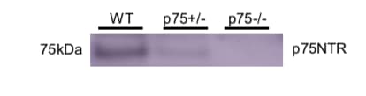
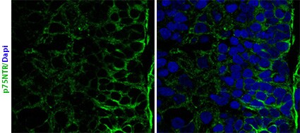
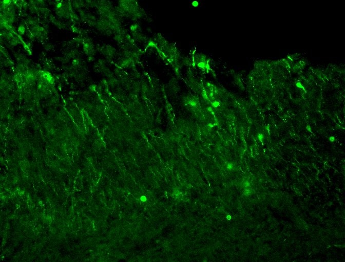
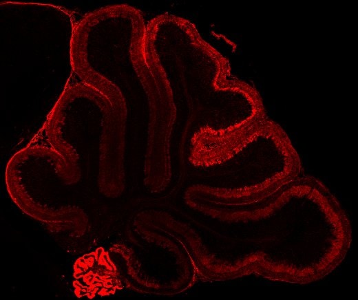
The low affinity nerve growth factor receptor (NGF R), also named p75 neurotrophin receptor, is a type I transmembrane protein that belongs to the tumor necrosis factor receptor family and has been designated TNFRSF16. NGF R cDNA encodes a 427 amino acid (aa) residue precursor protein with a 28 aa residue signal peptide, a 222 aa residue extracellular domain, a 22 aa residue transmembrane domain and a 155 aa residue intracellular domain. The extracellular region contains four cysteine-rich domains and binds NGF, BDNF, NT-3, and NT-4 approximately equally with low affinity. The cytoplasmic region of the receptor contains a subtype 2 death domain.

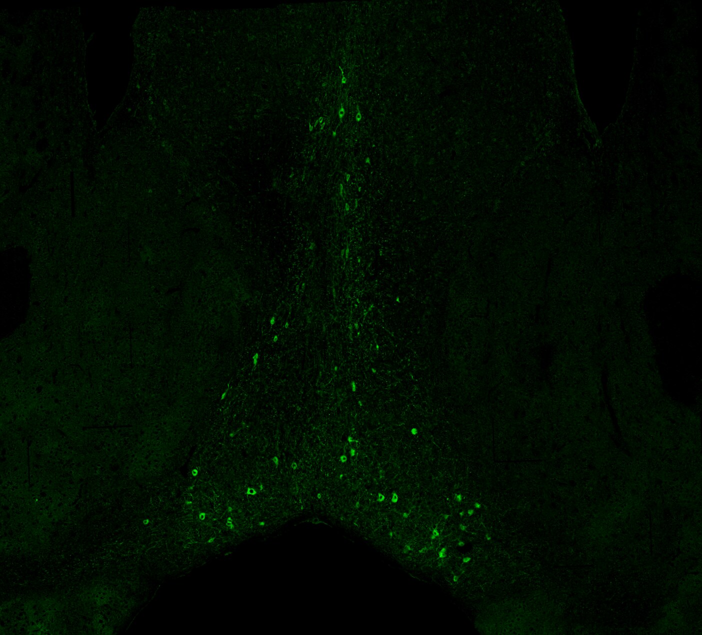
NGF R expression has been shown to occur widely during development and in the adult. Expression has been detected in both neuronal and non-neuronal cells. NGF R was originally reported to function as a positive regulator of TrkA activity. NGF R has also been shown to signal by itself. Depending on its cellular environment, NGF R has now been shown to regulate cell migration, gene expression and to mediate apoptosis. Recombinant NGF R Fc chimera binds NGF with high affinity and is a potent NGF antagonist. Naturally occurring truncated NGF R containing the extracellular domain and lacking the transmembrane or intracellular domain has been detected in vivo in urine, plasma, and in the amniotic fluid of humans and rats (1-3).

References

  1. Barker, P.A. and R.A. Murphy (1992) Molecular and Cellular Biochemistry 110:1.
  2. Bamji, A.X. et al. (1998) J. Cell Biol. 140:911.
  3. Feinstein, E. et al. (1995) Trends Biochem. Sci. 20:342.

Long Name

Nerve Growth Factor Receptor

Alternate Names

CD271, NGF R, p75 NTR, TNFRSF16

Entrez Gene IDs

4804 (Human); 18053 (Mouse); 117089 (Rat)

Gene Symbol

NGFR

UniProt

Additional NGFR/TNFRSF16 Products

Product Documents for Mouse NGFR/TNFRSF16 Antibody

Certificate of Analysis

To download a Certificate of Analysis, please enter a lot or batch number in the search box below.

Note: Certificate of Analysis not available for kit components.

Product Specific Notices for Mouse NGFR/TNFRSF16 Antibody

For research use only

Citations for Mouse NGFR/TNFRSF16 Antibody

Customer Reviews for Mouse NGFR/TNFRSF16 Antibody (6)

4.7 out of 5
6 Customer Ratings
5 Stars
67%
4 Stars
33%
3 Stars
0%
2 Stars
0%
1 Stars
0%

Have you used Mouse NGFR/TNFRSF16 Antibody?

Submit a review and receive an Amazon gift card!

$25/€18/£15/$25CAN/¥2500 Yen for a review with an image

$10/€7/£6/$10CAN/¥1110 Yen for a review without an image

Submit a review
Amazon Gift Card

Customer Images


Showing  1 - 5 of 6 reviews Showing All
Filter By:
  • Mouse NGFR/TNFRSF16 Antibody
    Name: Valeria Avdoshina
    Application: Western Blot
    Sample Tested: Brain (hippocampus) tissue
    Species: Mouse
    Verified Customer | Posted 11/18/2019
    2 microgram per mL Primary antibody dilution.
    Mouse NGFR/TNFRSF16 Antibody AF1157
  • Mouse NGFR/TNFRSF16 Antibody
    Name: Valeria Avdoshina
    Application: Immunohistochemistry
    Sample Tested: Brain (forebrain)
    Species: Mouse
    Verified Customer | Posted 11/18/2019
    Primary antibody dilution: 5 microgram per mL
    Mouse NGFR/TNFRSF16 Antibody AF1157
  • Mouse NGF R/TNFRSF16 Antibody
    Name: Anonymous
    Application: Immunohistochemistry
    Sample Tested: Cerebellum tissue
    Species: Mouse
    Verified Customer | Posted 11/02/2017
    Postnatal day 7 mouse cerebellar tissue.
    Mouse NGFR/TNFRSF16 Antibody AF1157
  • Mouse NGF R/TNFRSF16 Antibody
    Name: Anonymous
    Application: Immunohistochemistry
    Sample Tested: Brain (cortex) tissue
    Species: Mouse
    Verified Customer | Posted 10/17/2016
    PN18 mouse Closed Head Injury Dilution 1:500
    Mouse NGFR/TNFRSF16 Antibody AF1157
  • Mouse NGF R/TNFRSF16 Antibody
    Name: Anonymous
    Application: Immunohistochemistry
    Sample Tested: Brain (cerebral cortex)
    Species: Mouse
    Verified Customer | Posted 07/26/2016
    Frozen section. 20um thickness. Antibody dilution 1/500.
    Mouse NGFR/TNFRSF16 Antibody AF1157
  • Mouse NGF R/TNFRSF16 Antibody
    Name: Juan Zanin
    Application: Immunohistochemistry
    Sample Tested: Cerebellum tissue
    Species: Rat and Mouse
    Verified Customer | Posted 07/13/2016
    Cryostat Frozen section. 20um ticknes. Antibody dilution 1/500 ON 4C.
    Mouse NGFR/TNFRSF16 Antibody AF1157

There are no reviews that match your criteria.

Showing  1 - 5 of 6 reviews Showing All

Protocols

Find general support by application which include: protocols, troubleshooting, illustrated assays, videos and webinars.

FAQs

No product specific FAQs exist for this product.

View all FAQs for Antibodies