Key Product Details

Validated by

Biological Validation

Species Reactivity

Validated:

Mouse, Rat

Cited:

Human, Mouse, Transgenic Mouse

Applications

Validated:

Immunohistochemistry, Western Blot, Simple Western

Cited:

Immunohistochemistry, Immunohistochemistry-Paraffin, Western Blot, Flow Cytometry, Immunocytochemistry, In vivo assay, Functional Assay

Label

Unconjugated

Antibody Source

Polyclonal Goat IgG
Loading...

Product Specifications

Immunogen

Mouse myeloma cell line NS0-derived recombinant mouse P-Selectin/CD62P
Trp42-Ala709
Accession # Q01102

Specificity

Detects mouse P-Selectin/CD62P in direct ELISAs. Detects mouse and rat P-Selectin/CD62P in Western blot. In direct ELISAs, less than 15% cross-reactivity with recombinant human P-Selectin/CD62P is observed.

Clonality

Polyclonal

Host

Goat

Isotype

IgG

Scientific Data Images for Mouse/Rat P‑Selectin/CD62P Antibody

Detection of Mouse and Rat P-Selectin/CD62P antibody by Western Blot.

Detection of Mouse and Rat P-Selectin/CD62P by Western Blot.

Western blot shows lysates of mouse platelets and rat platelets. PVDF membrane was probed with 2 µg/mL of Goat Anti-Mouse/Rat P-Selectin/CD62P Antigen Affinity-purified Polyclonal Antibody (Catalog # AF737) followed by HRP-conjugated Anti-Goat IgG Secondary Antibody (Catalog # HAF017). A specific band was detected for P-Selectin/CD62P at approximately 140-150 kDa (as indicated). This experiment was conducted under reducing conditions and using Immunoblot Buffer Group 1.
P-Selectin/CD62P antibody in Mouse Liver by Immunohistochemistry (IHC-Fr).

P‑Selectin/CD62P in Mouse Liver.

P-Selectin/CD62P was detected in perfusion fixed frozen sections of mouse liver using Goat Anti-Mouse/Rat P-Selectin/CD62P Antigen Affinity-purified Polyclonal Antibody (Catalog # AF737) at 5 µg/mL for 1 hour at room temperature followed by incubation with the Anti-Goat IgG VisUCyte™ HRP Polymer Antibody (Catalog # VC004). Tissue was stained using DAB (brown) and counterstained with hematoxylin (blue). Specific staining was localized to endothelial cells. View our protocol for IHC Staining with VisUCyte HRP Polymer Detection Reagents.
Detection of Rat P-Selectin/CD62P antibody by Simple WesternTM.

Detection of Rat P-Selectin/CD62P by Simple WesternTM.

Simple Western lane view shows lysates of rat platelets, loaded at 0.2 mg/mL. A specific band was detected for P-Selectin/CD62P at approximately 197 kDa (as indicated) using 20 µg/mL of Goat Anti-Mouse P-Selectin/CD62P Antigen Affinity-purified Polyclonal Antibody (Catalog # AF737) followed by 1:50 dilution of HRP-conjugated Anti-Goat IgG Secondary Antibody (Catalog # HAF109). This experiment was conducted under reducing conditions and using the 12-230 kDa separation system.
Detection of Mouse P-Selectin/CD62P by Immunocytochemistry/ Immunofluorescence

Detection of Mouse P-Selectin/CD62P by Immunocytochemistry/ Immunofluorescence

In-vivo targeting of BYL719-loaded nanoparticles prepared with either fucoidan (Fi) or dextran sulfate (Dex).(a) Representative ex vivo fluorescence images of mice organs 24 h after i.v. administration of FiBYL719 or DexBYL719 nanoparticles, and pre-treated with anti-P-selectin antibody (Ab). (b) Nanoparticle biodistribution in organs and tumour, calculated from ex vivo fluorescence images shown in a as total fluorescence efficiency divided by organ weight (n=3). (c) Representative immunofluorescence staining for CD-31 (red), P-selectin (green) and DAPI (blue) in Cal-33 xenografts before and after a single dose of ionizing radiation (4 Gy). Scale bars, 50 μm. (d) Quantification of double-staining positive endothelial cells per tumour shown in c (n=3). (e) In vivo fluorescence imaging of Cal-33 xenograft-bearing mice 24 h after treatment with FiBYL719 or 4 Gy RT followed by FiBYL719. (f) Quantification of total fluorescence efficiency of tumours shown e (n=10). (g) Representative immunofluorescence stains of tumour sections for P-selectin (green), NIR (red) and DAPI (blue) from H22 xenografts 24 h after treatments. Scale bars, 50 μm. In b,d,f error bars indicate mean±s.e.m. *P<0.05, **P<0.01; by Mann–Whitney U-test in b or one-way ANOVA with post hoc Tukey test in d,f. Image collected and cropped by CiteAb from the following open publication (https://www.nature.com/articles/ncomms14292), licensed under a CC-BY license. Not internally tested by R&D Systems.
Detection of Mouse P-Selectin/CD62P by Immunocytochemistry/ Immunofluorescence

Detection of Mouse P-Selectin/CD62P by Immunocytochemistry/ Immunofluorescence

In-vivo targeting of BYL719-loaded nanoparticles prepared with either fucoidan (Fi) or dextran sulfate (Dex).(a) Representative ex vivo fluorescence images of mice organs 24 h after i.v. administration of FiBYL719 or DexBYL719 nanoparticles, and pre-treated with anti-P-selectin antibody (Ab). (b) Nanoparticle biodistribution in organs and tumour, calculated from ex vivo fluorescence images shown in a as total fluorescence efficiency divided by organ weight (n=3). (c) Representative immunofluorescence staining for CD-31 (red), P-selectin (green) and DAPI (blue) in Cal-33 xenografts before and after a single dose of ionizing radiation (4 Gy). Scale bars, 50 μm. (d) Quantification of double-staining positive endothelial cells per tumour shown in c (n=3). (e) In vivo fluorescence imaging of Cal-33 xenograft-bearing mice 24 h after treatment with FiBYL719 or 4 Gy RT followed by FiBYL719. (f) Quantification of total fluorescence efficiency of tumours shown e (n=10). (g) Representative immunofluorescence stains of tumour sections for P-selectin (green), NIR (red) and DAPI (blue) from H22 xenografts 24 h after treatments. Scale bars, 50 μm. In b,d,f error bars indicate mean±s.e.m. *P<0.05, **P<0.01; by Mann–Whitney U-test in b or one-way ANOVA with post hoc Tukey test in d,f. Image collected and cropped by CiteAb from the following open publication (https://www.nature.com/articles/ncomms14292), licensed under a CC-BY license. Not internally tested by R&D Systems.
Detection of P-Selectin/CD62P by Immunohistochemistry

Detection of P-Selectin/CD62P by Immunohistochemistry

Expression of P-selectin in different brain regions following GMH. P14 brain sections stained for P-selectin (CD62p) and quantified in 3 specific brain regions of each experimental group as indicated (a, b, c). One-way ANOVA with Turkey’s correction for multiple comparisons. ****p < 0.0001. Error bars = mean ± SEM. d 40 × representative images of P-selectin staining (green) and DAPI (blue) within each brain region for naïve and GMH animals Image collected and cropped by CiteAb from the following open publication (https://pubmed.ncbi.nlm.nih.gov/37322469), licensed under a CC-BY license. Not internally tested by R&D Systems.
Detection of P-Selectin/CD62P by Immunohistochemistry

Detection of P-Selectin/CD62P by Immunohistochemistry

Expression of P-selectin in different brain regions following GMH. P14 brain sections stained for P-selectin (CD62p) and quantified in 3 specific brain regions of each experimental group as indicated (a, b, c). One-way ANOVA with Turkey’s correction for multiple comparisons. ****p < 0.0001. Error bars = mean ± SEM. d 40 × representative images of P-selectin staining (green) and DAPI (blue) within each brain region for naïve and GMH animals Image collected and cropped by CiteAb from the following open publication (https://pubmed.ncbi.nlm.nih.gov/37322469), licensed under a CC-BY license. Not internally tested by R&D Systems.

Applications for Mouse/Rat P‑Selectin/CD62P Antibody

Application
Recommended Usage

Immunohistochemistry

5-15 µg/mL
Sample:

Perfusion fixed frozen sections of mouse liver

Simple Western

20 µg/mL
Sample: Rat platelets

Western Blot

2 µg/mL
Sample: Mouse platelets and Rat platelets

Reviewed Applications

Read 1 review rated 5 using AF737 in the following applications:

Formulation, Preparation, and Storage

Purification

Antigen Affinity-purified

Reconstitution

Reconstitute at 0.2 mg/mL in sterile PBS. For liquid material, refer to CoA for concentration.


Loading...

Formulation

Lyophilized from a 0.2 μm filtered solution in PBS with Trehalose. See Certificate of Analysis for details.
*Small pack size (-SP) is supplied either lyophilized or as a 0.2 µm filtered solution in PBS.

Shipping

Lyophilized product is shipped at ambient temperature. Liquid small pack size (-SP) is shipped with polar packs. Upon receipt, store immediately at the temperature recommended below.

Stability & Storage

Use a manual defrost freezer and avoid repeated freeze-thaw cycles.
  • 12 months from date of receipt, -20 to -70 °C as supplied.
  • 1 month, 2 to 8 °C under sterile conditions after reconstitution.
  • 6 months, -20 to -70 °C under sterile conditions after reconstitution.

Calculators

The reconstitution calculator allows you to quickly calculate the volume of a reagent to reconstitute your vial. Simply enter the mass of reagent and the target concentration and the calculator will determine the rest.

=
÷

Background: P-Selectin/CD62P

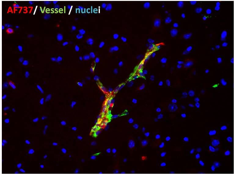
Mouse P-Selectin (GMP-140, LECAM-3, PADGEM, CD62P), a member of the Selectin family, is a cell surface glycoprotein expressed by activated platelets and endothelial cells. P-Selectin is translocated to the cell surface within minutes, from alpha granules of platelets or Weibel-Palade bodies of endothelial cells, following stimulation with thrombin, histamine, PMA or peroxides. P-Selectin binds to a 106 kDa protein present on myeloid cells, neutrophils, monocytes and lymphocytes, termed PSGL-1 (P-Selectin glycoprotein ligand-1).

P-Selectin plays a role in the adhesion of leukocytes and neutrophils to the endothelium. Acting in cooperation with L-Selectin, P-Selectin mediates the initial interaction of circulating leukocytes with endothelial cells that produces a characteristic ‘rolling’ of the leukocytes on the endothelium. This initial interaction is followed by a stronger interaction involving E-Selectin, and later ICAM-1 and VCAM-1, that leads eventually to extravasation of the white blood cell through the blood vessel wall into the extracellular matrix tissue.

Mouse P-Selectin cDNA encodes a 768 amino acid (aa) residue type I transmembrane protein with a 41 aa signal peptide, a 668 aa extracellular domain, a transmembrane domain and a short (35 aa) cytoplasmic domain. The extracellular domain has an NH2-terminal C-type lectin domain and an EGF-like domain followed by a series of complement factor A repeat homology domains. The extracellular domains of human and mouse P-Selectin share approximately 73% sequence homology.

References

  1. Kansas, G.S. (1996) Blood 88:3259.
  2. McEver, R.P. and R.D. Cummings (1997) J. Clin. Invest. 100:485.

Alternate Names

CD62P, GMP140, GRMP, PADGEM, PSEL, SELP

Entrez Gene IDs

6403 (Human); 20344 (Mouse)

Gene Symbol

SELP

UniProt

Additional P-Selectin/CD62P Products

Product Documents for Mouse/Rat P‑Selectin/CD62P Antibody

Certificate of Analysis

To download a Certificate of Analysis, please enter a lot or batch number in the search box below.

Note: Certificate of Analysis not available for kit components.

Product Specific Notices for Mouse/Rat P‑Selectin/CD62P Antibody

For research use only

Citations for Mouse/Rat P‑Selectin/CD62P Antibody

Customer Reviews for Mouse/Rat P‑Selectin/CD62P Antibody (1)

5 out of 5
1 Customer Rating
5 Stars
100%
4 Stars
0%
3 Stars
0%
2 Stars
0%
1 Stars
0%

Have you used Mouse/Rat P‑Selectin/CD62P Antibody?

Submit a review and receive an Amazon gift card!

$25/€18/£15/$25CAN/¥2500 Yen for a review with an image

$10/€7/£6/$10CAN/¥1110 Yen for a review without an image

Submit a review
Amazon Gift Card

Customer Images


Showing  1 - 1 of 1 review Showing All
Filter By:
  • Mouse/Rat P-Selectin/CD62P Antibody
    Name: Anonymous
    Application: Immunohistochemistry
    Sample Tested: Spinal cord and Brain tissue
    Species: Mouse
    Verified Customer | Posted 01/22/2018
    dilution: 1/1000
    Mouse/Rat P‑Selectin/CD62P Antibody AF737

There are no reviews that match your criteria.

Showing  1 - 1 of 1 review Showing All

Protocols

Find general support by application which include: protocols, troubleshooting, illustrated assays, videos and webinars.

FAQs

No product specific FAQs exist for this product.

View all FAQs for Antibodies
Loading...