Key Product Details

Validated by

Biological Validation

Species Reactivity

Validated:

Mouse, Rat

Cited:

Human, Mouse, Rat, Avian - Chicken, Transgenic Mouse

Applications

Validated:

Immunohistochemistry, Western Blot

Cited:

Immunohistochemistry, Immunohistochemistry-Frozen, Western Blot, Neutralization, Immunocytochemistry/Immunofluorescence

Label

Unconjugated

Antibody Source

Polyclonal Goat IgG
Loading...

Product Specifications

Immunogen

Mouse myeloma cell line NS0-derived recombinant mouse Tie‑2
Ala23-Lys744
Accession # CAA47857

Specificity

Detects mouse and rat Tie-2 in direct ELISAs and Western blots. In direct ELISAs, approximately 35% cross-reactivity with recombinant human (rh) Tie‑2 is observed and less than 1% cross-reactivity with recombinant zebrafish Tie-2 and rhTie-1 is observed.

Clonality

Polyclonal

Host

Goat

Isotype

IgG

Scientific Data Images for Mouse/Rat Tie‑2 Antibody

Tie-2 antibody in Human Intestine Tissue by Immunohistochemistry (IHC-P).

Tie-2 in Mouse Intestine Tissue.

Tie-2 was detected in immersion fixed paraffin-embedded sections of mouse intestine tissue using Goat Anti-Mouse/Rat Tie-2 Antigen Affinity-purified Polyclonal Antibody (Catalog # AF762) at 1 µg/mL for 1 hour at room temperature followed by incubation with the Anti-Goat IgG VisUCyte™ HRP Polymer Antibody (Catalog # VC004). Before incubation with the primary antibody, tissue was subjected to heat-induced epitope retrieval using Antigen Retrieval Reagent-Basic (Catalog # CTS013). Tissue was stained using DAB (brown) and counterstained with hematoxylin (blue). Specific staining was localized to cell surface on endothelial cells. View our protocol for IHC Staining with VisUCyte HRP Polymer Detection Reagents.

Detection of Mouse Tie-2 by Western Blot

Detection of Mouse Tie-2 by Western Blot

Pericytes express functional Tie2.(a) Microarray-based expression screening of brain pericytes (BP), placenta pericytes (PP), pancreas pericytes (PA), lung pericytes (LP), muscle pericytes (MP) and human umbilical vein endothelial cells (HUVEC [HU]). (b) Semi-quantitative PCR of TEK (Tie2), endothelial marker genes (PECAM1, CDH5) and pericyte marker genes (CSPG4, CD248) in HUVEC and BP; house keeping gene: GAPDH. (c) Representative images showing expression of Tie2 in HUVEC and BP. (d) Histogram of membrane-bound Tie2 expression in HUVEC and BP compared to isotype control measured by flow cytometry. (e,g) Western blot (WB) analysis of tyrosine phosphorylation (pTyr) and total Tie2 after Tie2 immunoprecipitation (IP) in BP (e) and HUVEC (g) upon stimulation with recombinant human (rh) Ang1 compared to unstimulated (us) cells. (f,h) Quantification of the ratio of phosphorylated Tie2 (pTie2) relative to total Tie2 protein in BP (f) and HUVEC (h) normalized to us control (n=3). (i,k) Western blot analysis of AKT phosphorylation (Ser473) in control (shCtr) and Tie2-silenced (shTie2 I/shTie2 II) BP (i) and HUVEC (k) upon stimulation with rhAng1 or rhAng2. (j,l) Quantification of the ratio of pAKT to total AKT protein expression in BP (j) and HUVEC (l) normalized to unstimulated control (n=3). Representative western blot images are cropped versions and original images can be found in Supplementary Fig. 15. Scale bars: 20 μm (c). Data are shown as mean±s.d. Statistics were performed using Mann–Whitney U test (f,h) and one-way ANOVA (j,l). *P<0.05, **P<0.01. Image collected and cropped by CiteAb from the following open publication (https://pubmed.ncbi.nlm.nih.gov/28719590), licensed under a CC-BY license. Not internally tested by R&D Systems.
Detection of Mouse Tie-2 by Immunocytochemistry/ Immunofluorescence

Detection of Mouse Tie-2 by Immunocytochemistry/ Immunofluorescence

Endothelial changes after pericyte depletion. a–f Maximum intensity projection of confocal images from control and DTRiPC P6 retinas stained for IB4 (red) in combination with VEGF-A a, VEGFR2 b, VEGFR3 c, Tie2 d, Esm1 e, and Dll4 f (all in white), as indicated. Note local increase of VEGFR2, VEGFR3, and Esm1 (arrowheads in b, c, e) but not Tie2 or VEGF-A at the edge of the vessel plexus. Dll4 expression in DTRiPC sprouts is increased in some regions (arrowheads) but absent in others (arrows). Scale bar, 100 µm. g–j Quantitation of VEGF-A immunosignals area and intensity g, signal intensity for VEGFR2 h and VEGFR3 i and proportion of Esm1+ area with respect to vascular area j in the P6 control and DTRiPC angiogenic front. Error bars, s.e.m. p-values, Student’s t-test. Image collected and cropped by CiteAb from the following open publication (https://pubmed.ncbi.nlm.nih.gov/29146905), licensed under a CC-BY license. Not internally tested by R&D Systems.
Detection of Mouse Tie-2 by Western Blot

Detection of Mouse Tie-2 by Western Blot

Pericyte-derived Angpt1 controls alveologenesis. a RT-qPCR analysis of Angpt1 and Tie2/Tek expression in freshly sorted lung GFP+, CD31+ or EpCAM+ cells from P7 Pdgfrb(BAC)-CreERT2 R26-mT/mG mice. Data represents mean ± s.e.m. (n = 4 mice). b High magnification images of P10 Angpt1GFP lungs stained for GFP (green), PDGFR beta (red), and PDGFR alpha (blue). Arrows indicate GFP and PDGFR beta double positive pericytes. Scale bar, 15 µm. c RT-qPCR analysis of Angpt1 expression in freshly sorted PDGFR beta + cells from P7 Yap1,Wwtr1iPCKO and control lungs. Data represents mean ± s.e.m. (n = 4 mice, two-tailed unpaired t-test). dAngpt1 expression in cultured Verteporfin (VP)-treated (48 h) and control pericytes. Data represents mean ± s.e.m. (n = 4, Welch’s t-test). e Expression of the indicated transcripts in freshly sorted CD31+ cells from P7 Yap1,Wwtr1iPCKO and control lungs. Data represents mean ± s.e.m. (n = 4 mice, NS not significant, two-tailed unpaired t-test). f–h Western blot analysis of Angpt1 protein (f; n = 2 controls and 4 mutant mice) and of total and phospho-Tie2 (pTie2) in P12 Yap1,Wwtr1iPCKO and control total lung lysates (g, n = 3 controls and 5 mutants). Molecular weight marker (kDa) is indicated. Relative quantification of signals is shown in h. Two-tailed unpaired t-test. i Scheme showing the time points of tamoxifen administration and analysis for Angpt1iPCKO mice. j, k 3D reconstruction confocal images of P12 Angpt1iPCKO and littermate control lungs stained for AQP5 (green), PDGFR beta (red), and PECAM1 (blue). Panels in k show higher magnification of PECAM1 staining. Scale bar, 50 µm (j) and 30 µm (k). l Quantitation of airspace volume in P12 Angpt1iPCKO and littermate control lung sections with 3D reconstruction surface images. Data represents mean ± s.e.m. (n = 4 mice; p < 0.0001, two-tailed unpaired t-test). m 3D reconstruction confocal images of P12 Angpt1iPCKO and littermate control lungs stained for alpha SMA (red) and tropoelastin (blue). Scale bar, 50 µm. n Quantitation of staining intensity for alpha SMA or tropoelastin shown in m. Intensity was normalized to the size of the parenchymal region. Data represents mean ± s.e.m. (n = 4 mice; NS not significant, two-tailed unpaired t-test). o Schematic summary of findings. Pulmonary pericytes regulate ECs and alveolar epithelial cells via angiocrine factors such as angiopoietin-1 and HGF. Image collected and cropped by CiteAb from the following open publication (https://pubmed.ncbi.nlm.nih.gov/29934496), licensed under a CC-BY license. Not internally tested by R&D Systems.
Detection of Mouse Tie-2 by Western Blot

Detection of Mouse Tie-2 by Western Blot

Pericytes express functional Tie2.(a) Microarray-based expression screening of brain pericytes (BP), placenta pericytes (PP), pancreas pericytes (PA), lung pericytes (LP), muscle pericytes (MP) and human umbilical vein endothelial cells (HUVEC [HU]). (b) Semi-quantitative PCR of TEK (Tie2), endothelial marker genes (PECAM1, CDH5) and pericyte marker genes (CSPG4, CD248) in HUVEC and BP; house keeping gene: GAPDH. (c) Representative images showing expression of Tie2 in HUVEC and BP. (d) Histogram of membrane-bound Tie2 expression in HUVEC and BP compared to isotype control measured by flow cytometry. (e,g) Western blot (WB) analysis of tyrosine phosphorylation (pTyr) and total Tie2 after Tie2 immunoprecipitation (IP) in BP (e) and HUVEC (g) upon stimulation with recombinant human (rh) Ang1 compared to unstimulated (us) cells. (f,h) Quantification of the ratio of phosphorylated Tie2 (pTie2) relative to total Tie2 protein in BP (f) and HUVEC (h) normalized to us control (n=3). (i,k) Western blot analysis of AKT phosphorylation (Ser473) in control (shCtr) and Tie2-silenced (shTie2 I/shTie2 II) BP (i) and HUVEC (k) upon stimulation with rhAng1 or rhAng2. (j,l) Quantification of the ratio of pAKT to total AKT protein expression in BP (j) and HUVEC (l) normalized to unstimulated control (n=3). Representative western blot images are cropped versions and original images can be found in Supplementary Fig. 15. Scale bars: 20 μm (c). Data are shown as mean±s.d. Statistics were performed using Mann–Whitney U test (f,h) and one-way ANOVA (j,l). *P<0.05, **P<0.01. Image collected and cropped by CiteAb from the following open publication (https://pubmed.ncbi.nlm.nih.gov/28719590), licensed under a CC-BY license. Not internally tested by R&D Systems.

Applications for Mouse/Rat Tie‑2 Antibody

Application
Recommended Usage

Immunohistochemistry

1-15 µg/mL
Sample: Immersion fixed paraffin-embedded sections of mouse intestine tissue

Western Blot

0.1 µg/mL
Sample:

Recombinant Mouse Tie‑2 Fc Chimera (Catalog # 762-T2) and Recombinant Rat Tie-2 Fc Chimera (Catalog # 3874-T2)

Reviewed Applications

Read 3 reviews rated 4.7 using AF762 in the following applications:

Formulation, Preparation, and Storage

Purification

Antigen Affinity-purified

Reconstitution

Reconstitute at 0.2 mg/mL in sterile PBS. For liquid material, refer to CoA for concentration.


Loading...

Formulation

Lyophilized from a 0.2 μm filtered solution in PBS with Trehalose. *Small pack size (SP) is supplied either lyophilized or as a 0.2 µm filtered solution in PBS.

Shipping

Lyophilized product is shipped at ambient temperature. Liquid small pack size (-SP) is shipped with polar packs. Upon receipt, store immediately at the temperature recommended below.

Stability & Storage

Use a manual defrost freezer and avoid repeated freeze-thaw cycles.
  • 12 months from date of receipt, -20 to -70 °C as supplied.
  • 1 month, 2 to 8 °C under sterile conditions after reconstitution.
  • 6 months, -20 to -70 °C under sterile conditions after reconstitution.

Calculators

The reconstitution calculator allows you to quickly calculate the volume of a reagent to reconstitute your vial. Simply enter the mass of reagent and the target concentration and the calculator will determine the rest.

=
÷

Background: Tie-2

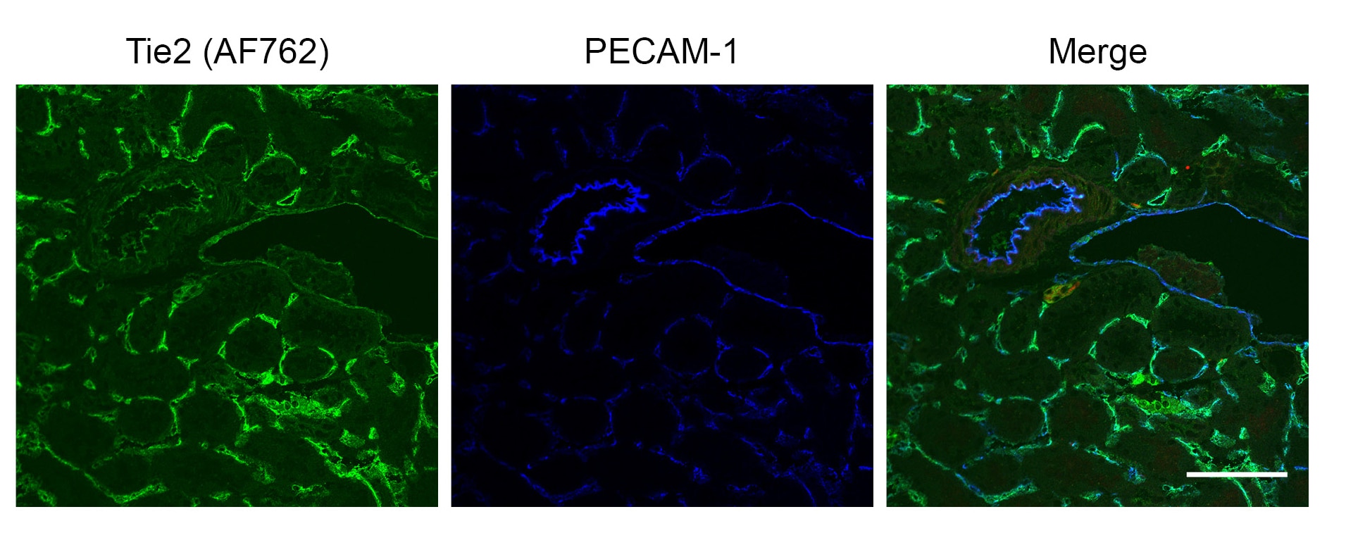
Tie-1/Tie (tyrosine kinase with Ig and EGF homology domains 1) and Tie-2/Tek comprise a receptor tyrosine kinase (RTK) subfamily with unique structural characteristics: two immunoglobulin-like domains flanking three epidermal growth factor (EGF)-like domains, followed by three fibronectin type III-like repeats in the extracellular region and a split tyrosine kinase domain in the cytoplasmic region. These receptors are expressed primarily on endothelial and hematopoietic progenitor cells and play critical roles in angiogenesis, vasculogenesis and hematopoiesis.

Two ligands, angiopoietin-1 (Ang-1) and angiopoietin-2 (Ang-2), which bind Tie-2 with high-affinity have been identified. Ang-2 has been reported to act as an antagonist for Ang-1. Mice engineered to overexpress Ang-2 or to lack Ang-1 or Tie-2 display similar angiogenesis defects.

References

  1. Partanen, J. and D.J. Dumont (1999) Curr. Top. Microbiol. Immunol. 237:159.
  2. Takakura, N. et al. (1998) Immunity 9:677.

Long Name

Tyrosine Kinase with Immunoglobulin and Epidermal Growth Factor Homology Domains 2

Alternate Names

CD202b, TEK, Tie2

Entrez Gene IDs

7010 (Human); 21687 (Mouse); 89804 (Rat); 396729 (Porcine); 403714 (Canine); 102122204 (Cynomolgus Monkey); 30747 (Zebrafish)

Gene Symbol

TEK

UniProt

Additional Tie-2 Products

Product Documents for Mouse/Rat Tie‑2 Antibody

Certificate of Analysis

To download a Certificate of Analysis, please enter a lot or batch number in the search box below.

Note: Certificate of Analysis not available for kit components.

Product Specific Notices for Mouse/Rat Tie‑2 Antibody

For research use only

Citations for Mouse/Rat Tie‑2 Antibody

Customer Reviews for Mouse/Rat Tie‑2 Antibody (3)

4.7 out of 5
3 Customer Ratings
5 Stars
67%
4 Stars
33%
3 Stars
0%
2 Stars
0%
1 Stars
0%

Have you used Mouse/Rat Tie‑2 Antibody?

Submit a review and receive an Amazon gift card!

$25/€18/£15/$25CAN/¥2500 Yen for a review with an image

$10/€7/£6/$10CAN/¥1110 Yen for a review without an image

Submit a review
Amazon Gift Card

Customer Images


Showing  1 - 3 of 3 reviews Showing All
Filter By:
  • Mouse/Rat Tie-2 Antibody
    Name: Hayk Simonyan
    Application: Immunocytochemistry
    Sample Tested: Mouse brain
    Species: Mouse
    Verified Customer | Posted 06/08/2018
    Paraventricular Nucleus of mouse brain. 3% PFA perfused, frozen brain with 10 um thickness. Ab dilution is 1:1000 for both.
    Mouse/Rat Tie‑2 Antibody AF762
  • Mouse/Rat Tie-2 Antibody
    Name: Hayk Simonyan
    Application: Immunohistochemistry
    Sample Tested: Adult mouse brain tissue
    Species: Mouse
    Verified Customer | Posted 05/12/2017
    IHC image of mouse brain cortex. Dilution 1:1000.
    Mouse/Rat Tie‑2 Antibody AF762
  • Mouse/Rat Tie-2 Antibody
    Name: Anonymous
    Application: Immunohistochemistry
    Sample Tested: Fresh-frozen kidney sections
    Species: Mouse
    Verified Customer | Posted 08/29/2016
    Unfixed cryosections were fixed in 95% EtOH followed by acetone before blocking and overnight incubation in 1:50 AF762 at 4C. Staining was detected with Alexafluor-488 labeled donkey anti-goat secondary AB. Strong staining was observed in mouse kidney sections.
    Mouse/Rat Tie‑2 Antibody AF762

There are no reviews that match your criteria.

Showing  1 - 3 of 3 reviews Showing All

Protocols

Find general support by application which include: protocols, troubleshooting, illustrated assays, videos and webinars.

FAQs

No product specific FAQs exist for this product.

View all FAQs for Antibodies