Scientific images may be resized, cropped, or adjusted for brightness or contrast for presentation purposes. These updates are limited to presentation and do not affect the underlying scientific interpretation of the data. Where available, additional source data may be provided upon request.
Key Product Details
Species Reactivity
Validated:
Cited:
Applications
Validated:
Cited:
Label
Antibody Source
Product Specifications
Immunogen
Ser27-Arg332
Accession # P14719
Specificity
Clonality
Host
Isotype
Endotoxin Level
Scientific Data Images for Mouse ST2/IL-33R Antibody
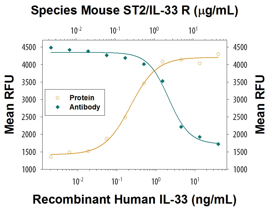
Cell Proliferation Induced by IL‑33 and Neutralization by Mouse ST2/IL-33 R Antibody.
Recombinant Human IL-33 (Catalog # 3625-IL) induces proliferation in the D10.G4.1 mouse helper T cell line in a dose-dependent manner (orange line), as measured by Resazurin (Catalog # AR002). Under these conditions, proliferation elicited by IL-33 is neutralized (green line) by increasing concentrations of Goat Anti-Mouse ST2/IL-33 R Antigen Affinity-purified Polyclonal Antibody (Catalog # AF1004). The ND50 is typically 1-6 µg/mL.
Mouse ST2 / IL-33 R ELISA Standard Curve
Recombinant Mouse ST2/IL-33R Fc Chimera (Catalog # 1004-MR) was serially diluted and captured by Rat Anti-Mouse ST2/IL-33R Monoclonal Antibody (Catalog # MAB10041) coated on a Clear Polystyrene Microplate (Catalog # DY990). Goat Anti-Mouse ST2/IL-33R Antigen Affinity-purified Polyclonal Antibody (Catalog # AF1004) was biotinylated and incubated with the protein captured on the plate. Detection of the standard curve was achieved by incubating Streptavidin-HRP (Catalog # DY998)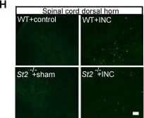
Detection of ST2/IL-33R by Immunohistochemistry
IL-33/ST2 signaling mediates mechanical hypersensitivity of INC pain model mice. (A) Experimental protocol denoting time points for the incision, treatment and behavioral observation. (B) Overlaid time courses showing PWT changes in control, INC+Iso IgG and INC+IL-33 neutralizing antibody (IL-33 Ab) groups. (C) Normalized area under the curve (AUC) analysis of each curve in panel B. (D) Overlaid time courses showing PWT changes in control, INC+Iso IgG and INC+ST2 neutralizing antibody (ST2 Ab) groups. (E) Normalized AUC analysis of each curve in panel D. (F) Overlaid time courses showing PWT changes in 4 groups of mice as indicated. (G) Normalized AUC analysis of each curve in panel F. (H) Representative pictures of c-Fos immunostaining in ipsilateral spinal cord dorsal horn of 4 groups of mice. Scale bar represents 100 μm. (I) Summarized data showing c-Fos+ cells per observational field of 4 groups. (J) Representative photos showing wound healing of WT+INC vs. St2-/-+INC group of mice on Day 1, 3, 5&7 after incision. (K) Summary of the width (in mm) of the incision on Day 3, 5&7 as in panel J. The suture was removed 1 day after the incision. **p<0.01 vs. control group. #p<0.05, ##p<0.01 vs. IL-33 Ab/ST2 Ab/WT+INC group as indicated. n=6 mice/group. Two-way ANOVA followed with Tukey's post-hoc test was used for panel B, D, F&K. One-way ANOVA followed with Tukey's post-hoc test was used for panel C, E, G&I. Image collected and cropped by CiteAb from the following open publication (https://pubmed.ncbi.nlm.nih.gov/39267790), licensed under a CC-BY license. Not internally tested by R&D Systems.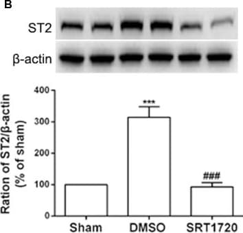
Detection of ST2/IL-33R by Western Blot
The increased expression of IL-33, ST2, TNF-alpha, and IL-1 beta were retarded by intrathecal injection of SIRT1 agonist SRT1720. (A,B) Application of SRT1720(i.t.) reduced the expression of IL-33 and its receptor ST2 after SNI surgery on day 7. (C,D) Inflammatory cytokines TNF-alpha and IL-1 beta were significantly downregulated by delivery of SRT1720(i.t.) on day 7, n = 4/group, ***p < 0.001 compared with the sham group, ###p < 0.001 compared with SNI+DMSO group. Image collected and cropped by CiteAb from the following open publication (https://pubmed.ncbi.nlm.nih.gov/32116550), licensed under a CC-BY license. Not internally tested by R&D Systems.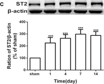
Detection of ST2/IL-33R by Western Blot
Spared nerve injury (SNI) induced mechanical allodynia and enhanced expression of interleukin (IL)-33 and ST2 in dorsal root ganglion (DRG) of rats. (A) SNI induced mechanical allodynia in the ipsilateral but not contralateral hind paw (n = 6), but the sham operation did not induce changes in bilateral hind paw(n = 6). (B,C) Compared with the sham rats, expression of IL-33 and its receptor ST2 increased in L4-L6 DRG since day 1 and persisted till day 14 after SNI (n = 4). *p < 0.05, ***p < 0.001 compared with the sham group. Image collected and cropped by CiteAb from the following open publication (https://pubmed.ncbi.nlm.nih.gov/32116550), licensed under a CC-BY license. Not internally tested by R&D Systems.Applications for Mouse ST2/IL-33R Antibody
Western Blot
Sample: Recombinant Mouse ST2/IL-33 R Fc Chimera (Catalog # 1004-MR)
Neutralization
Reviewed Applications
Read 2 reviews rated 2.5 using AF1004 in the following applications:
Formulation, Preparation, and Storage
Purification
Reconstitution
Reconstitute at 0.2 mg/mL in sterile PBS. For liquid material, refer to CoA for concentration.
Formulation
Shipping
Stability & Storage
- 12 months from date of receipt, -20 to -70 °C as supplied.
- 1 month, 2 to 8 °C under sterile conditions after reconstitution.
- 6 months, -20 to -70 °C under sterile conditions after reconstitution.
Calculators
Background: ST2/IL-33R
References
- Barksby, H.E. et al. (2007) Clin. Exp. Immunol. 149:217.
- Gadina, M. and C.A. Jefferies (2007) Science STKE 2007:pe31.
- Yanagisawa, K. et al. (1993) FEBS Lett. 318:83.
- Schmitz, J. et al. (2005) Immunity 23:479.
- Lecart, S. et al. (2002) Eur. J. Immunol. 32:2979.
- Brint, E.K. et al. (2004) Nat. Immunol. 5:373.
- Weinberg, E.O. et al. (2002) Circulation 106:2961.
- Sanada S. et al. (2007) J. Clin. Invest. 117:1538
- Palmer, G. et al. (2008) Cytokine 42:358.
- Chackerian, A.A. et al. (2007) J. Immunol. 179:2551.
- Allakhverdi, Z. et al. (2007) J. Immunol. 179:2051.
- Verri Jr., W.A. et al. (2008) Proc. Natl. Acad. Sci. 105:2723.
- Miller, A.M. et al. (2008) J. Exp. Med. 205:339.
- Hayakawa, H. et al. (2007) J. Biol. Chem. 282:26369.
Long Name
Alternate Names
Gene Symbol
UniProt
Additional ST2/IL-33R Products
Product Documents for Mouse ST2/IL-33R Antibody
Certificate of Analysis
To download a Certificate of Analysis, please enter a lot or batch number in the search box below.
Note: Certificate of Analysis not available for kit components.
Product Specific Notices for Mouse ST2/IL-33R Antibody
For research use only
Related Research Areas
Citations for Mouse ST2/IL-33R Antibody
 Powered by Bioz
Customer Reviews for Mouse ST2/IL-33R Antibody (2)
Have you used Mouse ST2/IL-33R Antibody?
Submit a review and receive an Amazon gift card!
$25/€18/£15/$25CAN/¥2500 for a review with an image
$10/€7/£6/$10CAN/¥1110 for a review without an image
Submit a review
Customer Images

-
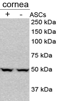
Application: Simple WesternSample Tested: Lung tissueSpecies: MouseVerified Customer | Posted 09/08/2025treated and untreated mouse lung homogenate, IL-33 protein expression using JESSchecked the protein expression of IL-33 in mouse lung homogenates
 Bio-Techne ResponseThis review reflects a new species or application tested on a primary antibody.
 -
Application: Western BlotSample Tested:Species: MouseVerified Customer | Posted 08/06/2016Review for ST2/IL-1 R4 Antibody (AF1004)
There are no reviews that match your criteria.
Protocols
Find general support by application which include: protocols, troubleshooting, illustrated assays, videos and webinars.
- Cellular Response to Hypoxia Protocols
- R&D Systems Quality Control Western Blot Protocol
- Troubleshooting Guide: Western Blot Figures
- Western Blot Conditions
- Western Blot Protocol
- Western Blot Protocol for Cell Lysates
- Western Blot Troubleshooting
- Western Blot Troubleshooting Guide
- View all Protocols, Troubleshooting, Illustrated assays and Webinars
Associated Pathways