Angiogensin I Converting Enzyme-2 (ACE-2), also called ACEH (ACE homolog), is a type I transmembrane zinc protease that cleaves angiotensins I and II to produce vasodilatory and anti-proliferative peptides. The balance between ACE-1 and ACE-2 activity is critical for maintaining cardiovascular, renal, and pulmonary function (1). ACE-2 also functions as the cellular uptake receptor for the SARS coronoavirus. Within the extracellular domain, human ACE-2 shares 83% aa sequence identity with mouse and rat ACE-2. Human ACE-2 has about 40% amino acid identity to the N- and C-terminal domains of human somatic ACE. The predicted human ACE-2 protein sequence consists of 805 amino acids, including a N-terminal signal peptide, a single catalytic domain, a C-terminal membrane anchor, and a short cytoplasmic tail. ACE-2 mRNA is found at high levels in testis, kidney and heart and at moderate levels in colon, small intestine and ovary. Classical ACE inhibitors such as captopril and lisinopril do not inhibit ACE-2 activity. Novel peptide inhibitors of ACE-2 do not inhibit ACE activity (2). Genetic data from Drosophila, mice and rats show that ACE-2 is an essential regulator of heart function in vivo (3). ACE-2 isoforms of 75 kDa and 120 kDa are differentially expressed between lung and kidney, respectively, and a shed soluble form is generated by TACE/ADAM17 mediated cleavage.

Key Product Details
Species Reactivity
Validated:
Cited:
Applications
Validated:
Cited:
Label
Antibody Source
Product Specifications
Immunogen
Gln18-Ser740 (predicted)
Accession # Q9BYF1
Specificity
Clonality
Host
Isotype
Scientific Data Images for ACE-2 Antibody (171606)
Detection of Human ACE‑2 by Western Blot.
Western blot shows lysates of NS0 mouse myeloma cell line and human kidney tissue. PVDF membrane was probed with 2 µg/mL of Mouse Anti-Human ACE-2 Monoclonal Antibody (Catalog # MAB933) followed by HRP-conjugated Anti-Mouse IgG Secondary Antibody (Catalog # HAF007). A specific band was detected for ACE-2 at approximately 110 kDa (as indicated). This experiment was conducted under reducing conditions and using Immunoblot Buffer Group 1.
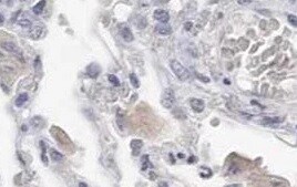
ACE‑2 in Human Kidney.
ACE-2 was detected in immersion fixed paraffin-embedded sections of human kidney using Mouse Anti-Human ACE-2 Monoclonal Antibody (Catalog # MAB933) at 15 µg/mL overnight at 4 °C. Before incubation with the primary antibody, tissue was subjected to heat-induced epitope retrieval using Antigen Retrieval Reagent-Basic (Catalog # CTS013). Tissue was stained using the Anti-Mouse HRP-DAB Cell & Tissue Staining Kit (brown; Catalog # CTS002) and counterstained with hematoxylin (blue). Specific staining was localized to cell surface of epithelial cells in convoluted tubules. View our protocol for Chromogenic IHC Staining of Paraffin-embedded Tissue Sections.
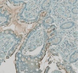
ACE‑2 in Hamster Lung.
ACE‑2 was detected in immersion fixed paraffin-embedded sections of hamster lung using Mouse Anti-Human ACE‑2 Monoclonal Antibody (Catalog # MAB933) at 10 µg/mL for 1 hour at room temperature followed by incubation with the Anti-Mouse IgG VisUCyte™ HRP Polymer Antibody (VC001). Before incubation with the primary antibody, tissue was subjected to heat-induced epitope retrieval using Antigen Retrieval Reagent-Basic (CTS013). Tissue was stained using DAB (brown) and counterstained with hematoxylin (blue). Specific staining was localized to respiratory bronchioles. Staining was performed our protocol for IHC Staining with VisUCyte HRP Polymer Detection Reagents.
Detection of Human ACE-2 by Immunohistochemistry
In microvasculature, ACE2 is putatively expressed in pericytes. (A) Representative image of human pancreatic Formalin-Fixed Paraffin Embedded (FFPE) section stained for ACE2 in case #301118. In panel-a, a representative image of a pancreatic section showing two adjacent lobules (blue and red dotted lines) with different staining for ACE2 in endothelial cells/pericytes. A specific segmentation of the two lobules with high (blue) (zoom-in, panel-b) and low or null expression of ACE2 (red) (zoom-in, panel-c) is shown, suggesting lobularity of ACE2 expression in exocrine endothelial cells/pericytes of human pancreas. Scale bar in panel-a: 100 µm. Scale bar in panels-b and -c: 30 µm. (B) Double immunofluorescence staining of ACE2 (green) and CD31 (red) in FFPE pancreas sections from Body01A of Case #110118 (panels-a to -d) and of Body01B of Case #141117 (panels-e to -i). Digital zoom-in overlay images are shown in panels-d, -h and -i. Scale bar in panels-d and -g: 100 μm. Image collected and cropped by CiteAb from the following publication (https://pubmed.ncbi.nlm.nih.gov/33281748), licensed under a CC-BY license. Not internally tested by R&D Systems.
Detection of Human ACE-2 by Immunohistochemistry
ACE2 staining pattern in human pancreas. Immunohistochemistry for ACE2 in human pancreatic tissue sections (case #110118) using R&D MAB933 antibody. ACE2 is markedly expressed in microvasculature associated cells (A, B) in some rare ductal cells (C, D) and in a subset of endocrine cells within pancreatic islets (E, F). Scale bars in (A, C, E) 150 µm. Scale bars in (B, D, F) 70 µm. Zoom-in images are reported in (B, D, F). Image collected and cropped by CiteAb from the following publication (https://pubmed.ncbi.nlm.nih.gov/33281748), licensed under a CC-BY license. Not internally tested by R&D Systems.
Detection of Human ACE-2 by Immunohistochemistry
In microvasculature, ACE2 is putatively expressed in pericytes. (A) Representative image of human pancreatic Formalin-Fixed Paraffin Embedded (FFPE) section stained for ACE2 in case #301118. In panel-a, a representative image of a pancreatic section showing two adjacent lobules (blue and red dotted lines) with different staining for ACE2 in endothelial cells/pericytes. A specific segmentation of the two lobules with high (blue) (zoom-in, panel-b) and low or null expression of ACE2 (red) (zoom-in, panel-c) is shown, suggesting lobularity of ACE2 expression in exocrine endothelial cells/pericytes of human pancreas. Scale bar in panel-a: 100 µm. Scale bar in panels-b and -c: 30 µm. (B) Double immunofluorescence staining of ACE2 (green) and CD31 (red) in FFPE pancreas sections from Body01A of Case #110118 (panels-a to -d) and of Body01B of Case #141117 (panels-e to -i). Digital zoom-in overlay images are shown in panels-d, -h and -i. Scale bar in panels-d and -g: 100 μm. Image collected and cropped by CiteAb from the following publication (https://pubmed.ncbi.nlm.nih.gov/33281748), licensed under a CC-BY license. Not internally tested by R&D Systems.
Detection of Human ACE-2 by Immunocytochemistry/Immunofluorescence
In microvasculature, ACE2 is putatively expressed in pericytes. (A) Representative image of human pancreatic Formalin-Fixed Paraffin Embedded (FFPE) section stained for ACE2 in case #301118. In panel-a, a representative image of a pancreatic section showing two adjacent lobules (blue and red dotted lines) with different staining for ACE2 in endothelial cells/pericytes. A specific segmentation of the two lobules with high (blue) (zoom-in, panel-b) and low or null expression of ACE2 (red) (zoom-in, panel-c) is shown, suggesting lobularity of ACE2 expression in exocrine endothelial cells/pericytes of human pancreas. Scale bar in panel-a: 100 µm. Scale bar in panels-b and -c: 30 µm. (B) Double immunofluorescence staining of ACE2 (green) and CD31 (red) in FFPE pancreas sections from Body01A of Case #110118 (panels-a to -d) and of Body01B of Case #141117 (panels-e to -i). Digital zoom-in overlay images are shown in panels-d, -h and -i. Scale bar in panels-d and -g: 100 μm. Image collected and cropped by CiteAb from the following publication (https://pubmed.ncbi.nlm.nih.gov/33281748), licensed under a CC-BY license. Not internally tested by R&D Systems.
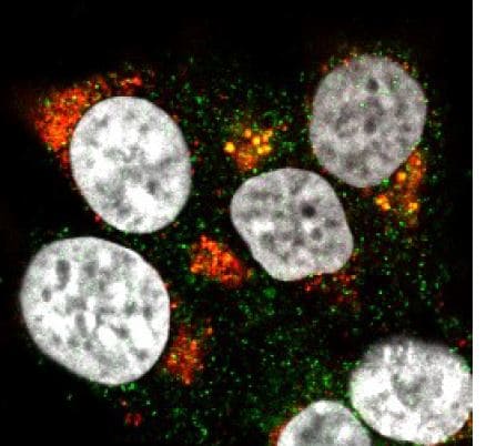
Detection of Chlorocebus sabaeus ACE-2 by Immunocytochemistry/ Immunofluorescence
Modulation of cell surface-expressed ATR1 and ACE2 molecules in Vero E6 cells infected with SARS-CoV-2 (24 h.p.i) and treated with Azilsartan (15 µM) for 72 hours by immunofluorescence microscopy. (A) Schematic flow of the analysis: Vero E6 cells were treated with various ARBs (the MTT assay previously defined non-cytotoxic concentrations: Azilosartan 15µM; Eprosartan 30µM; Irbesartan 60µM; Losartan 7µM; Olmesartan 15µM; Telmisartan 7µM; Valsartan 7µM) for 72 hours and were subsequently infected for analysis of ACE2 and ATR1 on treated and infected cells, 24 hours post-infection (h.p.i.). (B) The panel presents SARS-CoV-2 infected cells after incubation with Azilsartan (15 µM) and evaluation of fluorescence corresponding to the ATR1, ACE2, viral spike protein, and the nucleus of the cells. The merge of the images is displayed at the right of the panel. Images were acquired using a confocal microscope (Zeiss LSM 800) with a 63X/1.4 oil objective. (C) Quantitative representation of Mean Fluorescence corresponding to ATR1 and ACE2 molecules expression on VERO E6 cells treated or not treated with Azilsartan and SARS-CoV-2 in the cells. ***P < 0.001; ****P < 0.0001. Image collected and cropped by CiteAb from the following open publication (https://pubmed.ncbi.nlm.nih.gov/34178717), licensed under a CC-BY license. Not internally tested by R&D Systems.Applications for ACE-2 Antibody (171606)
Immunohistochemistry
Sample: Immersion fixed paraffin-embedded sections of human kidney and hamster lung.
Western Blot
Sample: NS0 mouse myeloma cell line and human kidney tissue
Reviewed Applications
Read 4 reviews rated 4.5 using MAB933 in the following applications:
Formulation, Preparation, and Storage
Purification
Reconstitution
Reconstitute at 0.5 mg/mL in sterile PBS. For liquid material, refer to CoA for concentration.
Formulation
Shipping
Stability & Storage
- 12 months from date of receipt, -20 to -70 °C as supplied.
- 1 month, 2 to 8 °C under sterile conditions after reconstitution.
- 6 months, -20 to -70 °C under sterile conditions after reconstitution.
Calculators
Background: ACE-2
References
- Tipnis, S.R. et al. (2000) J. Biol. Chem. 275:33238.
- Crackower, M.A. et al. (2002) Nature 417:822.
- Huang, L. et al. (2003) J. Biol. Chem. 278:15532.
Long Name
Alternate Names
Entrez Gene IDs
Gene Symbol
UniProt
Additional ACE-2 Products
Product Documents for ACE-2 Antibody (171606)
Certificate of Analysis
To download a Certificate of Analysis, please enter a lot or batch number in the search box below.
Note: Certificate of Analysis not available for kit components.
Product Specific Notices for ACE-2 Antibody (171606)
For research use only
Related Research Areas
Citations for ACE-2 Antibody (171606)
 Powered by Bioz
Customer Reviews for ACE-2 Antibody (171606) (4)
Have you used ACE-2 Antibody (171606)?
Submit a review and receive an Amazon gift card!
$25/€18/£15/$25CAN/¥2500 Yen for a review with an image
$10/€7/£6/$10CAN/¥1110 Yen for a review without an image
Submit a review
Customer Images
-
Application: Immunocytochemistry/ImmunofluorescenceSample Tested: Human cell lineSpecies: HumanVerified Customer | Posted 11/26/2021

-
Application: ImmunohistochemistrySample Tested: Lung tissueSpecies: HumanVerified Customer | Posted 10/19/2021
-
Application: ImmunohistochemistrySample Tested: Small intestineSpecies: HumanVerified Customer | Posted 08/04/2021
-
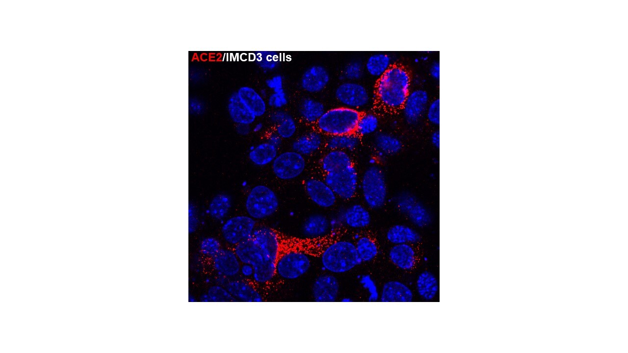
Application: Immunocytochemistry/ImmunofluorescenceSample Tested: Mouse IMCD3 cellsSpecies: Human ACE2Verified Customer | Posted 09/28/2020IMCD3 cells were transiently transfected with human ACE2. 48 hrs post transfection, the cells were fixed, then stain with the ACE2 antibody (1:250 dilution).
There are no reviews that match your criteria.
Protocols
Find general support by application which include: protocols, troubleshooting, illustrated assays, videos and webinars.
- Antigen Retrieval Protocol (PIER)
- Antigen Retrieval for Frozen Sections Protocol
- Appropriate Fixation of IHC/ICC Samples
- Cellular Response to Hypoxia Protocols
- Chromogenic IHC Staining of Formalin-Fixed Paraffin-Embedded (FFPE) Tissue Protocol
- Chromogenic Immunohistochemistry Staining of Frozen Tissue
- ClariTSA™ Fluorophore Kits
- Detection & Visualization of Antibody Binding
- Fluorescent IHC Staining of Frozen Tissue Protocol
- Graphic Protocol for Heat-induced Epitope Retrieval
- Graphic Protocol for the Preparation and Fluorescent IHC Staining of Frozen Tissue Sections
- Graphic Protocol for the Preparation and Fluorescent IHC Staining of Paraffin-embedded Tissue Sections
- Graphic Protocol for the Preparation of Gelatin-coated Slides for Histological Tissue Sections
- IHC Sample Preparation (Frozen sections vs Paraffin)
- Immunofluorescent IHC Staining of Formalin-Fixed Paraffin-Embedded (FFPE) Tissue Protocol
- Immunohistochemistry (IHC) and Immunocytochemistry (ICC) Protocols
- Immunohistochemistry Frozen Troubleshooting
- Immunohistochemistry Paraffin Troubleshooting
- Preparing Samples for IHC/ICC Experiments
- Preventing Non-Specific Staining (Non-Specific Binding)
- Primary Antibody Selection & Optimization
- Protocol for Heat-Induced Epitope Retrieval (HIER)
- Protocol for Making a 4% Formaldehyde Solution in PBS
- Protocol for VisUCyte™ HRP Polymer Detection Reagent
- Protocol for the Preparation & Fixation of Cells on Coverslips
- Protocol for the Preparation and Chromogenic IHC Staining of Frozen Tissue Sections
- Protocol for the Preparation and Chromogenic IHC Staining of Frozen Tissue Sections - Graphic
- Protocol for the Preparation and Chromogenic IHC Staining of Paraffin-embedded Tissue Sections
- Protocol for the Preparation and Chromogenic IHC Staining of Paraffin-embedded Tissue Sections - Graphic
- Protocol for the Preparation and Fluorescent IHC Staining of Frozen Tissue Sections
- Protocol for the Preparation and Fluorescent IHC Staining of Paraffin-embedded Tissue Sections
- Protocol for the Preparation of Gelatin-coated Slides for Histological Tissue Sections
- R&D Systems Quality Control Western Blot Protocol
- TUNEL and Active Caspase-3 Detection by IHC/ICC Protocol
- The Importance of IHC/ICC Controls
- Troubleshooting Guide: Immunohistochemistry
- Troubleshooting Guide: Western Blot Figures
- Western Blot Conditions
- Western Blot Protocol
- Western Blot Protocol for Cell Lysates
- Western Blot Troubleshooting
- Western Blot Troubleshooting Guide
- View all Protocols, Troubleshooting, Illustrated assays and Webinars
FAQs for ACE-2 Antibody (171606)
-
Q: Does Human/Hamster ACE-2 Antibody, Catalog# MAB933, detect mouse ACE2?
A: We have not evaluated cross-reactivity of this antibody to mouse ACE-2. Human ACE-2 shares 83% amino acid sequence identity with mouse ACE-2. We have one citation on our webpage where researcher reports using MAB933 to detect mouse ACE-2.
-
Q: Is Catalog # MAB933 kappa or lambda light chain?
A: MAB933 is kappa light chain.
-
Q: Does Human/Hamster ACE-2 Antibody, Catalog# MAB933, detect mouse ACE2?
A: We have not evaluated cross-reactivity of this antibody to mouse ACE-2. Human ACE-2 shares 83% amino acid sequence identity with mouse ACE-2. We have one citation on our webpage where researcher reports using MAB933 to detect mouse ACE-2.
-
Q: Is Catalog # MAB933 kappa or lambda light chain?
A: MAB933 is kappa light chain.
Associated Pathways

