Angiopoietin-1 (Ang-1) and Angiopoietin-2 (Ang-2) are two closely related secreted ligands which bind with similar affinity to Tie-2, a receptor tyrosine kinase with immunoglobulin and epidermal growth factor homology domains expressed primarily on endothelial cells and early hematopoietic cells. Tie-2 and angiopoietins have been shown to play critical roles in embryogenic angiogenesis and in maintaining the integrity of the adult vasculature (1). Ang-1 cDNA encodes a 498 amino acid (aa) precursor protein that contains a coiled-coiled domain near the amino-terminus and a fibrinogen-like domain at the C-terminus. Human Ang-1 shares approximately 97% and 60% aa sequence identity with mouse Ang‑1 and human Ang‑2, respectively (1, 2). Ang-1 activates Tie-2 signaling on endothelial cells to promote chemotaxis, cell survival, cell sprouting, vessel growth and stabilization (1, 3, 4). Ang-2 has alternatively been reported to be an antagonist for Ang-1-induced Tie-2 signaling as well as an agonist for Tie-2 signaling, depending on the cell context (5).
Human Angiopoietin-1 Antibody (171718)
R&D Systems | Catalog # MAB923

Key Product Details
Validated by
Biological Validation
Species Reactivity
Validated:
Human
Cited:
Human, Mouse, Rat, Porcine
Applications
Validated:
Immunohistochemistry, Western Blot
Cited:
Immunohistochemistry, Immunohistochemistry-Frozen, Western Blot, Neutralization, Immunocytochemistry, Immunoprecipitation, ELISA Detection, ELISA Development
Label
Unconjugated
Antibody Source
Monoclonal Mouse IgG2B Clone # 171718
Loading...
Product Specifications
Immunogen
Mouse myeloma cell line NS0-derived recombinant human Angiopoietin-1
Ser20-Phe498
Accession # Q15389
Ser20-Phe498
Accession # Q15389
Specificity
Detects human Angiopoietin-1 in direct ELISAs and Western blots. In direct ELISAs and Western blots, no cross-reactivity with recombinant human (rh) Angiopoietin-2, recombinant mouse Angiopoietin-like 3, rhAngiopoietin-4, or rhAngiopoietin-like 7 is observed.
Clonality
Monoclonal
Host
Mouse
Isotype
IgG2B
Scientific Data Images for Human Angiopoietin-1 Antibody (171718)
Angiopoietin‑1 in Human Prostate Cancer Tissue.
Angiopoietin-1 was detected in immersion fixed paraffin-embedded sections of human prostate cancer tissue using Mouse Anti-Human Angiopoietin-1 Monoclonal Antibody (Catalog # MAB923) at 15 µg/mL overnight at 4 °C. Tissue was stained using the Anti-Mouse HRP-DAB Cell & Tissue Staining Kit (brown; Catalog # CTS002) and counterstained with hematoxylin (blue). Specific staining was localized to cytoplasm in cancer cells. View our protocol for Chromogenic IHC Staining of Paraffin-embedded Tissue Sections.
Detection of Mouse Angiopoietin-1 by Western Blot
Pericyte-derived Angpt1 controls alveologenesis. a RT-qPCR analysis of Angpt1 and Tie2/Tek expression in freshly sorted lung GFP+, CD31+ or EpCAM+ cells from P7 Pdgfrb(BAC)-CreERT2 R26-mT/mG mice. Data represents mean ± s.e.m. (n = 4 mice). b High magnification images of P10 Angpt1GFP lungs stained for GFP (green), PDGFR beta (red), and PDGFR alpha (blue). Arrows indicate GFP and PDGFR beta double positive pericytes. Scale bar, 15 µm. c RT-qPCR analysis of Angpt1 expression in freshly sorted PDGFR beta + cells from P7 Yap1,Wwtr1iPCKO and control lungs. Data represents mean ± s.e.m. (n = 4 mice, two-tailed unpaired t-test). dAngpt1 expression in cultured Verteporfin (VP)-treated (48 h) and control pericytes. Data represents mean ± s.e.m. (n = 4, Welch’s t-test). e Expression of the indicated transcripts in freshly sorted CD31+ cells from P7 Yap1,Wwtr1iPCKO and control lungs. Data represents mean ± s.e.m. (n = 4 mice, NS not significant, two-tailed unpaired t-test). f–h Western blot analysis of Angpt1 protein (f; n = 2 controls and 4 mutant mice) and of total and phospho-Tie2 (pTie2) in P12 Yap1,Wwtr1iPCKO and control total lung lysates (g, n = 3 controls and 5 mutants). Molecular weight marker (kDa) is indicated. Relative quantification of signals is shown in h. Two-tailed unpaired t-test. i Scheme showing the time points of tamoxifen administration and analysis for Angpt1iPCKO mice. j, k 3D reconstruction confocal images of P12 Angpt1iPCKO and littermate control lungs stained for AQP5 (green), PDGFR beta (red), and PECAM1 (blue). Panels in k show higher magnification of PECAM1 staining. Scale bar, 50 µm (j) and 30 µm (k). l Quantitation of airspace volume in P12 Angpt1iPCKO and littermate control lung sections with 3D reconstruction surface images. Data represents mean ± s.e.m. (n = 4 mice; p < 0.0001, two-tailed unpaired t-test). m 3D reconstruction confocal images of P12 Angpt1iPCKO and littermate control lungs stained for alpha SMA (red) and tropoelastin (blue). Scale bar, 50 µm. n Quantit
Detection of Porcine Angiopoietin-1 by Immunohistochemistry
The Angpt2/Angpt1 balance is disturbed in atherogenic DM pigs.A. Representative illustrations of kidney sections stained with Angpt1 (upper panels; arrow: glomerulus; arrowhead: peritubular area) or Angpt2 (lower panels; arrow: glomerulus; arrowhead: tubular staining) in Controls, ATH, and DM+ATH pigs. B. Immunofluorescent double staining of representative kidney sections for desmin (green)/Angpt1 (red;left panel) and vWF (green/Angpt2 (red; middle/right panel). Insets: double positivity for vWF/Angpt2 staining in yellow. C: Quantitative analysis of renal expression of Angpt1, Angpt2 and Angpt2/Angpt1 ratio. D. Relative mRNA expression of Angpt1 and Angpt2. Data are shown as mean ± SEM. *P<0.05 compared to Controls or ATH pigs. Original magnification of A and B: x400. Image collected and cropped by CiteAb from the following publication (https://dx.plos.org/10.1371/journal.pone.0121555), licensed under a CC-BY license. Not internally tested by R&D Systems.Applications for Human Angiopoietin-1 Antibody (171718)
Application
Recommended Usage
Immunohistochemistry
8-25 µg/mL
Sample: Immersion fixed paraffin-embedded sections of human prostate cancer tissue
Sample: Immersion fixed paraffin-embedded sections of human prostate cancer tissue
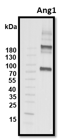
Western Blot
1 µg/mL
Sample: Recombinant Human Angiopoietin‑1 (Catalog # 923-AN)
Sample: Recombinant Human Angiopoietin‑1 (Catalog # 923-AN)
Reviewed Applications
Read 2 reviews rated 4.5 using MAB923 in the following applications:
Formulation, Preparation, and Storage
Purification
Protein A or G purified from hybridoma culture supernatant
Reconstitution
Reconstitute at 0.5 mg/mL in sterile PBS. For liquid material, refer to CoA for concentration.
Loading...
Formulation
Lyophilized from a 0.2 μm filtered solution in PBS with Trehalose. *Small pack size (SP) is supplied either lyophilized or as a 0.2 µm filtered solution in PBS.
Shipping
Lyophilized product is shipped at ambient temperature. Liquid small pack size (-SP) is shipped with polar packs. Upon receipt, store immediately at the temperature recommended below.
Stability & Storage
Use a manual defrost freezer and avoid repeated freeze-thaw cycles.
- 12 months from date of receipt, -20 to -70 °C as supplied.
- 1 month, 2 to 8 °C under sterile conditions after reconstitution.
- 6 months, -20 to -70 °C under sterile conditions after reconstitution.
Calculators
Background: Angiopoietin-1
References
- Jones, N. et al. (2001) Nat. Rev. Mol. Cell Biol. 2:257.
- Davis, S. et al. (1996) Cell 87:1161.
- Witzenbichler, B. et al. (1998) J. Biol. Chem. 273:18514.
- Papapetropoulos, A. et al. (1999) Lab. Inest. 79:213.
- Teichert-Kuliszewska, K. et al. (2001) Cardiovasc. Res. 49:659.
Alternate Names
ANGPT1
Gene Symbol
ANGPT1
UniProt
Additional Angiopoietin-1 Products
Product Documents for Human Angiopoietin-1 Antibody (171718)
Certificate of Analysis
To download a Certificate of Analysis, please enter a lot or batch number in the search box below.
Note: Certificate of Analysis not available for kit components.
Product Specific Notices for Human Angiopoietin-1 Antibody (171718)
For research use only
Citations for Human Angiopoietin-1 Antibody (171718)
 Powered by Bioz
Customer Reviews for Human Angiopoietin-1 Antibody (171718) (2)
4.5 out of 5
2 Customer Ratings
Have you used Human Angiopoietin-1 Antibody (171718)?
Submit a review and receive an Amazon gift card!
$25/€18/£15/$25CAN/¥2500 Yen for a review with an image
$10/€7/£6/$10CAN/¥1110 Yen for a review without an image
Submit a review
Customer Images
Showing
1
-
2 of
2 reviews
Showing All
Filter By:
-
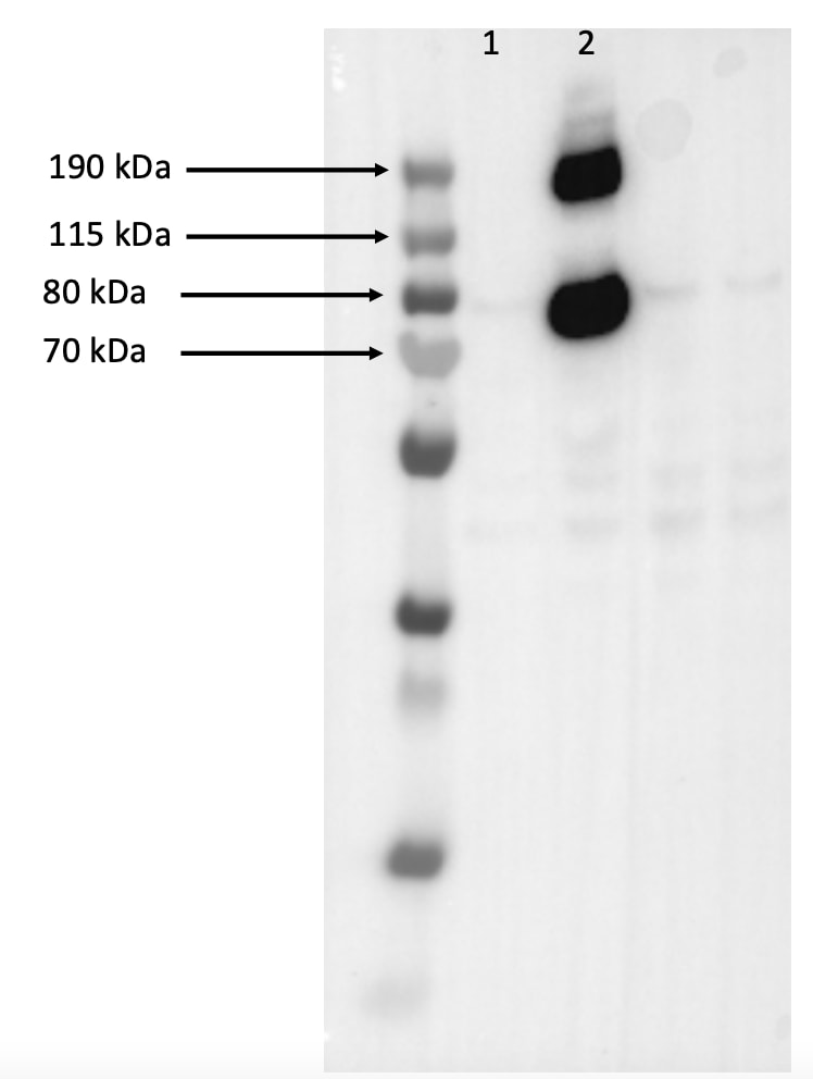
Application: Western BlotSample Tested: HUVEC human umbilical vein endothelial cellsSpecies: HumanVerified Customer | Posted 04/10/2024Lane 1 is control treated cells (water); lane 2 is ANGPT1 (923-AN) treated cells. Here, I was testing the ability of a new TIE2 antagonist and used rh ANGPT (923-AN0 as control). The antibody worked for my experiment.

-
Application: Western BlotSample Tested: Recombinant proteinSpecies: HumanVerified Customer | Posted 08/12/2019
There are no reviews that match your criteria.
Protocols
Find general support by application which include: protocols, troubleshooting, illustrated assays, videos and webinars.
- Antigen Retrieval Protocol (PIER)
- Antigen Retrieval for Frozen Sections Protocol
- Appropriate Fixation of IHC/ICC Samples
- Cellular Response to Hypoxia Protocols
- Chromogenic IHC Staining of Formalin-Fixed Paraffin-Embedded (FFPE) Tissue Protocol
- Chromogenic Immunohistochemistry Staining of Frozen Tissue
- ClariTSA™ Fluorophore Kits
- Detection & Visualization of Antibody Binding
- Fluorescent IHC Staining of Frozen Tissue Protocol
- Graphic Protocol for Heat-induced Epitope Retrieval
- Graphic Protocol for the Preparation and Fluorescent IHC Staining of Frozen Tissue Sections
- Graphic Protocol for the Preparation and Fluorescent IHC Staining of Paraffin-embedded Tissue Sections
- Graphic Protocol for the Preparation of Gelatin-coated Slides for Histological Tissue Sections
- IHC Sample Preparation (Frozen sections vs Paraffin)
- Immunofluorescent IHC Staining of Formalin-Fixed Paraffin-Embedded (FFPE) Tissue Protocol
- Immunohistochemistry (IHC) and Immunocytochemistry (ICC) Protocols
- Immunohistochemistry Frozen Troubleshooting
- Immunohistochemistry Paraffin Troubleshooting
- Preparing Samples for IHC/ICC Experiments
- Preventing Non-Specific Staining (Non-Specific Binding)
- Primary Antibody Selection & Optimization
- Protocol for Heat-Induced Epitope Retrieval (HIER)
- Protocol for Making a 4% Formaldehyde Solution in PBS
- Protocol for VisUCyte™ HRP Polymer Detection Reagent
- Protocol for the Preparation & Fixation of Cells on Coverslips
- Protocol for the Preparation and Chromogenic IHC Staining of Frozen Tissue Sections
- Protocol for the Preparation and Chromogenic IHC Staining of Frozen Tissue Sections - Graphic
- Protocol for the Preparation and Chromogenic IHC Staining of Paraffin-embedded Tissue Sections
- Protocol for the Preparation and Chromogenic IHC Staining of Paraffin-embedded Tissue Sections - Graphic
- Protocol for the Preparation and Fluorescent IHC Staining of Frozen Tissue Sections
- Protocol for the Preparation and Fluorescent IHC Staining of Paraffin-embedded Tissue Sections
- Protocol for the Preparation of Gelatin-coated Slides for Histological Tissue Sections
- R&D Systems Quality Control Western Blot Protocol
- TUNEL and Active Caspase-3 Detection by IHC/ICC Protocol
- The Importance of IHC/ICC Controls
- Troubleshooting Guide: Immunohistochemistry
- Troubleshooting Guide: Western Blot Figures
- Western Blot Conditions
- Western Blot Protocol
- Western Blot Protocol for Cell Lysates
- Western Blot Troubleshooting
- Western Blot Troubleshooting Guide
- View all Protocols, Troubleshooting, Illustrated assays and Webinars
Loading...