
Key Product Details
Species Reactivity
Validated:
Cited:
Applications
Validated:
Cited:
Label
Antibody Source
Product Specifications
Immunogen
Specificity
Clonality
Host
Isotype
Scientific Data Images for Human APRIL/TNFSF13 Antibody
Human APRIL/TNFSF13 ELISA Standard Curve.
Recombinant Human APRIL/TNFSF13 protein was serially diluted 2-fold and captured by Mouse Anti-Human APRIL/TNFSF13 Monoclonal Antibody (MAB8844) coated on a Clear Polystyrene Microplate (DY990). Goat Anti-Human APRIL/TNFSF13 Antigen Affinity-purified Polyclonal Antibody (Catalog # AF884) was biotinylated and incubated with the protein captured on the plate. Detection of the standard curve was achieved by incubating Streptavidin-HRP (DY998) followed by Substrate Solution (DY999) and stopping the enzymatic reaction with Stop Solution (DY994).
Detection of APRIL/TNFSF13 in U937 Human Cell Line by Flow Cytometry.
U937 human lymphoma cell line was stained with Goat Anti-Human APRIL/TNFSF13 Antigen Affinity-purified Polyclonal Antibody (Catalog # AF884, filled histogram) or control antibody staining (AB-108-C, open histogram) followed by anti-Goat IgG PE-conjugated Secondary Antibody (F0107). To facilitate intracellular staining, cells were fixed with Flow Cytometry Fixation Buffer (FC004) and permeabilized with Flow Cytometry Permeabilization/Wash Buffer I (FC005). View our protocol for Staining Intracellular Molecules.Applications for Human APRIL/TNFSF13 Antibody
CyTOF-ready
ELISA
This antibody functions as an ELISA detection antibody when paired with Mouse Anti-Human APRIL/TNFSF13 Monoclonal Antibody (Catalog # MAB8844).
This product is intended for assay development on various assay platforms requiring antibody pairs. We recommend the Human APRIL/TNFSF13 DuoSet ELISA Kit (Catalog # DY884B) for convenient development of a sandwich ELISA.
Flow Cytometry
Sample: U937 human lymphoma cell line fixed with Flow Cytometry Fixation Buffer (Catalog # FC004) and permeabilized with Flow Cytometry Permeabilization/Wash Buffer I (Catalog # FC005). View our protocol for Staining Intracellular Molecules.
Western Blot
Sample: Recombinant Human APRIL/TNFSF13 (Catalog # 884-AP)
Reviewed Applications
Read 1 review rated 3 using AF884 in the following applications:
Flow Cytometry Panel Builder
Bio-Techne Knows Flow Cytometry
Save time and reduce costly mistakes by quickly finding compatible reagents using the Panel Builder Tool.
Advanced Features
- Spectra Viewer - Custom analysis of spectra from multiple fluorochromes
- Spillover Popups - Visualize the spectra of individual fluorochromes
- Antigen Density Selector - Match fluorochrome brightness with antigen density

Formulation, Preparation, and Storage
Purification
Reconstitution
Reconstitute at 0.2 mg/mL in sterile PBS. For liquid material, refer to CoA for concentration.
Formulation
*Small pack size (-SP) is supplied either lyophilized or as a 0.2 µm filtered solution in PBS.
Shipping
Stability & Storage
- 12 months from date of receipt, -20 to -70 °C as supplied.
- 1 month, 2 to 8 °C under sterile conditions after reconstitution.
- 6 months, -20 to -70 °C under sterile conditions after reconstitution.
Calculators
Background: APRIL/TNFSF13
References
- Planelles, L. et al. (2008) Curr. Mol. Med. 8:829.
- Hahne, M. et al. (1998) J. Exp. Med. 188:1185.
- Lopez-Fraga, M. et al. (2001) EMBO Reports 2:945.
- Kelly, K. et al. (2000) Cancer Res. 60:1021.
- Roth, W. et al. (2001) Cell Death Differ. 8:403.
- Yu, G. et al. (2000) Nat. Immunol. 1:252.
- He, B. et al. (2004) J. Immunol. 172:3268.
- Schwaller, J. et al. (2007) Blood 109:331.
- Mhawech-Fauceglia, P. et al. (2008) Eur. J. Cancer 44:2097.
- Day, E.S. et al. (2005) Biochemistry 44:1919.
- Bossen, C. et al. (2006) J. Biol. Chem. 281:13964.
- Hendriks, J. et al. (2005) Cell Death Differ. 12:637.
- Ingold, K. et al. (2005) J. Exp. Med. 201:1375.
- Roschke, V. et al. (2002) J. Immunol. 169:4314.
- Pradet-Balade, B. et al. (2002) EMBO J. 21:5711.
Long Name
Alternate Names
Gene Symbol
Additional APRIL/TNFSF13 Products
Product Documents for Human APRIL/TNFSF13 Antibody
Certificate of Analysis
To download a Certificate of Analysis, please enter a lot or batch number in the search box below.
Note: Certificate of Analysis not available for kit components.
Product Specific Notices for Human APRIL/TNFSF13 Antibody
For research use only
Related Research Areas
Citations for Human APRIL/TNFSF13 Antibody
 Powered by Bioz
Customer Reviews for Human APRIL/TNFSF13 Antibody (1)
Have you used Human APRIL/TNFSF13 Antibody?
Submit a review and receive an Amazon gift card!
$25/€18/£15/$25CAN/¥2500 Yen for a review with an image
$10/€7/£6/$10CAN/¥1110 Yen for a review without an image
Submit a review
Customer Images
-
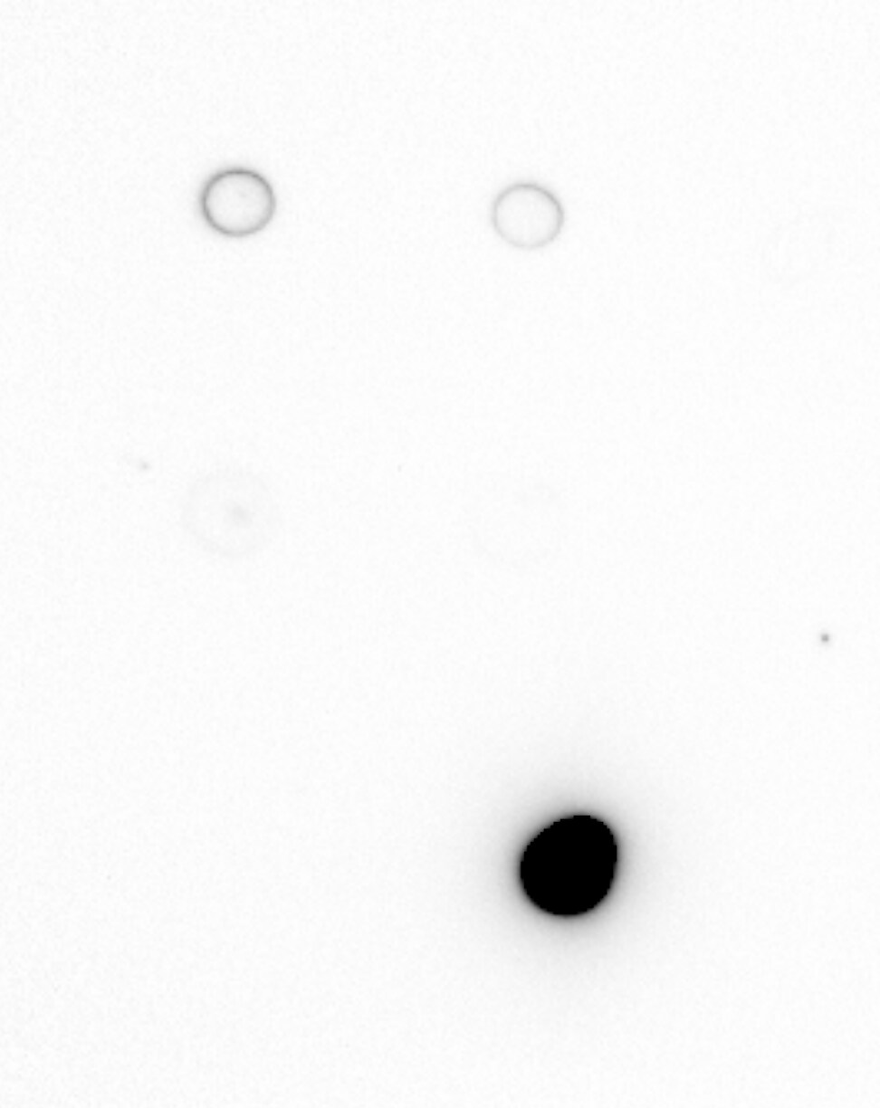
Application: ELISASample Tested: Recombinant proteinSpecies: HumanVerified Customer | Posted 09/09/2024dot blot showing compatibilityWhen I couldn't see signal with MAB8843, I contacted the Technical Team and they provided me with some aliquot of AF884. Again, my ELISA didn't produced expected signal based on my assay design. I then used dot blot to check for compatibility between AF884 and anti-goat secondary antibody. Attached is the dot blot image, showing compatibility.

There are no reviews that match your criteria.
Protocols
Find general support by application which include: protocols, troubleshooting, illustrated assays, videos and webinars.
- 7-Amino Actinomycin D (7-AAD) Cell Viability Flow Cytometry Protocol
- Cellular Response to Hypoxia Protocols
- ELISA Sample Preparation & Collection Guide
- ELISA Troubleshooting Guide
- Extracellular Membrane Flow Cytometry Protocol
- Flow Cytometry Protocol for Cell Surface Markers
- Flow Cytometry Protocol for Staining Membrane Associated Proteins
- Flow Cytometry Staining Protocols
- Flow Cytometry Troubleshooting Guide
- How to Run an R&D Systems DuoSet ELISA
- How to Run an R&D Systems Quantikine ELISA
- How to Run an R&D Systems Quantikine™ QuicKit™ ELISA
- Intracellular Flow Cytometry Protocol Using Alcohol (Methanol)
- Intracellular Flow Cytometry Protocol Using Detergents
- Intracellular Nuclear Staining Flow Cytometry Protocol Using Detergents
- Intracellular Staining Flow Cytometry Protocol Using Alcohol Permeabilization
- Intracellular Staining Flow Cytometry Protocol Using Detergents to Permeabilize Cells
- Propidium Iodide Cell Viability Flow Cytometry Protocol
- Protocol for Liperfluo
- Protocol for the Characterization of Human Th22 Cells
- Protocol for the Characterization of Human Th9 Cells
- Protocol: Annexin V and PI Staining by Flow Cytometry
- Protocol: Annexin V and PI Staining for Apoptosis by Flow Cytometry
- Quantikine HS ELISA Kit Assay Principle, Alkaline Phosphatase
- Quantikine HS ELISA Kit Principle, Streptavidin-HRP Polymer
- R&D Systems Quality Control Western Blot Protocol
- Sandwich ELISA (Colorimetric) – Biotin/Streptavidin Detection Protocol
- Sandwich ELISA (Colorimetric) – Direct Detection Protocol
- Troubleshooting Guide: ELISA
- Troubleshooting Guide: Fluorokine Flow Cytometry Kits
- Troubleshooting Guide: Western Blot Figures
- Western Blot Conditions
- Western Blot Protocol
- Western Blot Protocol for Cell Lysates
- Western Blot Troubleshooting
- Western Blot Troubleshooting Guide
- View all Protocols, Troubleshooting, Illustrated assays and Webinars
Associated Pathways
