Human Brg1 Antibody (2135A)
R&D Systems | Catalog # MAB5738
Recombinant Monoclonal Antibody.

Key Product Details
Validated by
Knockout/Knockdown
Species Reactivity
Validated:
Human
Cited:
Human
Applications
Validated:
Knockout Validated, Immunohistochemistry, Western Blot
Cited:
Western Blot
Label
Unconjugated
Antibody Source
Recombinant Monoclonal Rabbit IgG Clone # 2135A
Loading...
Product Specifications
Immunogen
E. coli-derived recombinant human Brg1
Gln673-Asn774
Accession # P51532
Gln673-Asn774
Accession # P51532
Specificity
Detects human Brg1 in direct ELISAs and Western blots.
Clonality
Monoclonal
Host
Rabbit
Isotype
IgG
Scientific Data Images for Human Brg1 Antibody (2135A)
Detection of Human Brg1 by Western Blot.
Western blot shows lysates of HeLa human cervical epithelial carcinoma cell line and K562 human chronic myelogenous leukemia cell line. Gels were loaded with 30 µg of cytoplasmic (Cyto) and 15 µg of nuclear extracts (Nuc). PVDF membrane was probed with 0.5 µg/mL of Rabbit Anti-Human Brg1 Monoclonal Antibody (Catalog # MAB5738) followed by HRP-conjugated Anti-Rabbit IgG Secondary Antibody (Catalog # HAF008). A specific band was detected for Brg1 at approximately 220 kDa (as indicated). This experiment was conducted under reducing conditions and using Immunoblot Buffer Group 1.
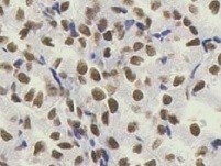
Brg1 in Human Brain.
Brg1 was detected in immersion fixed paraffin-embedded sections of human brain (cerebellum) using Rabbit Anti-Human Brg1 Monoclonal Antibody (Catalog # MAB5738) at 1 µg/mL for 1 hour at room temperature followed by incubation with the Anti-Rabbit IgG VisUCyte™ HRP Polymer Antibody (Catalog # VC003). Tissue was stained using DAB (brown) and counterstained with hematoxylin (blue). Specific staining was localized to neuronal nuclei. View our protocol for IHC Staining with VisUCyte HRP Polymer Detection Reagents.
Western Blot Shows Human Brg1 Specificity by Using Knockout Cell Line.
Western blot shows lysates of HEK293T human embryonic kidney parental cell line and Brg1 knockout HEK293T cell line (KO). PVDF membrane was probed with 0.5 µg/mL of Rabbit Anti-Human Brg1 Monoclonal Antibody (Catalog # MAB5738) followed by HRP-conjugated Anti-Rabbit IgG Secondary Antibody (Catalog # HAF008). A specific band was detected for Brg1 at approximately 250 kDa (as indicated) in the parental HEK293T cell line, but is not detectable in knockout HEK293T cell line. GAPDH (Catalog # MAB5718) is shown as a loading control. This experiment was conducted under reducing conditions and using Immunoblot Buffer Group 1.Applications for Human Brg1 Antibody (2135A)
Application
Recommended Usage
Immunohistochemistry
1-25 µg/mL
Sample: Immersion fixed paraffin-embedded sections of human brain (cerebellum)
Sample: Immersion fixed paraffin-embedded sections of human brain (cerebellum)
Knockout Validated
Brg1
is specifically detected in HEK293T human embryonic kidney parental cell line but is not detectable in
Brg1 knockout HEK293T cell line.
Western Blot
0.5 µg/mL
Sample: HeLa human cervical epithelial carcinoma cell line and K562 human chronic myelogenous leukemia cell line
Sample: HeLa human cervical epithelial carcinoma cell line and K562 human chronic myelogenous leukemia cell line
Reviewed Applications
Read 1 review rated 5 using MAB5738 in the following applications:
Formulation, Preparation, and Storage
Purification
Protein A or G purified from cell culture supernatant
Reconstitution
Reconstitute at 0.5 mg/mL in sterile PBS. For liquid material, refer to CoA for concentration.
Loading...
Formulation
Lyophilized from a 0.2 μm filtered solution in PBS with Trehalose. *Small pack size (SP) is supplied either lyophilized or as a 0.2 µm filtered solution in PBS.
Shipping
Lyophilized product is shipped at ambient temperature. Liquid small pack size (-SP) is shipped with polar packs. Upon receipt, store immediately at the temperature recommended below.
Stability & Storage
Use a manual defrost freezer and avoid repeated freeze-thaw cycles.
- 12 months from date of receipt, -20 to -70 °C as supplied.
- 1 month, 2 to 8 °C under sterile conditions after reconstitution.
- 6 months, -20 to -70 °C under sterile conditions after reconstitution.
Calculators
Background: Brg1
Long Name
BRM/SWI2-related gene 1
Alternate Names
SMARCA4, SNF2, SNF2-beta, SNF2L4, SNF2LB, SWI2
Gene Symbol
SMARCA4
UniProt
Additional Brg1 Products
Product Documents for Human Brg1 Antibody (2135A)
Certificate of Analysis
To download a Certificate of Analysis, please enter a lot or batch number in the search box below.
Note: Certificate of Analysis not available for kit components.
Product Specific Notices for Human Brg1 Antibody (2135A)
For research use only
Related Research Areas
Citations for Human Brg1 Antibody (2135A)
 Powered by Bioz
Customer Reviews for Human Brg1 Antibody (2135A) (1)
5 out of 5
1 Customer Rating
Have you used Human Brg1 Antibody (2135A)?
Submit a review and receive an Amazon gift card!
$25/€18/£15/$25CAN/¥2500 Yen for a review with an image
$10/€7/£6/$10CAN/¥1110 Yen for a review without an image
Submit a review
Customer Images
Showing
1
-
1 of
1 review
Showing All
Filter By:
-
Application: ImmunohistochemistrySample Tested: Kidney tissueSpecies: HumanVerified Customer | Posted 01/20/2022

There are no reviews that match your criteria.
Protocols
Find general support by application which include: protocols, troubleshooting, illustrated assays, videos and webinars.
- Antigen Retrieval Protocol (PIER)
- Antigen Retrieval for Frozen Sections Protocol
- Appropriate Fixation of IHC/ICC Samples
- Cellular Response to Hypoxia Protocols
- Chromogenic IHC Staining of Formalin-Fixed Paraffin-Embedded (FFPE) Tissue Protocol
- Chromogenic Immunohistochemistry Staining of Frozen Tissue
- ClariTSA™ Fluorophore Kits
- Detection & Visualization of Antibody Binding
- Fluorescent IHC Staining of Frozen Tissue Protocol
- Graphic Protocol for Heat-induced Epitope Retrieval
- Graphic Protocol for the Preparation and Fluorescent IHC Staining of Frozen Tissue Sections
- Graphic Protocol for the Preparation and Fluorescent IHC Staining of Paraffin-embedded Tissue Sections
- Graphic Protocol for the Preparation of Gelatin-coated Slides for Histological Tissue Sections
- IHC Sample Preparation (Frozen sections vs Paraffin)
- Immunofluorescent IHC Staining of Formalin-Fixed Paraffin-Embedded (FFPE) Tissue Protocol
- Immunohistochemistry (IHC) and Immunocytochemistry (ICC) Protocols
- Immunohistochemistry Frozen Troubleshooting
- Immunohistochemistry Paraffin Troubleshooting
- Preparing Samples for IHC/ICC Experiments
- Preventing Non-Specific Staining (Non-Specific Binding)
- Primary Antibody Selection & Optimization
- Protocol for Heat-Induced Epitope Retrieval (HIER)
- Protocol for Making a 4% Formaldehyde Solution in PBS
- Protocol for VisUCyte™ HRP Polymer Detection Reagent
- Protocol for the Preparation & Fixation of Cells on Coverslips
- Protocol for the Preparation and Chromogenic IHC Staining of Frozen Tissue Sections
- Protocol for the Preparation and Chromogenic IHC Staining of Frozen Tissue Sections - Graphic
- Protocol for the Preparation and Chromogenic IHC Staining of Paraffin-embedded Tissue Sections
- Protocol for the Preparation and Chromogenic IHC Staining of Paraffin-embedded Tissue Sections - Graphic
- Protocol for the Preparation and Fluorescent IHC Staining of Frozen Tissue Sections
- Protocol for the Preparation and Fluorescent IHC Staining of Paraffin-embedded Tissue Sections
- Protocol for the Preparation of Gelatin-coated Slides for Histological Tissue Sections
- R&D Systems Quality Control Western Blot Protocol
- TUNEL and Active Caspase-3 Detection by IHC/ICC Protocol
- The Importance of IHC/ICC Controls
- Troubleshooting Guide: Immunohistochemistry
- Troubleshooting Guide: Western Blot Figures
- Western Blot Conditions
- Western Blot Protocol
- Western Blot Protocol for Cell Lysates
- Western Blot Troubleshooting
- Western Blot Troubleshooting Guide
- View all Protocols, Troubleshooting, Illustrated assays and Webinars

