
Key Product Details
Species Reactivity
Human
Applications
Western Blot, Immunocytochemistry
Label
Unconjugated
Antibody Source
Monoclonal Mouse IgG2B Clone # 883926
Loading...
Product Specifications
Immunogen
E. coli-derived recombinant human c-Maf
Met1-Phe372
Accession # O75444
Met1-Phe372
Accession # O75444
Specificity
Detects human c-Maf in direct ELISA and Western Blot.
Clonality
Monoclonal
Host
Mouse
Isotype
IgG2B
Scientific Data Images for Human c-Maf Antibody (883926)
Detection of Human c-Maf by Western Blot.
Western blot shows lysates of RPMI 8226 human multiple myeloma cell line. Gels were loaded with 40 µg of cytoplasmic (Cyto) and 20 µg of nuclear (Nuc) extracts. PVDF membrane was probed with 2 µg/mL of Mouse Anti-Human c-Maf Monoclonal Antibody (Catalog # MAB8227) followed by HRP-conjugated Anti-Mouse IgG Secondary Antibody (Catalog # HAF018). A specific band was detected for c-Maf at approximately 48 kDa (as indicated). This experiment was conducted under reducing conditions and using Immunoblot Buffer Group 1.
c-Maf in RPMI 8226 Human Cell Line.
c-Maf was detected in immersion fixed RPMI 8226 human multiple myeloma cell line using Mouse Anti-Human c-Maf Monoclonal Antibody (Catalog # MAB8227) at 10 µg/mL for 3 hours at room temperature. Cells were stained using the NorthernLights™ 557-conjugated Anti-Mouse IgG Secondary Antibody (red, upper panel; Catalog # NL007) and counterstained with DAPI (blue, lower panel). Specific staining was localized to nuclei. View our protocol for Fluorescent ICC Staining of Non-adherent Cells.Applications for Human c-Maf Antibody (883926)
Application
Recommended Usage
Immunocytochemistry
8-25 µg/mL
Sample: Immersion fixed RPMI 8226 human multiple myeloma cell line
Sample: Immersion fixed RPMI 8226 human multiple myeloma cell line
Western Blot
2 µg/mL
Sample: RPMI 8226 human multiple myeloma cell line
Sample: RPMI 8226 human multiple myeloma cell line
Reviewed Applications
Read 2 reviews rated 3.5 using MAB8227 in the following applications:
Formulation, Preparation, and Storage
Purification
Protein A or G purified from hybridoma culture supernatant
Reconstitution
Reconstitute at 0.5 mg/mL in sterile PBS. For liquid material, refer to CoA for concentration.
Loading...
Formulation
Lyophilized from a 0.2 μm filtered solution in PBS with Trehalose. *Small pack size (SP) is supplied either lyophilized or as a 0.2 µm filtered solution in PBS.
Shipping
Lyophilized product is shipped at ambient temperature. Liquid small pack size (-SP) is shipped with polar packs. Upon receipt, store immediately at the temperature recommended below.
Stability & Storage
Use a manual defrost freezer and avoid repeated freeze-thaw cycles.
- 12 months from date of receipt, -20 to -70 °C as supplied.
- 1 month, 2 to 8 °C under sterile conditions after reconstitution.
- 6 months, -20 to -70 °C under sterile conditions after reconstitution.
Calculators
Background: c-Maf
Long Name
v-maf Musculoaponeurotic Fibrosarcoma Oncogene Homolog (Avian)
Alternate Names
CCA4, cMaf, MAF2
Gene Symbol
MAF
UniProt
Additional c-Maf Products
Product Documents for Human c-Maf Antibody (883926)
Certificate of Analysis
To download a Certificate of Analysis, please enter a lot or batch number in the search box below.
Note: Certificate of Analysis not available for kit components.
Product Specific Notices for Human c-Maf Antibody (883926)
For research use only
Citations for Human c-Maf Antibody (883926)
 Powered by Bioz
Customer Reviews for Human c-Maf Antibody (883926) (2)
3.5 out of 5
2 Customer Ratings
Have you used Human c-Maf Antibody (883926)?
Submit a review and receive an Amazon gift card!
$25/€18/£15/$25CAN/¥2500 Yen for a review with an image
$10/€7/£6/$10CAN/¥1110 Yen for a review without an image
Submit a review
Customer Images
Showing
1
-
2 of
2 reviews
Showing All
Filter By:
-
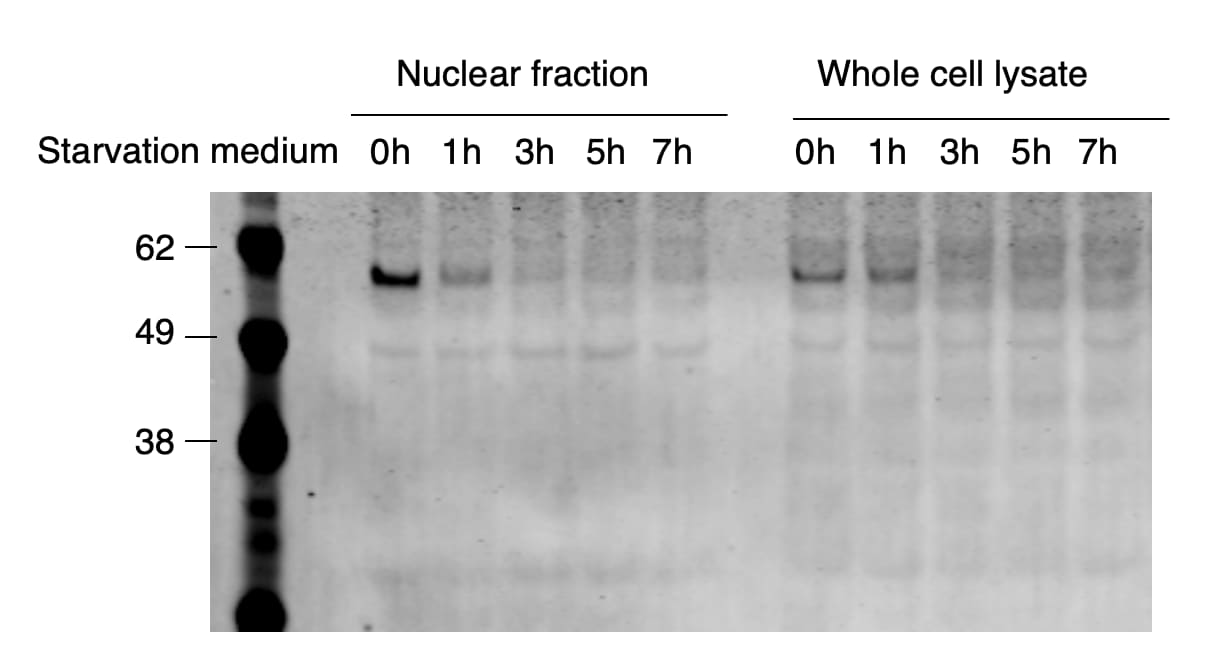
Application: Western BlotSample Tested: HEK293T human embryonic kidney cell lineSpecies: HumanVerified Customer | Posted 06/27/2020As hc-Maf mostly localizes in the nucleus, prepping nuclear fraction and performing WB might help if the WB signal from the total lysate is too low.

-
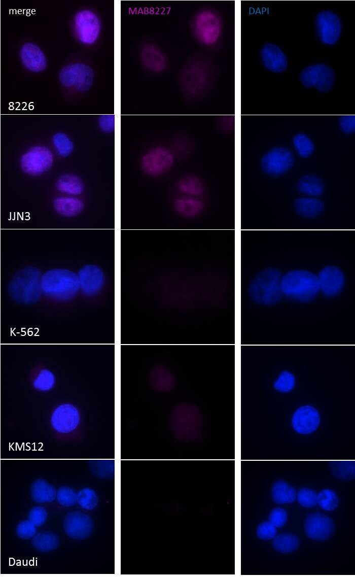
Application: Immunocytochemistry/ImmunofluorescenceSample Tested: RPMI-8226Species: HumanVerified Customer | Posted 10/24/2016ICC/IF on RPMI-8226, JJN3, K-562, KMS12 & Daudi with MAB8227Immersion-fixed cells; permeabilized Triton X-100 5 min; blocked 1 hour 10% donkey serum; 1/30 O/N @4oC; Donkey F(ab’)2 anti-mouse IgG (H+L) Alexa Fluor 647 Abcam #ab150103 1 hour at rt; Zeiss AxioImager Cy5 filter cube 500 ms exposure
There are no reviews that match your criteria.
Protocols
Find general support by application which include: protocols, troubleshooting, illustrated assays, videos and webinars.
- Appropriate Fixation of IHC/ICC Samples
- Cellular Response to Hypoxia Protocols
- ClariTSA™ Fluorophore Kits
- Detection & Visualization of Antibody Binding
- ICC Cell Smear Protocol for Suspension Cells
- ICC Immunocytochemistry Protocol Videos
- ICC for Adherent Cells
- Immunocytochemistry (ICC) Protocol
- Immunocytochemistry Troubleshooting
- Immunofluorescence of Organoids Embedded in Cultrex Basement Membrane Extract
- Immunohistochemistry (IHC) and Immunocytochemistry (ICC) Protocols
- Preparing Samples for IHC/ICC Experiments
- Preventing Non-Specific Staining (Non-Specific Binding)
- Primary Antibody Selection & Optimization
- Protocol for VisUCyte™ HRP Polymer Detection Reagent
- Protocol for the Fluorescent ICC Staining of Cell Smears - Graphic
- Protocol for the Fluorescent ICC Staining of Cultured Cells on Coverslips - Graphic
- Protocol for the Preparation and Fluorescent ICC Staining of Cells on Coverslips
- Protocol for the Preparation and Fluorescent ICC Staining of Non-adherent Cells
- Protocol for the Preparation and Fluorescent ICC Staining of Stem Cells on Coverslips
- Protocol for the Preparation of a Cell Smear for Non-adherent Cell ICC - Graphic
- R&D Systems Quality Control Western Blot Protocol
- TUNEL and Active Caspase-3 Detection by IHC/ICC Protocol
- The Importance of IHC/ICC Controls
- Troubleshooting Guide: Western Blot Figures
- Western Blot Conditions
- Western Blot Protocol
- Western Blot Protocol for Cell Lysates
- Western Blot Troubleshooting
- Western Blot Troubleshooting Guide
- View all Protocols, Troubleshooting, Illustrated assays and Webinars
Loading...
Associated Pathways
