CD14 is a 55 kDa cell surface glycoprotein that is preferentially expressed on monocytes/macrophages. The human CD14 cDNA encodes a 375 amino acid (aa) residue precursor protein with a 19 aa signal peptide and a C-terminal hydrophobic region characteristic for glycosylphosphatidyinositol (GPI)-anchored proteins. Human CD14 has four potential N-linked glycosylation sites and also bears O-linked carbohydrates. The amino acid sequence of human CD14 is approximately 65% identical with the mouse, rat, rabbit, and bovine proteins. CD14 is a pattern recognition receptor that binds lipopolysaccharides (LPS) and a variety of ligands derived from different microbial sources. The binding of CD14 with LPS is catalyzed by LPS-binding protein (LBP). The toll-like-receptors have also been implicated in the transduction of CD14-LPS signals. Similar to other GPI-anchored proteins, soluble CD14 can be released from the cell surface by phosphatidyinositol-specific phospholipase C. Soluble CD14 has been detected in serum and body fluids. High concentrations of soluble CD14 have been shown to inhibit LPS-mediated responses. However, soluble CD14 can also potentiate LPS response in cells that do not express cell surface CD14.

Key Product Details
Species Reactivity
Validated:
Human
Cited:
Human
Applications
Validated:
Immunohistochemistry, Western Blot, Neutralization, Flow Cytometry, CyTOF-ready
Cited:
Western Blot, Neutralization, Flow Cytometry, Immunocytochemistry, ELISA Development
Label
Unconjugated
Antibody Source
Monoclonal Mouse IgG1 Clone # 134620
Loading...
Product Specifications
Immunogen
Chinese hamster ovary cell line CHO-derived recombinant human CD14
Thr20-Cys352
Accession # P08571
Thr20-Cys352
Accession # P08571
Specificity
Detects human CD14 in direct ELISAs and Western blots.
Clonality
Monoclonal
Host
Mouse
Isotype
IgG1
Endotoxin Level
<0.10 EU per 1 μg of the antibody by the LAL method.
Scientific Data Images for Human CD14 Antibody (134620)
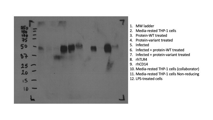
Detection of Human CD14 by Western Blot.
Western blot shows lysates of human peripheral blood mononuclear cells (PBMC). PVDF membrane was probed with 2 µg/mL of Mouse Anti-Human CD14 Monoclonal Antibody (Catalog # MAB3832) followed by HRP-conjugated Anti-Mouse IgG Secondary Antibody (HAF018). A specific band was detected for CD14 at approximately 55 kDa (as indicated). This experiment was conducted under non-reducing conditions and using Western Blot Buffer Group 1.
Detection of CD14 in Human PBMCs by Flow Cytometry.
Human peripheral blood mononuclear cells (PBMCs) were stained with Mouse Anti-Human CD14 Monoclonal Antibody (Catalog # MAB3832) followed by Goat anti-Mouse IgG APC-conjugated Secondary Antibody (Catalog # F0101B). View our protocol for Staining Membrane-associated Proteins.
Detection of CD14 in human tonsil.
CD14 was detected in immersion fixed paraffin-embedded sections of human tonsil using Mouse Anti-Human CD14 Monoclonal Antibody (Catalog # MAB3832) at 5 µg/mL for 1 hour at room temperature followed by incubation with the Anti-Mouse IgG VisUCyte™ HRP Polymer Antibody (Catalog # VC001). Before incubation with the primary antibody, tissue was subjected to heat-induced epitope retrieval using VisUCyte Antigen Retrieval Reagent-Basic (Catalog # VCTS021). Tissue was stained using DAB (brown) and counterstained with hematoxylin (blue). Specific staining was localized to cell surface in lymphocytes. View our protocol for IHC Staining with VisUCyte HRP Polymer Detection Reagents.Applications for Human CD14 Antibody (134620)
Application
Recommended Usage
CyTOF-ready
Ready to be labeled using established conjugation methods. No BSA or other carrier proteins that could interfere with conjugation.
Flow Cytometry
0.25 µg/106 cells
Sample: Human peripheral blood monocytes
Sample: Human peripheral blood monocytes
Immunohistochemistry
5-25 µg/mL
Sample: Immersion fixed paraffin-embedded sections of human tonsil
Sample: Immersion fixed paraffin-embedded sections of human tonsil
Western Blot
2 µg/mL
Sample: Human peripheral blood mononuclear cells (PBMC)
Sample: Human peripheral blood mononuclear cells (PBMC)
Neutralization
Measured by its ability to neutralize lipopolysacharide (LPS)-induced TNF‑ alpha secretion in the THP-1 human acute monocytic leukemia cell line. At 10 µg/mL, this anti-hCD14 antibody will neutralize >60% of 0.5 ng/mL LPS-induced TNF-alpha secretion.
Reviewed Applications
Read 2 reviews rated 4.5 using MAB3832 in the following applications:
Flow Cytometry Panel Builder
Bio-Techne Knows Flow Cytometry
Save time and reduce costly mistakes by quickly finding compatible reagents using the Panel Builder Tool.
Advanced Features
- Spectra Viewer - Custom analysis of spectra from multiple fluorochromes
- Spillover Popups - Visualize the spectra of individual fluorochromes
- Antigen Density Selector - Match fluorochrome brightness with antigen density

Formulation, Preparation, and Storage
Purification
Protein A or G purified from hybridoma culture supernatant
Reconstitution
Reconstitute at 0.5 mg/mL in sterile PBS. For liquid material, refer to CoA for concentration.
Loading...
Formulation
Lyophilized from a 0.2 μm filtered solution in PBS with Trehalose. *Small pack size (SP) is supplied either lyophilized or as a 0.2 µm filtered solution in PBS.
Shipping
Lyophilized product is shipped at ambient temperature. Liquid small pack size (-SP) is shipped with polar packs. Upon receipt, store immediately at the temperature recommended below.
Stability & Storage
Use a manual defrost freezer and avoid repeated freeze-thaw cycles.
- 12 months from date of receipt, -20 to -70 °C as supplied.
- 1 month, 2 to 8 °C under sterile conditions after reconstitution.
- 6 months, -20 to -70 °C under sterile conditions after reconstitution.
Calculators
Background: CD14
References
- Wright, S.D. et al. (1990) Science 249:1431.
- Pugin, J. et al. (1993) Proc. Natl. Acad. Sci. USA 90:2744.
- Beutler, B. (2000) Current Opinion in Immunology 12:20.
- Stelter, F. (2000) Chem. Immunol. 74:25.
Alternate Names
CD14
Gene Symbol
CD14
UniProt
Additional CD14 Products
Product Documents for Human CD14 Antibody (134620)
Certificate of Analysis
To download a Certificate of Analysis, please enter a lot or batch number in the search box below.
Note: Certificate of Analysis not available for kit components.
Product Specific Notices for Human CD14 Antibody (134620)
For research use only
Citations for Human CD14 Antibody (134620)
 Powered by Bioz
Customer Reviews for Human CD14 Antibody (134620) (2)
4.5 out of 5
2 Customer Ratings
Have you used Human CD14 Antibody (134620)?
Submit a review and receive an Amazon gift card!
$25/€18/£15/$25CAN/¥2500 Yen for a review with an image
$10/€7/£6/$10CAN/¥1110 Yen for a review without an image
Submit a review
Customer Images
Showing
1
-
2 of
2 reviews
Showing All
Filter By:
-
Application: Western BlotSample Tested: THP-1 human acute monocytic leukemia cell lineSpecies: HumanVerified Customer | Posted 10/12/2021Cells lysed in NP-40 lysis buffer. TBST-150mM NaCl. Blocked with 5% milk TBST. Antibody diluted in 2% milk TBST 1:100 (5ug/ml). Product works better under non-reducing conditions.

-
Application: ELISASample Tested: wheat extractsSpecies: wheatVerified Customer | Posted 03/29/2021we use this antibody to see interactions with CD14 recombinant protein, it showed a very good affinity towards CD14 protein in direct ELISA.
There are no reviews that match your criteria.
Protocols
Find general support by application which include: protocols, troubleshooting, illustrated assays, videos and webinars.
- 7-Amino Actinomycin D (7-AAD) Cell Viability Flow Cytometry Protocol
- Antigen Retrieval Protocol (PIER)
- Antigen Retrieval for Frozen Sections Protocol
- Appropriate Fixation of IHC/ICC Samples
- Cellular Response to Hypoxia Protocols
- Chromogenic IHC Staining of Formalin-Fixed Paraffin-Embedded (FFPE) Tissue Protocol
- Chromogenic Immunohistochemistry Staining of Frozen Tissue
- ClariTSA™ Fluorophore Kits
- Detection & Visualization of Antibody Binding
- Extracellular Membrane Flow Cytometry Protocol
- Flow Cytometry Protocol for Cell Surface Markers
- Flow Cytometry Protocol for Staining Membrane Associated Proteins
- Flow Cytometry Staining Protocols
- Flow Cytometry Troubleshooting Guide
- Fluorescent IHC Staining of Frozen Tissue Protocol
- Graphic Protocol for Heat-induced Epitope Retrieval
- Graphic Protocol for the Preparation and Fluorescent IHC Staining of Frozen Tissue Sections
- Graphic Protocol for the Preparation and Fluorescent IHC Staining of Paraffin-embedded Tissue Sections
- Graphic Protocol for the Preparation of Gelatin-coated Slides for Histological Tissue Sections
- IHC Sample Preparation (Frozen sections vs Paraffin)
- Immunofluorescent IHC Staining of Formalin-Fixed Paraffin-Embedded (FFPE) Tissue Protocol
- Immunohistochemistry (IHC) and Immunocytochemistry (ICC) Protocols
- Immunohistochemistry Frozen Troubleshooting
- Immunohistochemistry Paraffin Troubleshooting
- Intracellular Flow Cytometry Protocol Using Alcohol (Methanol)
- Intracellular Flow Cytometry Protocol Using Detergents
- Intracellular Nuclear Staining Flow Cytometry Protocol Using Detergents
- Intracellular Staining Flow Cytometry Protocol Using Alcohol Permeabilization
- Intracellular Staining Flow Cytometry Protocol Using Detergents to Permeabilize Cells
- Preparing Samples for IHC/ICC Experiments
- Preventing Non-Specific Staining (Non-Specific Binding)
- Primary Antibody Selection & Optimization
- Propidium Iodide Cell Viability Flow Cytometry Protocol
- Protocol for Heat-Induced Epitope Retrieval (HIER)
- Protocol for Liperfluo
- Protocol for Making a 4% Formaldehyde Solution in PBS
- Protocol for VisUCyte™ HRP Polymer Detection Reagent
- Protocol for the Characterization of Human Th22 Cells
- Protocol for the Characterization of Human Th9 Cells
- Protocol for the Preparation & Fixation of Cells on Coverslips
- Protocol for the Preparation and Chromogenic IHC Staining of Frozen Tissue Sections
- Protocol for the Preparation and Chromogenic IHC Staining of Frozen Tissue Sections - Graphic
- Protocol for the Preparation and Chromogenic IHC Staining of Paraffin-embedded Tissue Sections
- Protocol for the Preparation and Chromogenic IHC Staining of Paraffin-embedded Tissue Sections - Graphic
- Protocol for the Preparation and Fluorescent IHC Staining of Frozen Tissue Sections
- Protocol for the Preparation and Fluorescent IHC Staining of Paraffin-embedded Tissue Sections
- Protocol for the Preparation of Gelatin-coated Slides for Histological Tissue Sections
- Protocol: Annexin V and PI Staining by Flow Cytometry
- Protocol: Annexin V and PI Staining for Apoptosis by Flow Cytometry
- R&D Systems Quality Control Western Blot Protocol
- TUNEL and Active Caspase-3 Detection by IHC/ICC Protocol
- The Importance of IHC/ICC Controls
- Troubleshooting Guide: Fluorokine Flow Cytometry Kits
- Troubleshooting Guide: Immunohistochemistry
- Troubleshooting Guide: Western Blot Figures
- Western Blot Conditions
- Western Blot Protocol
- Western Blot Protocol for Cell Lysates
- Western Blot Troubleshooting
- Western Blot Troubleshooting Guide
- View all Protocols, Troubleshooting, Illustrated assays and Webinars
Loading...
Associated Pathways


