cIAP-2 (also known as MIHC and HIAP-1) is a member of the inhibitor of apoptosis (IAP) family of proteins that inhibit the proteolytic activity of mature caspases. cIAP-2 has 3 BIR (baculovirus inhibitor of apoptosis) domains, a RING finger domain, and a caspase recruitment domain (CARD). cIAP-2 inhibits caspases through the direct interaction of its BIR domain with the active caspase. Caspase activity may be restored through interactions with the Reaper like motif on mitochondrial proteins such as SMAC/Diablo or HtrA2/Omi. cIAP-2 is reported to be cleaved by HtrA2/Omi.

Key Product Details
Species Reactivity
Validated:
Human
Cited:
Human
Applications
Validated:
Immunohistochemistry, Western Blot, Simple Western
Cited:
Western Blot, Flow Cytometry, Immunocytochemistry
Label
Unconjugated
Antibody Source
Polyclonal Goat IgG
Loading...
Product Specifications
Immunogen
E. coli-derived recombinant human cIAP‑2/HIAP‑1
Asn2-Ser604
Accession # U45878
Asn2-Ser604
Accession # U45878
Specificity
Detects human cIAP‑2/HIAP‑1 in Western blots. In Western blots, less than 1% cross-reactivity with cIAP-1 is observed.
Clonality
Polyclonal
Host
Goat
Isotype
IgG
Scientific Data Images for Human cIAP-2/HIAP-1 Antibody
cIAP-2/HIAP-1 in Human Colon Tissue.
cIAP-2/HIAP-1 was detected in immersion fixed paraffin-embedded sections of human colon tissue using Goat Anti-Human cIAP-2/HIAP-1 Antigen Affinity-purified Polyclonal Antibody (Catalog # AF8171) at 1 µg/mL for 1 hour at room temperature followed by incubation with the Anti-Goat IgG VisUCyte™ HRP Polymer Antibody (Catalog # VC004). Before incubation with the primary antibody, tissue was subjected to heat-induced epitope retrieval using Antigen Retrieval Reagent-Basic (Catalog # CTS013). Tissue was stained using DAB (brown) and counterstained with hematoxylin (blue). Specific staining was localized to colon glands. View our protocol for IHC Staining with VisUCyte HRP Polymer Detection Reagents.
Detection of Human cIAP‑2/HIAP‑1 by Western Blot.
Western blot shows lysates of Raji human Burkitt's lymphoma cell line and Daudi human Burkitt's lymphoma cell line. PVDF membrane was probed with 0.5 µg/mL of Goat Anti-Human cIAP-2/HIAP-1 Antigen Affinity-purified Polyclonal Antibody (Catalog # AF8171) followed by HRP-conjugated Anti-Goat IgG Secondary Antibody (Catalog # HAF109). A specific band was detected for cIAP-2/HIAP-1 at approximately 68 kDa (as indicated). This experiment was conducted under reducing conditions and using Immunoblot Buffer Group 5.
Detection of Human cIAP‑2/HIAP‑1 by Simple WesternTM.
Simple Western lane view shows lysates of Raji human Burkitt's lymphoma cell line and Daudi human Burkitt's lymphoma cell line, loaded at 0.2 mg/mL. A specific band was detected for cIAP-2/HIAP-1 at approximately 70 kDa (as indicated) using 5 µg/mL of Goat Anti-Human cIAP-2/HIAP-1 Antigen Affinity-purified Polyclonal Antibody (Catalog # AF8171) followed by 1:50 dilution of HRP-conjugated Anti-Goat IgG Secondary Antibody (Catalog # HAF109). This experiment was conducted under reducing conditions and using the 12-230 kDa separation system.
Detection of Human cIAP-2/HIAP-1 by Western Blot
IAP expression in glioblastoma cell lines. Expression levels of cIAP1, cIAP2, XIAP and ML-IAP were analyzed by western blotting and quantified in U87MG and GL261 adherent GBM cell lines, and in GBM6 and GBM9 spheres. Expression level of beta -actin served as loading control. The four GBM cell lines expressed heterogeneously cIAP1, cIAP2, XIAP and ML-IAP. A representative experiment of four experiments is shown. Quantification was performed using ImageJ software (National Institutes of Health, Bethesda, MD, USA) and data presented were normalized to beta -actin expression Image collected and cropped by CiteAb from the following publication (https://pubmed.ncbi.nlm.nih.gov/27490930), licensed under a CC-BY license. Not internally tested by R&D Systems.
Detection of Human cIAP-2/HIAP-1 by Immunocytochemistry/Immunofluorescence
Prognostic value of cIAP1, cIAP2, XIAP and ML-IAP protein expression in human glioblastomas (cohorts 1 and 2). (a) cIAP1-, cIAP2-, XIAP- and ML-IAP-positive stainings in GBM. IAPs were heterogeneously expressed by tumor cells in GBM samples (Table 1). Stainings were diffused with a stronger punctuated positivity into the cytoplasm. Black arrows highlight cIAP2-positive nuclei. Scale bars, 50 μm. (b) Correlation of ML-IAP protein expression with PFS and OS in cohort 1. The cutoff was 35% and was determined by performing a ROC curve. ML-IAP expression of ⩾35% was correlated with a poor prognosis. (c) Correlation of ML-IAP protein expression with PFS and OS in cohort 2. The cutoff was the same as that for cohort 1 analysis (35%). ML-IAP expression of ⩾35% was correlated with a poor prognosis Image collected and cropped by CiteAb from the following publication (https://pubmed.ncbi.nlm.nih.gov/27490930), licensed under a CC-BY license. Not internally tested by R&D Systems.
Detection of Human cIAP-2/HIAP-1 by Western Blot
Apoptosis and IAP expression upon SMAC mimetic GDC-0152 treatment in glioblastoma cell lines. (a) Apoptosis (SubG0/G1) of DMSO control and GDC-0152-treated cells was determined by flow cytometry of propidium iodide-stained nuclei and percentage of apoptosis is shown. U87MG and GL261 cell lines were treated for 72 h and GBM6 and GBM9 cell lines were treated for 8 days at the indicated concentrations. At these respective time points, percentage of U87MG cells dead by apoptosis, percentage of GL261 cells, percentage of GBM6 cells and percentage of GBM9 cells. Data are expressed as mean+S.E.M. Three independent experiments were performed for the GL261 cell lines and five for the U87MG, GBM6 and GBM9 cell lines. *P<0.05; **P<0.01; ***P<0.005. (b) Expression levels of cIAP1, cIAP2, XIAP and ML-IAP were analyzed by western blotting. Cell lines were treated with 1 μM of GDC-0152. U87MG and GL261 were treated for 72 h and GBM6 and GBM9 cell lines for 8 days. In all GBM cell lines GDC-0152 decreased IAP expression. Expression level of beta -actin served as loading control. A representative experiment of three experiments is shown Image collected and cropped by CiteAb from the following publication (https://pubmed.ncbi.nlm.nih.gov/27490930), licensed under a CC-BY license. Not internally tested by R&D Systems.
Detection of Human cIAP-2/HIAP-1 by Western Blot
9-cis-RA induces the expression of cIAP2 in breast cancer cells in a cell context dependent manner. (A, B, C) Multiplex RNase protections assays (RPAs) to monitor the effect of 9-cis-RA on the expression of death receptor, death ligands, IAP and TRAF family members in four different breast cancer cell lines. Breast cancer cells were treated for the indicated time with 9-cis-RA at a concentration of 10-6 M. (D) Western blot of whole cell extracts of 9-cis-RA-treated T47D cells and H3396 cells for 0, 12, 24, 48, 72 and 96 hours with anti-cIAP2. The nonspecific signal (n. sp.) confirms equal loading. As a positive control, breast cancer cells were treated with 50 μg/ml of hTNF alpha for 24 and 48 hours. (E) Reversibility of 9-cis-RA-induced cIAP2 gene expression. T47D cells were treated either in the absence or presence of 1 μM 9- cis-RA and after 3 days, total RNA was extracted. In parallel flasks, the medium was removed, and cells were washed and treated with either fresh control medium or medium with 10-6 M of 9-cis-RA and grown for additional 3, 6 and 9 days. Media and ligands were renewed every 3 days. RNA was isolated and analyzed by RPA as described in (A). Equal loading was confirmed by GAPDH RNA level. The images shown are from one representative experiment performed twice with similar results. Image collected and cropped by CiteAb from the following open publication (http://molecular-cancer.biomedcentral.com/articles/10.1186/1476-4598-9-…), licensed under a CC-BY license. Not internally tested by R&D Systems.
Detection of Human cIAP-2/HIAP-1 by Western Blot
Suppression of cIAP2 expression is not sufficient to abrogate 9-cis-RA inhibition of etoposide-induced apoptosis in T47D cells. (A) T47D cells were transfected with either scrambled-siRNA or cIAP2-siRNA and pretreated with or without 9-cis-RA for 30 h, followed by treatment with etoposide 100 μM for 24 h. Cell lysates were analyzed by western blot for the expression of cleaved caspase-3, cIAP2 and beta -actin using specific antibodies. The images shown are from one representative experiment performed three times with similar results. (B) T47D cells were transfected with either scrambled-siRNA (white bars) or cIAP2-siRNA (black bars). After 24 h, lipid-siRNA complexes were removed from media and cells were pretreated with or without 1 μM 9-cis-RA for 30 h, followed by treatment with etoposide 100 μM for 72 h. The percentage of apoptotic cells was determined by FACS analysis after staining with propidium iodide. The values represent the mean ± SD of three experiments performed in duplicate. Asterisks denote statistically significant differences against the corresponding untreated cells. Image collected and cropped by CiteAb from the following open publication (http://molecular-cancer.biomedcentral.com/articles/10.1186/1476-4598-9-…), licensed under a CC-BY license. Not internally tested by R&D Systems.
Detection of Human cIAP-2/HIAP-1 by Western Blot
9-cis-RA induces the expression of cIAP2 in breast cancer cells in a cell context dependent manner. (A, B, C) Multiplex RNase protections assays (RPAs) to monitor the effect of 9-cis-RA on the expression of death receptor, death ligands, IAP and TRAF family members in four different breast cancer cell lines. Breast cancer cells were treated for the indicated time with 9-cis-RA at a concentration of 10-6 M. (D) Western blot of whole cell extracts of 9-cis-RA-treated T47D cells and H3396 cells for 0, 12, 24, 48, 72 and 96 hours with anti-cIAP2. The nonspecific signal (n. sp.) confirms equal loading. As a positive control, breast cancer cells were treated with 50 μg/ml of hTNF alpha for 24 and 48 hours. (E) Reversibility of 9-cis-RA-induced cIAP2 gene expression. T47D cells were treated either in the absence or presence of 1 μM 9- cis-RA and after 3 days, total RNA was extracted. In parallel flasks, the medium was removed, and cells were washed and treated with either fresh control medium or medium with 10-6 M of 9-cis-RA and grown for additional 3, 6 and 9 days. Media and ligands were renewed every 3 days. RNA was isolated and analyzed by RPA as described in (A). Equal loading was confirmed by GAPDH RNA level. The images shown are from one representative experiment performed twice with similar results. Image collected and cropped by CiteAb from the following open publication (http://molecular-cancer.biomedcentral.com/articles/10.1186/1476-4598-9-…), licensed under a CC-BY license. Not internally tested by R&D Systems.
Detection of Human cIAP-2/HIAP-1 by Western Blot
Over-expression of the super-repressor of NF-kappa B activation, I kappa B alpha -SR(S32A/S36A), leads to significant abrogation of retinoic acid-mediated inhibition of etoposide-induced apoptosis. (A) T47D-vector and T47D-I kappa B alpha SR cells were untreated or incubated for the indicated time with hTNF alpha (50 ng/ml). Extracts were analyzed by western blotting with an I kappa B alpha antibody. Equal loading was confirmed with an anti-beta -actin antibody. The images shown are from one representative experiment performed three times with similar results. (B) T47D-vector or T47D-I kappa B alpha SR cells were untreated or treated with 9-cis-RA for 48 h and expression of cIAP2 and beta -actin were analysed by Reverse-Transcriptase Polymerase Reaction and real time PCR. The values represent the mean ± SD of three different experiments performed in duplicate. (C) T47D-vector or T47D-I kappa B alpha SR cells were pretreated with or without 9-cis-RA for 30 h, followed by treatment with etoposide 100 μM as previously indicated. At the indicated times, cell lysates were analyzed by western blot for the expression of cleaved caspase-3, cIAP2 and beta -actin using specific antibodies. The images shown are from one representative experiment performed three times with similar results. (D) T47D-vector or T47D-I kappa B alpha SR cells were pretreated with or without 9-cis-RA for 30 h, followed by treatment with etoposide 100 μM for 72 h. The percentage of apoptotic cells was determined by FACS analysis after staining with propidium iodide. The values represent the mean ± SD of three independent experiments performed in duplicate. Asterisks denote the existence of statistically significant differences between the indicated groups; N.S.: not significant (Student's t-test). Image collected and cropped by CiteAb from the following open publication (http://molecular-cancer.biomedcentral.com/articles/10.1186/1476-4598-9-…), licensed under a CC-BY license. Not internally tested by R&D Systems.Applications for Human cIAP-2/HIAP-1 Antibody
Application
Recommended Usage
Immunohistochemistry
1-15 µg/mL
Sample: Immersion fixed paraffin-embedded sections of human colon tissue
Sample: Immersion fixed paraffin-embedded sections of human colon tissue
Simple Western
5 µg/mL
Sample: Raji human Burkitt's lymphoma cell line and Daudi human Burkitt's lymphoma cell line
Sample: Raji human Burkitt's lymphoma cell line and Daudi human Burkitt's lymphoma cell line
Western Blot
0.5 µg/mL
Sample: Raji human Burkitt's lymphoma cell line and Daudi human Burkitt's lymphoma cell line
Sample: Raji human Burkitt's lymphoma cell line and Daudi human Burkitt's lymphoma cell line
Reviewed Applications
Read 1 review rated 5 using AF8171 in the following applications:
Formulation, Preparation, and Storage
Purification
Antigen Affinity-purified
Reconstitution
Reconstitute at 0.2 mg/mL in sterile PBS. For liquid material, refer to CoA for concentration.
Loading...
Formulation
Lyophilized from a 0.2 μm filtered solution in PBS with Trehalose. *Small pack size (SP) is supplied either lyophilized or as a 0.2 µm filtered solution in PBS.
Shipping
Lyophilized product is shipped at ambient temperature. Liquid small pack size (-SP) is shipped with polar packs. Upon receipt, store immediately at the temperature recommended below.
Stability & Storage
Use a manual defrost freezer and avoid repeated freeze-thaw cycles.
- 12 months from date of receipt, -20 to -70 °C as supplied.
- 1 month, 2 to 8 °C under sterile conditions after reconstitution.
- 6 months, -20 to -70 °C under sterile conditions after reconstitution.
Calculators
Background: cIAP-2/HIAP-1
References
- Roy, N. et al. (1997) EMBO J. 23:6914.
- Deveraux, Q. et al. (1997) Nature 388:300.
- Deveraux, Q. and J. Reed (1999) Genes & Develop. 13:239.
- Srinivasula, S.M. et al. (2003) J. Biol. Chem. 278:31469.
- Yang, Q-H. et al. (2003) Genes Dev. 17:1487.
Alternate Names
BIRC3, cIAP2, HIAP-1, MIHC
Entrez Gene IDs
330 (Human)
Gene Symbol
BIRC3
UniProt
Additional cIAP-2/HIAP-1 Products
Product Documents for Human cIAP-2/HIAP-1 Antibody
Certificate of Analysis
To download a Certificate of Analysis, please enter a lot or batch number in the search box below.
Note: Certificate of Analysis not available for kit components.
Product Specific Notices for Human cIAP-2/HIAP-1 Antibody
For research use only
Related Research Areas
Citations for Human cIAP-2/HIAP-1 Antibody
 Powered by Bioz
Customer Reviews for Human cIAP-2/HIAP-1 Antibody (1)
5 out of 5
1 Customer Rating
Have you used Human cIAP-2/HIAP-1 Antibody?
Submit a review and receive an Amazon gift card!
$25/€18/£15/$25CAN/¥2500 Yen for a review with an image
$10/€7/£6/$10CAN/¥1110 Yen for a review without an image
Submit a review
Customer Images
Showing
1
-
1 of
1 review
Showing All
Filter By:
-
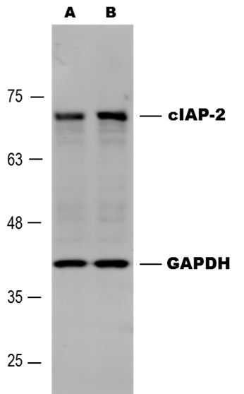
Application: Western BlotSample Tested: MDA-MB-231 human breast cancer cell lineSpecies: HumanVerified Customer | Posted 03/27/2017Western blot analysis of Human cIAP-2/HIAP-1 in MDA-MB-231 cells untreated (A) and treated with 10 uM Paclitaxel for 4 hours (B). Goat anti-Human cIAP-2/HIAP-1antibody (#AF8171) was used at 0.5 ug/mL, followed by Donkey anti-Goat secondary antibody at 1:5,000 dilution.

There are no reviews that match your criteria.
Protocols
Find general support by application which include: protocols, troubleshooting, illustrated assays, videos and webinars.
- Antigen Retrieval Protocol (PIER)
- Antigen Retrieval for Frozen Sections Protocol
- Appropriate Fixation of IHC/ICC Samples
- Cellular Response to Hypoxia Protocols
- Chromogenic IHC Staining of Formalin-Fixed Paraffin-Embedded (FFPE) Tissue Protocol
- Chromogenic Immunohistochemistry Staining of Frozen Tissue
- ClariTSA™ Fluorophore Kits
- Detection & Visualization of Antibody Binding
- Fluorescent IHC Staining of Frozen Tissue Protocol
- Graphic Protocol for Heat-induced Epitope Retrieval
- Graphic Protocol for the Preparation and Fluorescent IHC Staining of Frozen Tissue Sections
- Graphic Protocol for the Preparation and Fluorescent IHC Staining of Paraffin-embedded Tissue Sections
- Graphic Protocol for the Preparation of Gelatin-coated Slides for Histological Tissue Sections
- IHC Sample Preparation (Frozen sections vs Paraffin)
- Immunofluorescent IHC Staining of Formalin-Fixed Paraffin-Embedded (FFPE) Tissue Protocol
- Immunohistochemistry (IHC) and Immunocytochemistry (ICC) Protocols
- Immunohistochemistry Frozen Troubleshooting
- Immunohistochemistry Paraffin Troubleshooting
- Preparing Samples for IHC/ICC Experiments
- Preventing Non-Specific Staining (Non-Specific Binding)
- Primary Antibody Selection & Optimization
- Protocol for Heat-Induced Epitope Retrieval (HIER)
- Protocol for Making a 4% Formaldehyde Solution in PBS
- Protocol for VisUCyte™ HRP Polymer Detection Reagent
- Protocol for the Preparation & Fixation of Cells on Coverslips
- Protocol for the Preparation and Chromogenic IHC Staining of Frozen Tissue Sections
- Protocol for the Preparation and Chromogenic IHC Staining of Frozen Tissue Sections - Graphic
- Protocol for the Preparation and Chromogenic IHC Staining of Paraffin-embedded Tissue Sections
- Protocol for the Preparation and Chromogenic IHC Staining of Paraffin-embedded Tissue Sections - Graphic
- Protocol for the Preparation and Fluorescent IHC Staining of Frozen Tissue Sections
- Protocol for the Preparation and Fluorescent IHC Staining of Paraffin-embedded Tissue Sections
- Protocol for the Preparation of Gelatin-coated Slides for Histological Tissue Sections
- R&D Systems Quality Control Western Blot Protocol
- TUNEL and Active Caspase-3 Detection by IHC/ICC Protocol
- The Importance of IHC/ICC Controls
- Troubleshooting Guide: Immunohistochemistry
- Troubleshooting Guide: Western Blot Figures
- Western Blot Conditions
- Western Blot Protocol
- Western Blot Protocol for Cell Lysates
- Western Blot Troubleshooting
- Western Blot Troubleshooting Guide
- View all Protocols, Troubleshooting, Illustrated assays and Webinars
Loading...
Associated Pathways

