
Key Product Details
Validated by
Biological Validation
Species Reactivity
Validated:
Human
Cited:
Human, Mouse
Applications
Validated:
Western Blot, Immunocytochemistry
Cited:
Immunocytochemistry, Immunoprecipitation
Label
Unconjugated
Antibody Source
Monoclonal Mouse IgG2B Clone # 762203
Loading...
Product Specifications
Immunogen
E. coli-derived recombinant human COMMD1
Ser37-Ser135
Accession # Q8N668
Ser37-Ser135
Accession # Q8N668
Specificity
Detects human COMMD1 in direct ELISAs.
In direct ELISAs, no cross-reactivity
with recombinant human (rh) Attractin or rhCaspr1 is observed.
Clonality
Monoclonal
Host
Mouse
Isotype
IgG2B
Scientific Data Images for Human COMMD1 Antibody (762203)
Detection of Human COMMD1 by Western Blot.
Western blot shows lysates of U2OS human osteosarcoma cell line, HepG2 human hepatocellular carcinoma cell line, and human placenta tissue. PVDF membrane was probed with 0.2 µg/mL of Mouse Anti-Human COMMD1 Monoclonal Antibody (Catalog # MAB7526) followed by HRP-conjugated Anti-Mouse IgG Secondary Antibody (Catalog # HAF018). A specific band was detected for COMMD1 at approximately 20 kDa (as indicated). This experiment was conducted under reducing conditions and using Immunoblot Buffer Group 1.
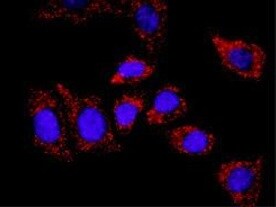
COMMD1 in U2OS Human Cell Line.
COMMD1 was detected in immersion fixed U2OS human osteosarcoma cell line using Mouse Anti-Human COMMD1 Monoclonal Antibody (Catalog # MAB7526) at 8 µg/mL for 3 hours at room temperature. Cells were stained using the NorthernLights™ 557-conjugated Anti-Mouse IgG Secondary Antibody (red; Catalog # NL007) and counterstained with DAPI (blue). Specific staining was localized to cytoplasm and nuclei. View our protocol for Fluorescent ICC Staining of Cells on Coverslips.
Detection of Mouse COMMD1 by Immunocytochemistry/Immunofluorescence
LDLR associates with COMMD1 and the WASH complex.(a) Human embryonic kidney 293T (HEK293T) cells were transfected with constructs expressing Flag-LDLR with either COMMD1-GST or GST alone. Interaction with COMMD1 was detected via pull-down assay using glutathione sepharose beads. (b) HEK293T cells were transfected with Flag-LDLR vector, and interaction with endogenous COMMD1 was detected by immunoprecipitation with rabbit anti-Flag-antibody. (c) Colocalization of LDLR (red) and COMMD1 (green) in Commd1f/f MEFs examined by immunofluorescence staining. Representative images are shown; scale bar, 5μm. LDLR (red) and COMMD1 (green) was stained in COMMD1-deficient MEFs (Commd1−/−) and imaged by confocal fluorescence microscopy. COMMD1 levels in Commd1f/f and in Commd1−/− MEFs determined by immunoblot analysis. (d) Liver of a WT chow-fed mouse was homogenized and loaded on a continuous 10–40% sucrose gradient. Fractions were separated by ultracentrifugation and immunoblotted using antibodies against COMMD1, LDLR, WASH1, FAM21, VPS35 and CCDC22. The figure represents results of three independent experiments. (e) HEK293T cells were transfected with Ha-COMMD1 construct together with GST alone, GST-LDLRct (GST-tagged cytosolic domain of LDLR) or GST-LDLRct Y807A (GST-tagged mutated cytosolic domain of LDLR). Pull-down assay was performed to study the interaction between LDLRct and COMMD1. (f) Lysates of Flag-LDLR-transfected HEK293T cells were used for immunoprecipitation assays. Immunoprecipitates were washed, separated by SDS–polyacrylamide gel electrophoresis and immunoblotted as indicated. Image collected and cropped by CiteAb from the following publication (https://www.nature.com/articles/ncomms10961), licensed under a CC-BY license. Not internally tested by R&D Systems.
Detection of Mouse COMMD1 by Immunocytochemistry/Immunofluorescence
COMMD1 deficiency impairs the function of LDLR.(a) LDLR (green), COMMD1 (red) and VPS35 (pink) were stained in Commd1f/f and Commd1−/− MEFs and imaged by confocal microscopy. Representative images are shown; scale bar, 5μm. (b) Quantification of the colocalization of LDLR with COMMD1, VPS35, EEA1 and LAMP1 was performed by the analysis of 30–40 cells. (c) Total and plasma membrane LDLR levels of Commd1f/f and Commd1−/− MEFs determined by biotinylation assay. Data represent three independent experiments, and (d) the relative levels of LDLR at the cell surface are quantified in all experiments. (e) In vitro LDL and transferrin uptake assay. Dil-labelled LDL (5 μg ml−1) or Alexa-633-labelled transferrin (5 μg ml−1) was added to serum-depleted medium and incubated with MEFs at 4 °C for 1 h and subsequently at 37 °C for 5 min. Dil-labelled LDL and Alexa-633-labelled transferrin uptake was measured by FACS analysis, and the relative uptake in triplicate is shown. The results are presented as mean±s.e.m.; significance was calculated relative to the control group by unpaired Student's t-test; *P<0.05, ***P<0.001. Image collected and cropped by CiteAb from the following publication (https://www.nature.com/articles/ncomms10961), licensed under a CC-BY license. Not internally tested by R&D Systems.
Detection of Mouse COMMD1 by Immunocytochemistry/Immunofluorescence
The WASH complex is essential for endosomal sorting of LDLR.(a) Cellular localization of LDLR (green), COMMD1 (red) and LAMP1 (pink) in Wash1f/f and Wash1−/− MEFs was determined by immunofluorescence staining. Representative images are shown; scale bar, 5 μm. Relative colocalization of (b) LDLR and (c) COMMD1 with VPS35, EEA1 and LAMP1 was quantified. (d) LDLR, WASH1, VPS35 and COMMD1 levels in Wash1f/f and Wash1−/− MEFs analysed by western blot. (e) Representative images (n=3) of immunoblot analysis of total LDLR levels in Wash1f/f and Wash1−/− MEFs treated with bafilomycin A (100 nM) for 0, 4 and 6 h. (f) Densitometry revealed the relative levels of LDLR in bafilomycin A-treated cells (n=3). (g) Representative images (n=3) of LDLR on the surface of Wash1f/f and Wash1−/− MEFs determined by surface biotinylation assay. (h) Densitometry revealed relative LDLR surface levels (n=3). (i) Wash1f/f and Wash1−/− MEFs were incubated with DiI-LDL for 30 min and imaged by fluorescence microscope. (j) Fluorescence intensity was quantified using ImageJ software and was normalized to the number of DAPI nuclei per image; >30 cells per condition were recorded. The results are presented as mean±s.e.m.; significance was calculated relative to the control group by unpaired Student's t-test; ***P<0.001. Image collected and cropped by CiteAb from the following publication (https://www.nature.com/articles/ncomms10961), licensed under a CC-BY license. Not internally tested by R&D Systems.Applications for Human COMMD1 Antibody (762203)
Application
Recommended Usage
Immunocytochemistry
8-25 µg/mL
Sample: Immersion fixed U2OS human osteosarcoma cell line
Sample: Immersion fixed U2OS human osteosarcoma cell line
Western Blot
0.2 µg/mL
Sample: U2OS human osteosarcoma cell line, HepG2 human hepatocellular carcinoma cell line, and human placenta tissue
Sample: U2OS human osteosarcoma cell line, HepG2 human hepatocellular carcinoma cell line, and human placenta tissue
Reviewed Applications
Read 1 review rated 5 using MAB7526 in the following applications:
Formulation, Preparation, and Storage
Purification
Protein A or G purified from hybridoma culture supernatant
Reconstitution
Sterile PBS to a final concentration of 0.5 mg/mL. For liquid material, refer to CoA for concentration.
Loading...
Formulation
Lyophilized from a 0.2 μm filtered solution in PBS with Trehalose. *Small pack size (SP) is supplied either lyophilized or as a 0.2 µm filtered solution in PBS.
Shipping
Lyophilized product is shipped at ambient temperature. Liquid small pack size (-SP) is shipped with polar packs. Upon receipt, store immediately at the temperature recommended below.
Stability & Storage
Use a manual defrost freezer and avoid repeated freeze-thaw cycles.
- 12 months from date of receipt, -20 to -70 °C as supplied.
- 1 month, 2 to 8 °C under sterile conditions after reconstitution.
- 6 months, -20 to -70 °C under sterile conditions after reconstitution.
Calculators
Background: COMMD1
Long Name
Copper Metabolism [MURR1] Domain Containing 1
Alternate Names
MURR1
Gene Symbol
COMMD1
UniProt
Additional COMMD1 Products
Product Documents for Human COMMD1 Antibody (762203)
Certificate of Analysis
To download a Certificate of Analysis, please enter a lot or batch number in the search box below.
Note: Certificate of Analysis not available for kit components.
Product Specific Notices for Human COMMD1 Antibody (762203)
For research use only
Related Research Areas
Citations for Human COMMD1 Antibody (762203)
 Powered by Bioz
Customer Reviews for Human COMMD1 Antibody (762203) (1)
5 out of 5
1 Customer Rating
Have you used Human COMMD1 Antibody (762203)?
Submit a review and receive an Amazon gift card!
$25/€18/£15/$25CAN/¥2500 Yen for a review with an image
$10/€7/£6/$10CAN/¥1110 Yen for a review without an image
Submit a review
Customer Images
Showing
1
-
1 of
1 review
Showing All
Filter By:
-
Application: Immunocytochemistry/ImmunofluorescenceSample Tested: HT-29 cells and input sample type hereSpecies: HumanVerified Customer | Posted 12/24/2021

There are no reviews that match your criteria.
Protocols
Find general support by application which include: protocols, troubleshooting, illustrated assays, videos and webinars.
- Appropriate Fixation of IHC/ICC Samples
- Cellular Response to Hypoxia Protocols
- ClariTSA™ Fluorophore Kits
- Detection & Visualization of Antibody Binding
- ICC Cell Smear Protocol for Suspension Cells
- ICC Immunocytochemistry Protocol Videos
- ICC for Adherent Cells
- Immunocytochemistry (ICC) Protocol
- Immunocytochemistry Troubleshooting
- Immunofluorescence of Organoids Embedded in Cultrex Basement Membrane Extract
- Immunohistochemistry (IHC) and Immunocytochemistry (ICC) Protocols
- Preparing Samples for IHC/ICC Experiments
- Preventing Non-Specific Staining (Non-Specific Binding)
- Primary Antibody Selection & Optimization
- Protocol for VisUCyte™ HRP Polymer Detection Reagent
- Protocol for the Fluorescent ICC Staining of Cell Smears - Graphic
- Protocol for the Fluorescent ICC Staining of Cultured Cells on Coverslips - Graphic
- Protocol for the Preparation and Fluorescent ICC Staining of Cells on Coverslips
- Protocol for the Preparation and Fluorescent ICC Staining of Non-adherent Cells
- Protocol for the Preparation and Fluorescent ICC Staining of Stem Cells on Coverslips
- Protocol for the Preparation of a Cell Smear for Non-adherent Cell ICC - Graphic
- R&D Systems Quality Control Western Blot Protocol
- TUNEL and Active Caspase-3 Detection by IHC/ICC Protocol
- The Importance of IHC/ICC Controls
- Troubleshooting Guide: Western Blot Figures
- Western Blot Conditions
- Western Blot Protocol
- Western Blot Protocol for Cell Lysates
- Western Blot Troubleshooting
- Western Blot Troubleshooting Guide
- View all Protocols, Troubleshooting, Illustrated assays and Webinars
Loading...
Associated Pathways
