CXCL9, a member of the alpha subfamily of chemokines that lack the ELR domain, was initially identified as a lymphokine-activated gene in mouse macrophages. Human CXCL9 was subsequently cloned using mouse MIG cDNA as a probe. The CXCL9 gene is induced in macrophages and in primary glial cells of the central nervous system specifically in response to IFN-gamma. CXCL9 has been shown to be a chemoattractant for activated T-lymphocytes and TIL but not for neutrophils or monocytes. The human CXCL9 cDNA encodes a 125 amino acid (aa) residue precursor protein with a 22 aa residue signal peptide that is cleaved to yield a 103 aa residue mature protein. CXCL9 has an extended carboxy-terminus containing greater than 50% basic aa residues and is larger than most other chemokines. The carboxy-terminal residues of CXCL9 are prone to proteolytic cleavage resulting in size heterogeneity of natural and recombinant CXCL9. CXCL9 with large carboxy-terminal deletions have been shown to have diminished activity in the calcium flux assay. A chemokine receptor (CXCR3) specific for CXCL9 and IP-10 has been cloned and shown to be highly expressed in IL-2-activated T-lymphocytes. The E. coli-expressed CXCL9 preparations produced at R&D Systems have been shown to contain greater than 80% full length CXCL9.
Human CXCL9/MIG Antibody (49106)
R&D Systems | Catalog # MAB392

Key Product Details
Validated by
Biological Validation
Species Reactivity
Validated:
Human
Cited:
Human
Applications
Validated:
Western Blot, ELISA Capture (Matched Antibody Pair), Neutralization, Intracellular Staining by Flow Cytometry, Immunocytochemistry, CyTOF-ready
Cited:
Immunohistochemistry, Immunohistochemistry-Paraffin, Immunohistochemistry-Frozen, Western Blot, Neutralization, Flow Cytometry, Immunocytochemistry, Immunodepletion, Luminex Development
Label
Unconjugated
Antibody Source
Monoclonal Mouse IgG1 Clone # 49106
Loading...
Product Specifications
Immunogen
E. coli-derived recombinant human CXCL9/MIG
Thr23-Thr125
Accession # Q07325
Thr23-Thr125
Accession # Q07325
Specificity
Detects human CXCL9/MIG in ELISAs and Western blots. In ELISAs, does not cross-react with recombinant mouse (rm) CXCL9, recombinant human CXCL10.
Clonality
Monoclonal
Host
Mouse
Isotype
IgG1
Endotoxin Level
<0.10 EU per 1 μg of the antibody by the LAL method.
Scientific Data Images for Human CXCL9/MIG Antibody (49106)
Chemotaxis Induced by CXCL9/MIG and Neutralization by Human CXCL9/MIG Antibody.
Recombinant Human CXCL9/MIG (Catalog # 392-MG) chemoattracts the BaF3 mouse pro-B cell line transfected with mouse CXCR3 in a dose-dependent manner (orange line). The amount of cells that migrated through to the lower chemotaxis chamber was measured by Resazurin (Catalog # AR002). Chemotaxis elicited by Recombinant Human CXCL9/MIG (0.25 µg/mL) is neutralized (green line) by increasing concentrations of Mouse Anti-Human CXCL9/MIG Monoclonal Antibody (Catalog # MAB392). The ND50 is typically 0.5-4 µg/mL.
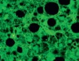
CXCL9/MIG in THP‑1 Human Cell Line.
CXCL9/MIG was detected in immersion fixed THP‑1 human acute monocytic leukemia cell line treated with IFN gamma (positive staining) and THP‑1 human acute monocytic leukemia cell line (untreated; negative staining) using Mouse Anti-Human CXCL9/MIG Monoclonal Antibody (Catalog # MAB392) at 25 µg/mL for 3 hours at room temperature. Cells were stained using the NorthernLights™ 557-conjugated Anti-Mouse IgG Secondary Antibody (red; NL007) and counterstained with DAPI (blue). Specific staining was localized to cytoplasm. Staining was performed using our protocol for Fluorescent ICC Staining of Non-adherent Cells.
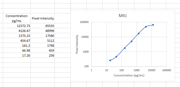
Human CXCL9 / MIG ELISA Standard Curve
Recombinant Human CXCL9/MIG (Catalog # 392-MG) was serially diluted and captured by Mouse Anti-Human CXCL9/MIG Monoclonal Antibody (Catalog # MAB392) coated on a Clear Polystyrene Microplate (Catalog # DY990). Goat Anti-Human CXCL9/MIG Antigen Affinity-purified Polyclonal Antibody (Catalog # AF392) was biotinylated and incubated with the protein captured on the plate. Detection of the standard curve was achieved by incubating Streptavidin-HRP (Catalog # DY998)Applications for Human CXCL9/MIG Antibody (49106)
Application
Recommended Usage
CyTOF-ready
Ready to be labeled using established conjugation methods. No BSA or other carrier proteins that could interfere with conjugation.
Immunocytochemistry
8-25 µg/mL
Sample: Immersion fixed THP‑1 human acute monocytic leukemia cell line treated with IFN gamma
Sample: Immersion fixed THP‑1 human acute monocytic leukemia cell line treated with IFN gamma
Intracellular Staining by Flow Cytometry
2.5 µg/106 cells
Sample: THP-1 cells treated with Recombinant Human IFN‑ gamma (Catalog # 285‑IF), fixed with paraformaldehyde, and permeabilized with saponin
Sample: THP-1 cells treated with Recombinant Human IFN‑ gamma (Catalog # 285‑IF), fixed with paraformaldehyde, and permeabilized with saponin
Western Blot
1 µg/mL
Sample: Recombinant Human CXCL9/MIG (Catalog # 392-MG)
under non-reducing conditions only
Sample: Recombinant Human CXCL9/MIG (Catalog # 392-MG)
under non-reducing conditions only
Neutralization
Measured by its ability to neutralize CXCL9/MIG-induced chemotaxis in the BaF3 mouse pro‑B cell line transfected with mouse CXCR3. The Neutralization Dose (ND50) is typically 0.5-4 µg/mL in the presence of 0.25 µg/mL Recombinant Human CXCL9/MIG.
Human CXCL9/MIG Sandwich Immunoassay
Please Note: Optimal dilutions of this antibody should be experimentally determined.
Reviewed Applications
Read 3 reviews rated 5 using MAB392 in the following applications:
Flow Cytometry Panel Builder
Bio-Techne Knows Flow Cytometry
Save time and reduce costly mistakes by quickly finding compatible reagents using the Panel Builder Tool.
Advanced Features
- Spectra Viewer - Custom analysis of spectra from multiple fluorochromes
- Spillover Popups - Visualize the spectra of individual fluorochromes
- Antigen Density Selector - Match fluorochrome brightness with antigen density

Formulation, Preparation, and Storage
Purification
Protein A or G purified from hybridoma culture supernatant
Reconstitution
Reconstitute at 0.5 mg/mL in sterile PBS. For liquid material, refer to CoA for concentration.
Loading...
Formulation
Lyophilized from a 0.2 μm filtered solution in PBS with Trehalose. *Small pack size (SP) is supplied either lyophilized or as a 0.2 µm filtered solution in PBS.
Shipping
Lyophilized product is shipped at ambient temperature. Liquid small pack size (-SP) is shipped with polar packs. Upon receipt, store immediately at the temperature recommended below.
Stability & Storage
Use a manual defrost freezer and avoid repeated freeze-thaw cycles.
- 12 months from date of receipt, -20 to -70 °C as supplied.
- 1 month, 2 to 8 °C under sterile conditions after reconstitution.
- 6 months, -20 to -70 °C under sterile conditions after reconstitution.
Calculators
Background: CXCL9/MIG
References
- Loetscher, M. et al. (1996) J. Exp. Med. 184:963.
- Liao, F. et al. (1995) J. Exp. Med. 182:1301.
- Vanguri, P. (1995) J. Neuroimmunol. 56:35.
Alternate Names
MIG
Gene Symbol
CXCL9
UniProt
Additional CXCL9/MIG Products
Product Documents for Human CXCL9/MIG Antibody (49106)
Certificate of Analysis
To download a Certificate of Analysis, please enter a lot or batch number in the search box below.
Note: Certificate of Analysis not available for kit components.
Product Specific Notices for Human CXCL9/MIG Antibody (49106)
For research use only
Citations for Human CXCL9/MIG Antibody (49106)
 Powered by Bioz
Customer Reviews for Human CXCL9/MIG Antibody (49106) (3)
5 out of 5
3 Customer Ratings
Have you used Human CXCL9/MIG Antibody (49106)?
Submit a review and receive an Amazon gift card!
$25/€18/£15/$25CAN/¥2500 Yen for a review with an image
$10/€7/£6/$10CAN/¥1110 Yen for a review without an image
Submit a review
Customer Images
Showing
1
-
3 of
3 reviews
Showing All
Filter By:
-
Application: ELISASample Tested: SerumSpecies: PrimateVerified Customer | Posted 11/14/2022Worked as a capture antibody in our non-human primate ELISA

-
Application: Immunocytochemistry/ImmunofluorescenceSample Tested: macrophagesSpecies: HumanVerified Customer | Posted 09/04/2021CXCL9 antibody showed positive macrophages in hepatitis liver tissue.
-
Application: ELISASample Tested: Serum and PlasmaSpecies: HumanVerified Customer | Posted 12/06/2017This antibody was used as a capture in a sandwich ELISA for MIG. The detection antibody was BAM392. The standard was 392-MG-010. Sensitivity was around 5 pg/ml.
There are no reviews that match your criteria.
Protocols
Find general support by application which include: protocols, troubleshooting, illustrated assays, videos and webinars.
- 7-Amino Actinomycin D (7-AAD) Cell Viability Flow Cytometry Protocol
- Appropriate Fixation of IHC/ICC Samples
- Cellular Response to Hypoxia Protocols
- ClariTSA™ Fluorophore Kits
- Detection & Visualization of Antibody Binding
- Extracellular Membrane Flow Cytometry Protocol
- Flow Cytometry Protocol for Cell Surface Markers
- Flow Cytometry Protocol for Staining Membrane Associated Proteins
- Flow Cytometry Staining Protocols
- Flow Cytometry Troubleshooting Guide
- ICC Cell Smear Protocol for Suspension Cells
- ICC Immunocytochemistry Protocol Videos
- ICC for Adherent Cells
- Immunocytochemistry (ICC) Protocol
- Immunocytochemistry Troubleshooting
- Immunofluorescence of Organoids Embedded in Cultrex Basement Membrane Extract
- Immunohistochemistry (IHC) and Immunocytochemistry (ICC) Protocols
- Intracellular Flow Cytometry Protocol Using Alcohol (Methanol)
- Intracellular Flow Cytometry Protocol Using Detergents
- Intracellular Nuclear Staining Flow Cytometry Protocol Using Detergents
- Intracellular Staining Flow Cytometry Protocol Using Alcohol Permeabilization
- Intracellular Staining Flow Cytometry Protocol Using Detergents to Permeabilize Cells
- Preparing Samples for IHC/ICC Experiments
- Preventing Non-Specific Staining (Non-Specific Binding)
- Primary Antibody Selection & Optimization
- Propidium Iodide Cell Viability Flow Cytometry Protocol
- Protocol for Liperfluo
- Protocol for VisUCyte™ HRP Polymer Detection Reagent
- Protocol for the Characterization of Human Th22 Cells
- Protocol for the Characterization of Human Th9 Cells
- Protocol for the Fluorescent ICC Staining of Cell Smears - Graphic
- Protocol for the Fluorescent ICC Staining of Cultured Cells on Coverslips - Graphic
- Protocol for the Preparation and Fluorescent ICC Staining of Cells on Coverslips
- Protocol for the Preparation and Fluorescent ICC Staining of Non-adherent Cells
- Protocol for the Preparation and Fluorescent ICC Staining of Stem Cells on Coverslips
- Protocol for the Preparation of a Cell Smear for Non-adherent Cell ICC - Graphic
- Protocol: Annexin V and PI Staining by Flow Cytometry
- Protocol: Annexin V and PI Staining for Apoptosis by Flow Cytometry
- R&D Systems Quality Control Western Blot Protocol
- TUNEL and Active Caspase-3 Detection by IHC/ICC Protocol
- The Importance of IHC/ICC Controls
- Troubleshooting Guide: Fluorokine Flow Cytometry Kits
- Troubleshooting Guide: Western Blot Figures
- Western Blot Conditions
- Western Blot Protocol
- Western Blot Protocol for Cell Lysates
- Western Blot Troubleshooting
- Western Blot Troubleshooting Guide
- View all Protocols, Troubleshooting, Illustrated assays and Webinars
Loading...
Associated Pathways
