CXCR3 is a G protein-coupled chemokine receptor that binds the alpha chemokines MIG (CXCL9), IP-10 (CXCL10), and I-TAC (CXCL11). CXCR3 is expressed on activated T cells, B cells, and NK cells.

Key Product Details
Validated by
Biological Validation
Species Reactivity
Validated:
Human
Cited:
Human, Mouse, Rat, Feline, Primate - Macaca mulatta (Rhesus Macaque)
Applications
Validated:
Immunohistochemistry, Neutralization, Flow Cytometry, CyTOF-ready
Cited:
Immunohistochemistry, Immunohistochemistry-Paraffin, Immunohistochemistry-Frozen, Western Blot, Neutralization, Flow Cytometry, Immunocytochemistry, Bioassay, Cell Culture, ELISA Detection, Functional Assay, Stimulation
Label
Unconjugated
Antibody Source
Monoclonal Mouse IgG1 Clone # 49801
Loading...
Product Specifications
Immunogen
NS0 mouse myeloma cell line transfected with human CXCR3
Met1-Leu368
Accession # P49682
Met1-Leu368
Accession # P49682
Specificity
Detects human CXCR3.
Clonality
Monoclonal
Host
Mouse
Isotype
IgG1
Endotoxin Level
<0.10 EU per 1 μg of the antibody by the LAL method.
Scientific Data Images for Human CXCR3 Antibody (49801)
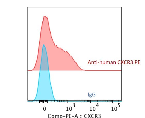
Detection of CXCR3 in Human PBMC Lymphocytes by Flow Cytometry.
Human PBMC lymphocytes were stained with (A) Mouse Anti-Human CXCR3 Monoclonal Antibody (Catalog # MAB160) or (B) Mouse IgG1 isotype control antibody (MAB002) followed by PE-conjugated anti-Mouse IgG Secondary Antibody (F0102B) and Mouse Anti-Human CD3 epsilon APC-conjugated Monoclonal Antibody (FAB100A).View our protocol for Staining Membrane-associated Proteins.
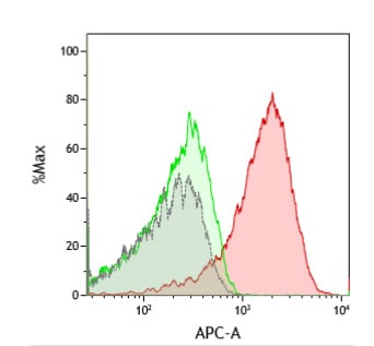
Detection of CXCR3 in HEK293 Human Cell Line Transfected with human CXCR3 and eGFP by Flow Cytometry.
HEK293 human embryonic kidney cell line transfected with (A) human CXCR3 or (B) irrelevant transfectants and eGFP was stained with and either Mouse Anti-Human CXCR3 Monoclonal Antibody (Catalog # MAB160) followed by Allophycocyanin-conjugated Anti-Mouse IgG Secondary Antibody (F0101B). Quadrant markers were set based on Mouse IgG1 Flow Cytometry Isotype Control (MAB002). View our protocol for Staining Membrane-associated Proteins.
CXCR3 in Human Tonsil.
CXCR3 was detected in immersion fixed paraffin-embedded sections of human tonsil using Human CXCR3 Monoclonal Antibody (Catalog # MAB160) at 3 µg/mL overnight at 4 °C. Tissue was stained using the Anti-Mouse HRP-DAB Cell & Tissue Staining Kit (brown; CTS002) and counter-stained with hematoxylin (blue). View our protocol for Chromogenic IHC Staining of Paraffin-embedded Tissue Sections.
Chemotaxis Induced by CXCL11/I‑TAC and Neutralization by Human CXCR3 Antibody.
Recombinant Human CXCL11/I-TAC (672-IT) chemoattracts the BaF3 mouse pro-B cell line transfected with human CXCR3 in a dose-dependent manner (orange line). The amount of cells that migrated through to the lower chemotaxis chamber was measured by Resazurin (AR002). Chemotaxis elicited by Recombinant Human CXCL11/I-TAC (7 ng/mL) is neutralized (green line) by increasing concentrations of Mouse Anti-Human CXCR3 Monoclonal Antibody (Catalog # MAB160). The ND50 is typically 0.3-1.5 µg/mL.
Detection of Human CXCR3 by Western Blot
Functional effects of IP-10 on PCCs(A) Expression of CXCR3 in PCCs, PSCs and HEK293-EBNA/CXCR3-B analyzed by Western Blot. alpha -tubulin was used as loading control. (B) Microphotographs of PCCs migrated through the Boyden chamber in response to 0.5% FBS or IP-10 (24 hr). (C-D) Viability of PCCs after IP-10 addition determined using the MTT assay. Low/high refers to cell seeding density. Data are representative of two/three independent experiments. Data in the histograms are presented as mean ± SD. Image collected and cropped by CiteAb from the following publication (https://www.oncotarget.com/lookup/doi/10.18632/oncotarget.2519), licensed under a CC-BY license. Not internally tested by R&D Systems.
Detection of Human CXCR3 by Immunocytochemistry/Immunofluorescence
Infiltration of CXCR3+ cells in the lamina propria of small intestine mucosa.a. Confocal immunofluorescence for CXCR3 was performed in sections of duodenal biopsies from controls (i), untreated celiac patients (ii), and treated celiac patients (iii). CXCR3 is shown in green, and nuclei are shown in red. Untreated celiac patients showed a higher number of positive cells infiltrating the LP. (Magnification, 630×). b. The number of CXCR3+ cells in the LP was higher in the duodenal mucosa of untreated celiac patients (n = 9) compared with control individuals (n = 6) and treated patients (n = 5) (unpaired t-test; p = 0.0089 and p = 0.0055, respectively). The positive cells in LP regions from sections of duodenal biopsies were counted using immunofluorescence microscopy. c. Representative flow cytometric analysis from the LP compartment of a duodenal sample of an untreated CD patient showing plasma cells (CD138+) that express CXCR3. d. Representative flow cytometric analysis from the LP compartment of a duodenal sample of an untreated CD patient showing LP lymphocytes (CD3+ or CD4+) that express CXCR3. Image collected and cropped by CiteAb from the following publication (https://dx.plos.org/10.1371/journal.pone.0089068), licensed under a CC-BY license. Not internally tested by R&D Systems.Applications for Human CXCR3 Antibody (49801)
Application
Recommended Usage
CyTOF-ready
Ready to be labeled using established conjugation methods. No BSA or other carrier proteins that could interfere with conjugation.
Flow Cytometry
0.25 µg/106 cells
Sample: Human PBMC lymphocytes and HEK293 human cell line transfected with human CXCR3 and eGFP
Sample: Human PBMC lymphocytes and HEK293 human cell line transfected with human CXCR3 and eGFP
Immunohistochemistry
8-25 µg/mL
Sample: Immersion fixed paraffin-embedded sections of human tonsil
Sample: Immersion fixed paraffin-embedded sections of human tonsil
Neutralization
Measured by its ability to neutralize CXCL11/I‑TAC-induced chemotaxis in the BaF3 mouse pro‑B cell line transfected with human CXCR3. The Neutralization Dose (ND50) is typically 0.3-1.5 µg/mL in the presence of 7 ng/mL Recombinant Human CXCL11/I‑TAC.
Reviewed Applications
Read 2 reviews rated 5 using MAB160 in the following applications:
Flow Cytometry Panel Builder
Bio-Techne Knows Flow Cytometry
Save time and reduce costly mistakes by quickly finding compatible reagents using the Panel Builder Tool.
Advanced Features
- Spectra Viewer - Custom analysis of spectra from multiple fluorochromes
- Spillover Popups - Visualize the spectra of individual fluorochromes
- Antigen Density Selector - Match fluorochrome brightness with antigen density

Formulation, Preparation, and Storage
Purification
Protein A or G purified from hybridoma culture supernatant
Reconstitution
Reconstitute at 0.5 mg/mL in sterile PBS. For liquid material, refer to CoA for concentration.
Loading...
Formulation
Lyophilized from a 0.2 μm filtered solution in PBS and NaCl with Trehalose. *Small pack size (SP) is supplied either lyophilized or as a 0.2 µm filtered solution in PBS.
Shipping
Lyophilized product is shipped at ambient temperature. Liquid small pack size (-SP) is shipped with polar packs. Upon receipt, store immediately at the temperature recommended below.
Stability & Storage
Use a manual defrost freezer and avoid repeated freeze-thaw cycles.
- 12 months from date of receipt, -20 to -70 °C as supplied.
- 1 month, 2 to 8 °C under sterile conditions after reconstitution.
- 6 months, -20 to -70 °C under sterile conditions after reconstitution.
Calculators
Background: CXCR3
Alternate Names
CD183, CXCR3, GPR9
Gene Symbol
CXCR3
UniProt
Additional CXCR3 Products
Product Documents for Human CXCR3 Antibody (49801)
Certificate of Analysis
To download a Certificate of Analysis, please enter a lot or batch number in the search box below.
Note: Certificate of Analysis not available for kit components.
Product Specific Notices for Human CXCR3 Antibody (49801)
For research use only
Citations for Human CXCR3 Antibody (49801)
 Powered by Bioz
Customer Reviews for Human CXCR3 Antibody (49801) (2)
5 out of 5
2 Customer Ratings
Have you used Human CXCR3 Antibody (49801)?
Submit a review and receive an Amazon gift card!
$25/€18/£15/$25CAN/¥2500 Yen for a review with an image
$10/€7/£6/$10CAN/¥1110 Yen for a review without an image
Submit a review
Customer Images
Showing
1
-
2 of
2 reviews
Showing All
Filter By:
-
Application: Flow CytometrySample Tested: Peripheral blood mononuclear cells (PBMCs)Species: HumanVerified Customer | Posted 01/24/2021

-
Application: Flow CytometrySample Tested: J45.01 human acute lymphoblastic leukemia T lymphocyte cell lineSpecies: HumanVerified Customer | Posted 11/15/2018
There are no reviews that match your criteria.
Protocols
Find general support by application which include: protocols, troubleshooting, illustrated assays, videos and webinars.
- 7-Amino Actinomycin D (7-AAD) Cell Viability Flow Cytometry Protocol
- Antigen Retrieval Protocol (PIER)
- Antigen Retrieval for Frozen Sections Protocol
- Appropriate Fixation of IHC/ICC Samples
- Cellular Response to Hypoxia Protocols
- Chromogenic IHC Staining of Formalin-Fixed Paraffin-Embedded (FFPE) Tissue Protocol
- Chromogenic Immunohistochemistry Staining of Frozen Tissue
- ClariTSA™ Fluorophore Kits
- Detection & Visualization of Antibody Binding
- Extracellular Membrane Flow Cytometry Protocol
- Flow Cytometry Protocol for Cell Surface Markers
- Flow Cytometry Protocol for Staining Membrane Associated Proteins
- Flow Cytometry Staining Protocols
- Flow Cytometry Troubleshooting Guide
- Fluorescent IHC Staining of Frozen Tissue Protocol
- Graphic Protocol for Heat-induced Epitope Retrieval
- Graphic Protocol for the Preparation and Fluorescent IHC Staining of Frozen Tissue Sections
- Graphic Protocol for the Preparation and Fluorescent IHC Staining of Paraffin-embedded Tissue Sections
- Graphic Protocol for the Preparation of Gelatin-coated Slides for Histological Tissue Sections
- IHC Sample Preparation (Frozen sections vs Paraffin)
- Immunofluorescent IHC Staining of Formalin-Fixed Paraffin-Embedded (FFPE) Tissue Protocol
- Immunohistochemistry (IHC) and Immunocytochemistry (ICC) Protocols
- Immunohistochemistry Frozen Troubleshooting
- Immunohistochemistry Paraffin Troubleshooting
- Intracellular Flow Cytometry Protocol Using Alcohol (Methanol)
- Intracellular Flow Cytometry Protocol Using Detergents
- Intracellular Nuclear Staining Flow Cytometry Protocol Using Detergents
- Intracellular Staining Flow Cytometry Protocol Using Alcohol Permeabilization
- Intracellular Staining Flow Cytometry Protocol Using Detergents to Permeabilize Cells
- Preparing Samples for IHC/ICC Experiments
- Preventing Non-Specific Staining (Non-Specific Binding)
- Primary Antibody Selection & Optimization
- Propidium Iodide Cell Viability Flow Cytometry Protocol
- Protocol for Heat-Induced Epitope Retrieval (HIER)
- Protocol for Liperfluo
- Protocol for Making a 4% Formaldehyde Solution in PBS
- Protocol for VisUCyte™ HRP Polymer Detection Reagent
- Protocol for the Characterization of Human Th22 Cells
- Protocol for the Characterization of Human Th9 Cells
- Protocol for the Preparation & Fixation of Cells on Coverslips
- Protocol for the Preparation and Chromogenic IHC Staining of Frozen Tissue Sections
- Protocol for the Preparation and Chromogenic IHC Staining of Frozen Tissue Sections - Graphic
- Protocol for the Preparation and Chromogenic IHC Staining of Paraffin-embedded Tissue Sections
- Protocol for the Preparation and Chromogenic IHC Staining of Paraffin-embedded Tissue Sections - Graphic
- Protocol for the Preparation and Fluorescent IHC Staining of Frozen Tissue Sections
- Protocol for the Preparation and Fluorescent IHC Staining of Paraffin-embedded Tissue Sections
- Protocol for the Preparation of Gelatin-coated Slides for Histological Tissue Sections
- Protocol: Annexin V and PI Staining by Flow Cytometry
- Protocol: Annexin V and PI Staining for Apoptosis by Flow Cytometry
- TUNEL and Active Caspase-3 Detection by IHC/ICC Protocol
- The Importance of IHC/ICC Controls
- Troubleshooting Guide: Fluorokine Flow Cytometry Kits
- Troubleshooting Guide: Immunohistochemistry
- View all Protocols, Troubleshooting, Illustrated assays and Webinars
Loading...
Associated Pathways

