Two distinct types of receptors that bind the pleiotropic cytokines IL-1 alpha and IL-1 beta have been described. The IL-1 receptor Type I is an 80 kDa transmembrane protein that is expressed predominantly by T cells, fibroblasts, and endothelial cells. IL-1 receptor Type II is a 68 kDa transmembrane protein found on B lymphocytes, neutrophils, monocytes, large granular leukocytes and endothelial cells. Both receptors are members of the immunoglobulin superfamily and show approximately 28% sequence identity in their extracellular domains. The two receptor types do not heterodimerize into a receptor complex. An IL-1 receptor accessory protein that can heterodimerize with the Type I receptor in the presence of IL-1 alpha or IL-1 beta but not IL-1ra, was identified (1). This Type I receptor complex appears to mediate all the known IL-1 biological responses. The receptor Type II has a short cytoplasmic domain and does not transduce IL-1 signals. In addition to the membrane-bound form of IL-1 RII, a naturally-occurring soluble form of IL-1 RII has been described. It has been suggested that the Type II receptor, either as the membrane-bound or as the soluble form, serves as a decoy for IL-1 and inhibits IL-1 action by blocking the binding of IL-1 to the signaling Type I receptor complex. Recombinant IL-1 soluble receptor Type I is a potent antagonist of IL-1 action.

Key Product Details
Species Reactivity
Human
Applications
Immunohistochemistry, Immunocytochemistry
Label
Unconjugated
Antibody Source
Monoclonal Mouse IgG2B Clone # 80937
Loading...
Product Specifications
Immunogen
Mouse myeloma cell line NS0-derived recombinant human Ephrin-A4
Leu26-Gly171
Accession # P52798
Leu26-Gly171
Accession # P52798
Specificity
Detects human Ephrin-A4 in direct ELISAs.
Clonality
Monoclonal
Host
Mouse
Isotype
IgG2B
Scientific Data Images for Human Ephrin-A4 Antibody (80937)
Ephrin‑A4 in MCF‑7 Human Cell Line.
Ephrin-A4 was detected in immersion fixed MCF-7 human breast cancer cell line using Mouse Anti-Human Ephrin-A4 Monoclonal Antibody (Catalog # MAB3692) at 8 µg/mL for 3 hours at room temperature. Cells were stained using the NorthernLights™ 557-conjugated Anti-Mouse IgG Secondary Antibody (red; Catalog # NL007) and counterstained with DAPI (blue). Specific staining was localized to cytoplasm. View our protocol for Fluorescent ICC Staining of Cells on Coverslips.
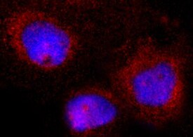
Ephrin‑A4 in U‑87 MG Human Cell Line.
Ephrin-A4 was detected in immersion fixed U-87 MG human glioblastoma/astrocytoma cell line using Mouse Anti-Human Ephrin-A4 Monoclonal Antibody (Catalog # MAB3692) at 8 µg/mL for 3 hours at room temperature. Cells were stained using the NorthernLights™ 557-conjugated Anti-Mouse IgG Secondary Antibody (red; Catalog # NL007) and counterstained with DAPI (blue). Specific staining was localized to cytoplasm. View our protocol for Fluorescent ICC Staining of Cells on Coverslips.
Ephrin‑A4 in Human Squamous Cell Carcinoma.
Ephrin-A4 was detected in immersion fixed paraffin-embedded sections of human squamous cell carcinoma using Mouse Anti-Human Ephrin-A4 Monoclonal Antibody (Catalog # MAB3692) at 5 µg/mL for 1 hour at room temperature followed by incubation with the Anti-Mouse IgG VisUCyte™ HRP Polymer Antibody (Catalog # VC001). Tissue was stained using DAB (brown) and counterstained with hematoxylin (blue). Specific staining was localized to cytoplasm in cancer cells. View our protocol for IHC Staining with VisUCyte HRP Polymer Detection Reagents.Applications for Human Ephrin-A4 Antibody (80937)
Application
Recommended Usage
Immunocytochemistry
5-25 µg/mL
Sample: Immersion fixed MCF‑7 human breast cancer cell line and U‑87 MG human glioblastoma/astrocytoma cell line
Sample: Immersion fixed MCF‑7 human breast cancer cell line and U‑87 MG human glioblastoma/astrocytoma cell line
Immunohistochemistry
5-25 µg/mL
Sample: Immersion fixed paraffin-embedded sections of human squamous cell carcinoma
Sample: Immersion fixed paraffin-embedded sections of human squamous cell carcinoma
Reviewed Applications
Read 1 review rated 5 using MAB3692 in the following applications:
Formulation, Preparation, and Storage
Purification
Protein A or G purified from hybridoma culture supernatant
Reconstitution
Reconstitute at 0.5 mg/mL in sterile PBS. For liquid material, refer to CoA for concentration.
Loading...
Formulation
Lyophilized from a 0.2 μm filtered solution in PBS with Trehalose. *Small pack size (SP) is supplied either lyophilized or as a 0.2 µm filtered solution in PBS.
Shipping
Lyophilized product is shipped at ambient temperature. Liquid small pack size (-SP) is shipped with polar packs. Upon receipt, store immediately at the temperature recommended below.
Stability & Storage
Use a manual defrost freezer and avoid repeated freeze-thaw cycles.
- 12 months from date of receipt, -20 to -70 °C as supplied.
- 1 month, 2 to 8 °C under sterile conditions after reconstitution.
- 6 months, -20 to -70 °C under sterile conditions after reconstitution.
Calculators
Background: Ephrin-A4
References
- Greenfeder, S. et al. (1995) J. Biol. Chem. 270:13757.
Alternate Names
EFL-4, EFNA4, EphrinA4, LERK-4
Gene Symbol
EFNA4
UniProt
Additional Ephrin-A4 Products
Product Documents for Human Ephrin-A4 Antibody (80937)
Certificate of Analysis
To download a Certificate of Analysis, please enter a lot or batch number in the search box below.
Note: Certificate of Analysis not available for kit components.
Product Specific Notices for Human Ephrin-A4 Antibody (80937)
For research use only
Related Research Areas
Customer Reviews for Human Ephrin-A4 Antibody (80937) (1)
5 out of 5
1 Customer Rating
Have you used Human Ephrin-A4 Antibody (80937)?
Submit a review and receive an Amazon gift card!
$25/€18/£15/$25CAN/¥2500 Yen for a review with an image
$10/€7/£6/$10CAN/¥1110 Yen for a review without an image
Submit a review
Customer Images
Showing
1
-
1 of
1 review
Showing All
Filter By:
-
Application: Immunocytochemistry/ImmunofluorescenceSample Tested: MCF-7 human breast cancer cell lineSpecies: HumanVerified Customer | Posted 07/09/2022

There are no reviews that match your criteria.
Protocols
Find general support by application which include: protocols, troubleshooting, illustrated assays, videos and webinars.
- Antigen Retrieval Protocol (PIER)
- Antigen Retrieval for Frozen Sections Protocol
- Appropriate Fixation of IHC/ICC Samples
- Cellular Response to Hypoxia Protocols
- Chromogenic IHC Staining of Formalin-Fixed Paraffin-Embedded (FFPE) Tissue Protocol
- Chromogenic Immunohistochemistry Staining of Frozen Tissue
- ClariTSA™ Fluorophore Kits
- Detection & Visualization of Antibody Binding
- Fluorescent IHC Staining of Frozen Tissue Protocol
- Graphic Protocol for Heat-induced Epitope Retrieval
- Graphic Protocol for the Preparation and Fluorescent IHC Staining of Frozen Tissue Sections
- Graphic Protocol for the Preparation and Fluorescent IHC Staining of Paraffin-embedded Tissue Sections
- Graphic Protocol for the Preparation of Gelatin-coated Slides for Histological Tissue Sections
- ICC Cell Smear Protocol for Suspension Cells
- ICC Immunocytochemistry Protocol Videos
- ICC for Adherent Cells
- IHC Sample Preparation (Frozen sections vs Paraffin)
- Immunocytochemistry (ICC) Protocol
- Immunocytochemistry Troubleshooting
- Immunofluorescence of Organoids Embedded in Cultrex Basement Membrane Extract
- Immunofluorescent IHC Staining of Formalin-Fixed Paraffin-Embedded (FFPE) Tissue Protocol
- Immunohistochemistry (IHC) and Immunocytochemistry (ICC) Protocols
- Immunohistochemistry Frozen Troubleshooting
- Immunohistochemistry Paraffin Troubleshooting
- Preparing Samples for IHC/ICC Experiments
- Preventing Non-Specific Staining (Non-Specific Binding)
- Primary Antibody Selection & Optimization
- Protocol for Heat-Induced Epitope Retrieval (HIER)
- Protocol for Making a 4% Formaldehyde Solution in PBS
- Protocol for VisUCyte™ HRP Polymer Detection Reagent
- Protocol for the Fluorescent ICC Staining of Cell Smears - Graphic
- Protocol for the Fluorescent ICC Staining of Cultured Cells on Coverslips - Graphic
- Protocol for the Preparation & Fixation of Cells on Coverslips
- Protocol for the Preparation and Chromogenic IHC Staining of Frozen Tissue Sections
- Protocol for the Preparation and Chromogenic IHC Staining of Frozen Tissue Sections - Graphic
- Protocol for the Preparation and Chromogenic IHC Staining of Paraffin-embedded Tissue Sections
- Protocol for the Preparation and Chromogenic IHC Staining of Paraffin-embedded Tissue Sections - Graphic
- Protocol for the Preparation and Fluorescent ICC Staining of Cells on Coverslips
- Protocol for the Preparation and Fluorescent ICC Staining of Non-adherent Cells
- Protocol for the Preparation and Fluorescent ICC Staining of Stem Cells on Coverslips
- Protocol for the Preparation and Fluorescent IHC Staining of Frozen Tissue Sections
- Protocol for the Preparation and Fluorescent IHC Staining of Paraffin-embedded Tissue Sections
- Protocol for the Preparation of Gelatin-coated Slides for Histological Tissue Sections
- Protocol for the Preparation of a Cell Smear for Non-adherent Cell ICC - Graphic
- TUNEL and Active Caspase-3 Detection by IHC/ICC Protocol
- The Importance of IHC/ICC Controls
- Troubleshooting Guide: Immunohistochemistry
- View all Protocols, Troubleshooting, Illustrated assays and Webinars
Loading...