Human ErbB2/Her2 Herstatin Isoform Antibody (416711)
R&D Systems | Catalog # MAB11291

Key Product Details
Species Reactivity
Human
Applications
Multiplex Immunofluorescence, Immunohistochemistry, COMET
Label
Unconjugated
Antibody Source
Monoclonal Mouse IgG2B Clone # 416711
Loading...
Product Specifications
Immunogen
S. frugiperda insect ovarian cell line Sf 21-derived recombinant human ErbB2/Her2 Herstatin Isoform
Thr23-Gly419
Accession # AAD56009
Thr23-Gly419
Accession # AAD56009
Specificity
Detects human ErbB2/Her2 Herstatin Isoform in direct ELISAs and Western blots. In direct ELISAs and Western blots, no cross-reactivity with recombinant human (rh) EGFR or rhErbB3 is observed.
Clonality
Monoclonal
Host
Mouse
Isotype
IgG2B
Scientific Data Images for Human ErbB2/Her2 Herstatin Isoform Antibody (416711)
Detection of HER2 in Human Breast Cancer via Multiplex Immunofluorescence staining on COMET™
HER2 was detected in immersion fixed paraffin-embedded sections of human breast cancer using Mouse Anti-Human ErbB2/HER2 Monoclonal Antibody (Catalog # MAB11291) at 12ug/mL at 37 ° Celsius for 4 minutes. Before incubation with the primary antibody, tissue underwent an all-in-one dewaxing and antigen retrieval preprocessing using PreTreatment Module (PT Module) and Dewax and HIER Buffer H (pH 9). Tissue was stained using the Alexa Fluor™ 647 Goat anti-Mouse IgG Secondary Antibody at 1:200 at 37 ° Celsius for 2 minutes. (Yellow; Lunaphore Catalog # DR647MS) and counterstained with DAPI (blue; Lunaphore Catalog # DR100). Specific staining was localized to the cytoplasm. Protocol available in COMET™ Panel Builder.
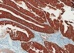
ErbB2/Her2 in Human Breast Cancer Tissue.
ErbB2/Her2 was detected in immersion fixed paraffin-embedded sections of human breast cancer tissue using Mouse Anti-Human ErbB2/Her2 Herstatin Isoform Monoclonal Antibody (Catalog # MAB11291) at 8 µg/mL overnight at 4 °C. Tissue was stained using the Anti-Mouse HRP-DAB Cell & Tissue Staining Kit (brown; Catalog # CTS002) and counterstained with hematoxylin (blue). Specific labeling was localized to the cytoplasm of cancer cells. View our protocol for Chromogenic IHC Staining of Paraffin-embedded Tissue Sections.
ErbB2/Her2 in Human Breast.
ErbB2/Her2 was detected in immersion fixed paraffin-embedded sections of human breast using Mouse Anti-Human ErbB2/Her2 Herstatin Isoform Monoclonal Antibody (Catalog # MAB11291) at 25 µg/mL overnight at 4 °C. Tissue was stained using the Anti-Mouse HRP-DAB Cell & Tissue Staining Kit (brown; Catalog # CTS002) and counterstained with hematoxylin (blue). View our protocol for Chromogenic IHC Staining of Paraffin-embedded Tissue Sections.Applications for Human ErbB2/Her2 Herstatin Isoform Antibody (416711)
Application
Recommended Usage
COMET
Optimal dilutions of this antibody should be experimentally determined.
Immunohistochemistry
8-25 µg/mL
Sample: Immersion fixed paraffin-embedded sections of human breast cancer tissue
Sample: Immersion fixed paraffin-embedded sections of human breast cancer tissue
Multiplex Immunofluorescence
12 µg/mL
Sample: Immersion fixed paraffin-embedded sections of human Breast Cancer
Sample: Immersion fixed paraffin-embedded sections of human Breast Cancer
Reviewed Applications
Read 4 reviews rated 4.8 using MAB11291 in the following applications:
Formulation, Preparation, and Storage
Purification
Protein A or G purified from hybridoma culture supernatant
Reconstitution
Reconstitute at 0.5 mg/mL in sterile PBS. For liquid material, refer to CoA for concentration.
Loading...
Formulation
Lyophilized from a 0.2 μm filtered solution in PBS with Trehalose. See Certificate of Analysis for details.
*Small pack size (-SP) is supplied either lyophilized or as a 0.2 µm filtered solution in PBS.
*Small pack size (-SP) is supplied either lyophilized or as a 0.2 µm filtered solution in PBS.
Shipping
Lyophilized product is shipped at ambient temperature. Liquid small pack size (-SP) is shipped with polar packs. Upon receipt, store immediately at the temperature recommended below.
Stability & Storage
Use a manual defrost freezer and avoid repeated freeze-thaw cycles.
- 12 months from date of receipt, -20 to -70 °C as supplied.
- 1 month, 2 to 8 °C under sterile conditions after reconstitution.
- 6 months, -20 to -70 °C under sterile conditions after reconstitution.
Calculators
Background: ErbB2/Her2
Long Name
Receptor Tyrosine Protein Kinase ErbB2
Alternate Names
CD340, HER2, Neu Oncogene, NGL, TKR1
Gene Symbol
ERBB2
UniProt
Additional ErbB2/Her2 Products
Product Documents for Human ErbB2/Her2 Herstatin Isoform Antibody (416711)
Certificate of Analysis
To download a Certificate of Analysis, please enter a lot or batch number in the search box below.
Note: Certificate of Analysis not available for kit components.
Product Specific Notices for Human ErbB2/Her2 Herstatin Isoform Antibody (416711)
For research use only
Related Research Areas
Citations for Human ErbB2/Her2 Herstatin Isoform Antibody (416711)
 Powered by Bioz
Customer Reviews for Human ErbB2/Her2 Herstatin Isoform Antibody (416711) (4)
4.8 out of 5
4 Customer Ratings
Have you used Human ErbB2/Her2 Herstatin Isoform Antibody (416711)?
Submit a review and receive an Amazon gift card!
$25/€18/£15/$25CAN/¥2500 Yen for a review with an image
$10/€7/£6/$10CAN/¥1110 Yen for a review without an image
Submit a review
Customer Images


Showing
1
-
4 of
4 reviews
Showing All
Filter By:
-
Application: ImmunohistochemistrySample Tested: Colorectal cancerSpecies: HumanVerified Customer | Posted 11/08/2021

-
Application: MicroarraySample Tested: EDTA PlasmaSpecies: HumanVerified Customer | Posted 12/13/2019Antibody was printed on custom arrays and incubated with fluorescently labeled human EDTA plasma
-
Application: MicroarraysSample Tested: EDTA PlasmaSpecies: HumanVerified Customer | Posted 03/11/2019
-
Application: ImmunocytochemistrySample Tested: Human BT474 Breast Cancer CellsSpecies: HumanVerified Customer | Posted 06/08/2015Novus mAb11291 Herstatin Antibody used for immunocytochemistry on BT474 breast cancer cells.
There are no reviews that match your criteria.
Protocols
Find general support by application which include: protocols, troubleshooting, illustrated assays, videos and webinars.
- Antigen Retrieval Protocol (PIER)
- Antigen Retrieval for Frozen Sections Protocol
- Appropriate Fixation of IHC/ICC Samples
- Cellular Response to Hypoxia Protocols
- Chromogenic IHC Staining of Formalin-Fixed Paraffin-Embedded (FFPE) Tissue Protocol
- Chromogenic Immunohistochemistry Staining of Frozen Tissue
- ClariTSA™ Fluorophore Kits
- Detection & Visualization of Antibody Binding
- Fluorescent IHC Staining of Frozen Tissue Protocol
- Graphic Protocol for Heat-induced Epitope Retrieval
- Graphic Protocol for the Preparation and Fluorescent IHC Staining of Frozen Tissue Sections
- Graphic Protocol for the Preparation and Fluorescent IHC Staining of Paraffin-embedded Tissue Sections
- Graphic Protocol for the Preparation of Gelatin-coated Slides for Histological Tissue Sections
- IHC Sample Preparation (Frozen sections vs Paraffin)
- Immunofluorescent IHC Staining of Formalin-Fixed Paraffin-Embedded (FFPE) Tissue Protocol
- Immunohistochemistry (IHC) and Immunocytochemistry (ICC) Protocols
- Immunohistochemistry Frozen Troubleshooting
- Immunohistochemistry Paraffin Troubleshooting
- Preparing Samples for IHC/ICC Experiments
- Preventing Non-Specific Staining (Non-Specific Binding)
- Primary Antibody Selection & Optimization
- Protocol for Heat-Induced Epitope Retrieval (HIER)
- Protocol for Making a 4% Formaldehyde Solution in PBS
- Protocol for VisUCyte™ HRP Polymer Detection Reagent
- Protocol for the Preparation & Fixation of Cells on Coverslips
- Protocol for the Preparation and Chromogenic IHC Staining of Frozen Tissue Sections
- Protocol for the Preparation and Chromogenic IHC Staining of Frozen Tissue Sections - Graphic
- Protocol for the Preparation and Chromogenic IHC Staining of Paraffin-embedded Tissue Sections
- Protocol for the Preparation and Chromogenic IHC Staining of Paraffin-embedded Tissue Sections - Graphic
- Protocol for the Preparation and Fluorescent IHC Staining of Frozen Tissue Sections
- Protocol for the Preparation and Fluorescent IHC Staining of Paraffin-embedded Tissue Sections
- Protocol for the Preparation of Gelatin-coated Slides for Histological Tissue Sections
- TUNEL and Active Caspase-3 Detection by IHC/ICC Protocol
- The Importance of IHC/ICC Controls
- Troubleshooting Guide: Immunohistochemistry
- View all Protocols, Troubleshooting, Illustrated assays and Webinars
Loading...
Associated Pathways


