ErbB4, also called Her4 (human epidermal growth factor receptor 4), is a type I membrane glycoprotein that is a member of the ErbB family of tyrosine kinase receptors. ErbB family members serve as receptors for the epidermal growth factor (EGF) family of growth factors. ErbB4 is expressed in normal skeletal muscle, heart, pituitary, brain and several breast carcinomas. ErbB4 ligands include the neuregulins, beta-cellulin and heparin-binding EGF-like growth factor (HB-EGF). Monomeric ErbB4 binds its ligands with low affinity. Typically, heterodimerization with ErbB2 forms the high affinity receptor complex. However, ErbB4 has also been shown to heterodimerize with both ErbB1 and ErbB3. It has been suggested that the identity of the ligand may influence the dimerization partner. Because ErbB3 contains a defective kinase domain, the kinase domain of ErbB2 is responsible for initiating the tyrosine phosphorylation signal through the heterodimeric receptor. It has been found that a discrete three amino acid signal in the ErbB3 cytoplasmic domain is critical for transactivation of ErbB2. Interestingly, this same three amino acid signal has been found in ErbB4 and ErbB1 (EGFR). Several ErbB4 isoforms exist. Two of these differ in the presence of juxtamembrane extracellular sequences which regulate the ability of TACE (TNF-alpha converting enzyme) to proteolytically cleave ErbB4 from the cell surface. These isoforms exhibit tissue-specific expression. Another isoform lacks the phosphoinositide 3-kinase activation sequence present in the ErbB4 cytoplasmic domain. Human ErbB4 consists of 1308 amino acids (aa) with a 25 aa signal sequence, a 626 aa extracellular domain, a 24 aa transmembrane region, and a 633 aa cytoplasmic domain. ErbB4 appears to play important roles in neuronal development, development of the heart and cancer.
Human ErbB4/Her4 Antibody (182803)
R&D Systems | Catalog # MAB1131

Key Product Details
Species Reactivity
Validated:
Human
Cited:
Human
Applications
Validated:
Immunohistochemistry, Western Blot, Immunocytochemistry
Cited:
Immunohistochemistry, Immunohistochemistry-Paraffin, Western Blot, Immunoprecipitation, Luminex Development
Label
Unconjugated
Antibody Source
Monoclonal Mouse IgG2B Clone # 182803
Loading...
Product Specifications
Immunogen
Mouse myeloma cell line NS0-derived recombinant human ErbB4/Her4
Gln26-Arg649
Accession # Q15303
Gln26-Arg649
Accession # Q15303
Specificity
Detects human ErbB4/Her4 in direct ELISAs and Western blots. In direct ELISAs and Western blots, no cross‑reactivity with recombinant human (rh) EGF R, rhErbB2, and rhErbB3 is observed.
Clonality
Monoclonal
Host
Mouse
Isotype
IgG2B
Scientific Data Images for Human ErbB4/Her4 Antibody (182803)
ErbB4/Her4 in MCF‑7 Human Cell Line.
ErbB4/Her4 was detected in immersion fixed MCF‑7 human breast cancer cell line using Mouse Anti-Human ErbB4/Her4 Monoclonal Antibody (Catalog # MAB1131) at 10 µg/mL for 3 hours at room temperature. Cells were stained using the NorthernLights™ 557-conjugated Anti-Mouse IgG Secondary Antibody (red; Catalog # NL007) and counterstained with DAPI (blue). Specific staining was localized to plasma membrane. View our protocol for Fluorescent ICC Staining of Cells on Coverslips.
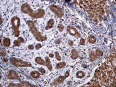
ErbB4/Her4 in Human Colon Cancer Tissue.
ErbB4/Her4 was detected in immersion fixed paraffin-embedded sections of human colon cancer tissue using Mouse Anti-Human ErbB4/Her4 Monoclonal Antibody (Catalog # MAB1131) at 15 µg/mL overnight at 4 °C. Tissue was stained using the Anti-Mouse HRP-DAB Cell & Tissue Staining Kit (brown; Catalog # CTS002) and counterstained with hematoxylin (blue). Specific staining was localized to smooth muscle cells. View our protocol for Chromogenic IHC Staining of Paraffin-embedded Tissue Sections.Applications for Human ErbB4/Her4 Antibody (182803)
Application
Recommended Usage
Immunocytochemistry
5-25 µg/mL
Sample: Immersion fixed MCF-7 human breast cancer cell line
Sample: Immersion fixed MCF-7 human breast cancer cell line
Immunohistochemistry
8-25 µg/mL
Sample: Immersion fixed paraffin-embedded sections of human colon cancer tissue
Sample: Immersion fixed paraffin-embedded sections of human colon cancer tissue
Western Blot
1 µg/mL
Sample: Recombinant Human ErbB4/Her4 Fc Chimera (Catalog # 1131-ER)
Sample: Recombinant Human ErbB4/Her4 Fc Chimera (Catalog # 1131-ER)
Reviewed Applications
Read 2 reviews rated 5 using MAB1131 in the following applications:
Formulation, Preparation, and Storage
Purification
Protein A or G purified from hybridoma culture supernatant
Reconstitution
Reconstitute at 0.5 mg/mL in sterile PBS. For liquid material, refer to CoA for concentration.
Loading...
Formulation
Lyophilized from a 0.2 μm filtered solution in PBS with Trehalose. *Small pack size (SP) is supplied either lyophilized or as a 0.2 µm filtered solution in PBS.
Shipping
Lyophilized product is shipped at ambient temperature. Liquid small pack size (-SP) is shipped with polar packs. Upon receipt, store immediately at the temperature recommended below.
Stability & Storage
Use a manual defrost freezer and avoid repeated freeze-thaw cycles.
- 12 months from date of receipt, -20 to -70 °C as supplied.
- 1 month, 2 to 8 °C under sterile conditions after reconstitution.
- 6 months, -20 to -70 °C under sterile conditions after reconstitution.
Calculators
Background: ErbB4/Her4
References
- Plowman, G.D. et al. (1993) Proc. Natl. Acad. Sci. USA 90:1746.
- Elenius, K. et al. (1997) J. Biol. Chem. 272:26761.
- Elenius, K. et al. (1999) Oncogene 18:2607.
- Rio, C. et al. (2000) J. Biol. Chem. 275:10379.
- Emkey, R. and C.R. Kahn (1997) J. Biol. Chem. 272:31172.
- Sundaresan, S. et al. (1998) Endocrinology 139:4756.
- Schaefer, G. et al. (1999) J. Biol. Chem. 274:859.
- Schlessinger, J. (2000) Cell 103:211.
- Daly, R.J. (1999) Growth Factors 16:255.
Long Name
Receptor Tyrosine Protein Kinase ErbB4
Alternate Names
HER4
Gene Symbol
ERBB4
UniProt
Additional ErbB4/Her4 Products
Product Documents for Human ErbB4/Her4 Antibody (182803)
Certificate of Analysis
To download a Certificate of Analysis, please enter a lot or batch number in the search box below.
Note: Certificate of Analysis not available for kit components.
Product Specific Notices for Human ErbB4/Her4 Antibody (182803)
For research use only
Related Research Areas
Citations for Human ErbB4/Her4 Antibody (182803)
 Powered by Bioz
Customer Reviews for Human ErbB4/Her4 Antibody (182803) (2)
5 out of 5
2 Customer Ratings
Have you used Human ErbB4/Her4 Antibody (182803)?
Submit a review and receive an Amazon gift card!
$25/€18/£15/$25CAN/¥2500 Yen for a review with an image
$10/€7/£6/$10CAN/¥1110 Yen for a review without an image
Submit a review
Customer Images
Showing
1
-
2 of
2 reviews
Showing All
Filter By:
-
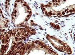
Application: ImmunohistochemistrySample Tested: Breast cancer tissueSpecies: HumanVerified Customer | Posted 04/20/2022

-
Application: ImmunohistochemistrySample Tested: Breast cancer tissueSpecies: HumanVerified Customer | Posted 10/22/2021
There are no reviews that match your criteria.
Protocols
Find general support by application which include: protocols, troubleshooting, illustrated assays, videos and webinars.
- Antigen Retrieval Protocol (PIER)
- Antigen Retrieval for Frozen Sections Protocol
- Appropriate Fixation of IHC/ICC Samples
- Cellular Response to Hypoxia Protocols
- Chromogenic IHC Staining of Formalin-Fixed Paraffin-Embedded (FFPE) Tissue Protocol
- Chromogenic Immunohistochemistry Staining of Frozen Tissue
- ClariTSA™ Fluorophore Kits
- Detection & Visualization of Antibody Binding
- Fluorescent IHC Staining of Frozen Tissue Protocol
- Graphic Protocol for Heat-induced Epitope Retrieval
- Graphic Protocol for the Preparation and Fluorescent IHC Staining of Frozen Tissue Sections
- Graphic Protocol for the Preparation and Fluorescent IHC Staining of Paraffin-embedded Tissue Sections
- Graphic Protocol for the Preparation of Gelatin-coated Slides for Histological Tissue Sections
- ICC Cell Smear Protocol for Suspension Cells
- ICC Immunocytochemistry Protocol Videos
- ICC for Adherent Cells
- IHC Sample Preparation (Frozen sections vs Paraffin)
- Immunocytochemistry (ICC) Protocol
- Immunocytochemistry Troubleshooting
- Immunofluorescence of Organoids Embedded in Cultrex Basement Membrane Extract
- Immunofluorescent IHC Staining of Formalin-Fixed Paraffin-Embedded (FFPE) Tissue Protocol
- Immunohistochemistry (IHC) and Immunocytochemistry (ICC) Protocols
- Immunohistochemistry Frozen Troubleshooting
- Immunohistochemistry Paraffin Troubleshooting
- Preparing Samples for IHC/ICC Experiments
- Preventing Non-Specific Staining (Non-Specific Binding)
- Primary Antibody Selection & Optimization
- Protocol for Heat-Induced Epitope Retrieval (HIER)
- Protocol for Making a 4% Formaldehyde Solution in PBS
- Protocol for VisUCyte™ HRP Polymer Detection Reagent
- Protocol for the Fluorescent ICC Staining of Cell Smears - Graphic
- Protocol for the Fluorescent ICC Staining of Cultured Cells on Coverslips - Graphic
- Protocol for the Preparation & Fixation of Cells on Coverslips
- Protocol for the Preparation and Chromogenic IHC Staining of Frozen Tissue Sections
- Protocol for the Preparation and Chromogenic IHC Staining of Frozen Tissue Sections - Graphic
- Protocol for the Preparation and Chromogenic IHC Staining of Paraffin-embedded Tissue Sections
- Protocol for the Preparation and Chromogenic IHC Staining of Paraffin-embedded Tissue Sections - Graphic
- Protocol for the Preparation and Fluorescent ICC Staining of Cells on Coverslips
- Protocol for the Preparation and Fluorescent ICC Staining of Non-adherent Cells
- Protocol for the Preparation and Fluorescent ICC Staining of Stem Cells on Coverslips
- Protocol for the Preparation and Fluorescent IHC Staining of Frozen Tissue Sections
- Protocol for the Preparation and Fluorescent IHC Staining of Paraffin-embedded Tissue Sections
- Protocol for the Preparation of Gelatin-coated Slides for Histological Tissue Sections
- Protocol for the Preparation of a Cell Smear for Non-adherent Cell ICC - Graphic
- R&D Systems Quality Control Western Blot Protocol
- TUNEL and Active Caspase-3 Detection by IHC/ICC Protocol
- The Importance of IHC/ICC Controls
- Troubleshooting Guide: Immunohistochemistry
- Troubleshooting Guide: Western Blot Figures
- Western Blot Conditions
- Western Blot Protocol
- Western Blot Protocol for Cell Lysates
- Western Blot Troubleshooting
- Western Blot Troubleshooting Guide
- View all Protocols, Troubleshooting, Illustrated assays and Webinars
Loading...
Associated Pathways


