Human Erythropoietin R Antibody (38409)
R&D Systems | Catalog # MAB307

Key Product Details
Species Reactivity
Validated:
Human
Cited:
Human, Rat
Applications
Validated:
Western Blot, Flow Cytometry, Immunoprecipitation
Cited:
Immunohistochemistry, Western Blot, Neutralization, Flow Cytometry, Immunocytochemistry, Immunoprecipitation
Label
Unconjugated
Antibody Source
Monoclonal Mouse IgG2B Clone # 38409
Loading...
Product Specifications
Immunogen
Mouse myeloma cell line NS0-derived recombinant human Erythropoietin R
Pro26-Pro250
Accession # P19235
Pro26-Pro250
Accession # P19235
Specificity
Detects human Erythropoietin R in direct ELISAs and Western blots. In direct ELISAs, no cross-reactivity with recombinant mouse Erythropoietin R is observed.
Clonality
Monoclonal
Host
Mouse
Isotype
IgG2B
Scientific Data Images for Human Erythropoietin R Antibody (38409)
Detection of Human Erythropoietin R by Western Blot.
Western blot shows lysates of UT-7 human acute myeloid leukemia cell line. PVDF membrane was probed with 2 µg/mL of Mouse Anti-Human Erythropoietin R Monoclonal Antibody (Catalog # MAB307) followed by HRP-conjugated Anti-Mouse IgG Secondary Antibody (Catalog # HAF007). For additional reference, PVDF membrane was probed with 1 µg/mL of Goat Anti-Human Erythropoietin R Antigen Affinity-purified Polyclonal Antibody (Catalog # AF-322-PB) followed by HRP-conjugated Anti-Goat IgG Secondary Antibody (Catalog # HAF109). A specific band was detected for Erythropoietin R at approximately 70 kDa (as indicated). This experiment was conducted under reducing conditions and using Immunoblot Buffer Group 1.
Detection of Erythropoietin R in TF‑1 Human Cell Line by Flow Cytometry.
TF-1 human erythroleukemic cell line was stained with Mouse Anti-Human Erythropoietin R Monoclonal Antibody (Catalog # MAB307, filled histogram) or isotype control antibody (MAB0041, open histogram) followed by anti-Mouse IgG Allophycocyanin-conjugated Secondary Antibody (F0101B). Staining was performed using our Staining Membrane-associated Proteins protocol.
Detection of Human Erythropoietin R by Western Blot
sEpoR characterization in uremic serum.1a. Soluble EpoR is detectable in serum from dialysis patients by western blot. Human serum was subjected to immunoprecipitation with goat anti-human erythropoietin receptor antibody (R&D Systems, AF-322-PB) followed by western blotting with mouse monoclonal anti-human erythropoietin receptor (R&D Systems, MAB307). Both antibodies recognize the extracellular domain of the receptor. Lanes 1–6 are serum from 6 representative dialysis patients, lane 7 is blank and lane 8 is recombinant sEpoR (Sigma Aldrich E0643, Saint Louis MI). Shown in the serum samples is a band of expected molecular weight of approximately 27 kDa. The control sEpoR with Fc tag is consistent with the manufacturers reported molecular weight of 32 kDa. 1b. Soluble EpoR is also detected using the same dialysis patient serum samples by performing immunoprecipitation in reverse order. In this experiment immunoprecipitation was done with mouse monoclonal anti-human erythropoietin receptor (R&D Systems, MAB307) followed by western blotting with goat anti-human erythropoietin receptor (R&D Systems, AF-322-PB). Lanes 1 to 3 are serum from 3 dialysis patients, and lane 4 is recombinant sEpoR-Fc (Sigma, 307) as positive control. Image collected and cropped by CiteAb from the following publication (https://pubmed.ncbi.nlm.nih.gov/20169072), licensed under a CC-BY license. Not internally tested by R&D Systems.
Detection of Mouse Erythropoietin R by Western Blot
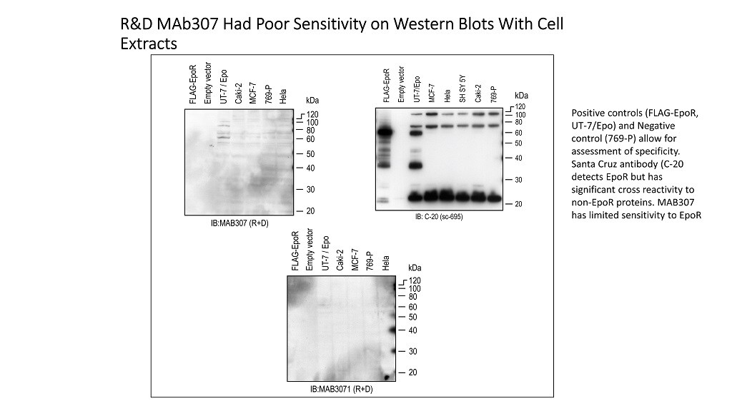
The 59 kDa protein detected by M-20 In MCF-7 cells is not bound by other anti-EpoR antibodies.The indicated lysates were immunoprecipitated (IP) then the immunoblotted (IB) with the indicated antibodies: ab10653 (abcam Inc), Mab307 (R&D systems), C-20 & M-20 (Santa Cruz Inc) or A-82 (Amgen Inc). COS cell lysates expressing a FLAG-tagged version of EpoR (FLAG-EpoR) [6] and UT-7/Epo cells served as EpoR positive controls. 769-P cells served as the EpoR negative control. (A) Westerns were immunoprecipitated (IP) with ab10653 or M-20 followed by immunoblotting (IB) with M-20. The position of full-length 59 kDa EpoR in positive controls is indicated by the arrow. Positions of molecular weight markers (kDa) are shown. Bands detected in 769-P lysates are non-EpoR cross-reacting proteins and include antibody chains that were not removed completely or protein G that leached from beads. Note the detection of a 59 kDa band with MCF-7 cells with the M:20/M:20 combination but not with the ab10653/IB:M-20 combination. (B) IP:IB combinations with the indicated antibodies were subjected to western analysis. The western slice containing the 59 kDa EpoR band from each combination is shown. Note the 59 kDa bands detected in EpoR positive controls but not 769-P cells. Only the M-20:M-20 combination detected a 59 kDa band in MCF-7 cells. Image collected and cropped by CiteAb from the following publication (https://dx.plos.org/10.1371/journal.pone.0068083), licensed under a CC-BY license. Not internally tested by R&D Systems.Applications for Human Erythropoietin R Antibody (38409)
Application
Recommended Usage
Flow Cytometry
0.25 µg/106 cells
Sample: TF-1 human erythroleukemic cell line
Sample: TF-1 human erythroleukemic cell line
Immunoprecipitation
Khankin, E.V. et al. (2010) PLoS One 5: e9246.
Western Blot
2 µg/mL
Sample: UT‑7 human acute myeloid leukemia cell line
Sample: UT‑7 human acute myeloid leukemia cell line
Reviewed Applications
Read 1 review rated 1 using MAB307 in the following applications:
Flow Cytometry Panel Builder
Bio-Techne Knows Flow Cytometry
Save time and reduce costly mistakes by quickly finding compatible reagents using the Panel Builder Tool.
Advanced Features
- Spectra Viewer - Custom analysis of spectra from multiple fluorochromes
- Spillover Popups - Visualize the spectra of individual fluorochromes
- Antigen Density Selector - Match fluorochrome brightness with antigen density

Formulation, Preparation, and Storage
Purification
Protein A or G purified from ascites
Reconstitution
Reconstitute at 0.5 mg/mL in sterile PBS. For liquid material, refer to CoA for concentration.
Loading...
Formulation
Lyophilized from a 0.2 μm filtered solution in PBS with Trehalose. *Small pack size (SP) is supplied either lyophilized or as a 0.2 µm filtered solution in PBS.
Shipping
Lyophilized product is shipped at ambient temperature. Liquid small pack size (-SP) is shipped with polar packs. Upon receipt, store immediately at the temperature recommended below.
Stability & Storage
Use a manual defrost freezer and avoid repeated freeze-thaw cycles.
- 12 months from date of receipt, -20 to -70 °C as supplied.
- 1 month, 2 to 8 °C under sterile conditions after reconstitution.
- 6 months, -20 to -70 °C under sterile conditions after reconstitution.
Calculators
Background: Erythropoietin R
Long Name
Erythropoietin Receptor
Alternate Names
EpoR
Gene Symbol
EPOR
UniProt
Additional Erythropoietin R Products
Product Documents for Human Erythropoietin R Antibody (38409)
Certificate of Analysis
To download a Certificate of Analysis, please enter a lot or batch number in the search box below.
Note: Certificate of Analysis not available for kit components.
Product Specific Notices for Human Erythropoietin R Antibody (38409)
For research use only
Related Research Areas
Citations for Human Erythropoietin R Antibody (38409)
 Powered by Bioz
Customer Reviews for Human Erythropoietin R Antibody (38409) (1)
1 out of 5
1 Customer Rating
Have you used Human Erythropoietin R Antibody (38409)?
Submit a review and receive an Amazon gift card!
$25/€18/£15/$25CAN/¥2500 Yen for a review with an image
$10/€7/£6/$10CAN/¥1110 Yen for a review without an image
Submit a review
Customer Images
Showing
1
-
1 of
1 review
Showing All
Filter By:
-
Application: Western BlotSample Tested: UT-7 human acute myeloid leukemia cell lineSpecies: HumanVerified Customer | Posted 09/10/2016Sensitivity and specificity was assessed by western. Results indicate Mab307 cannot be used by western because of limited sensitivity to EpoR. By FLOW cytometry, MAB307 gave false-positive staining of negative control cell types.
 Bio-Techne ResponseR&D Systems Technical Service is following up.
There are no reviews that match your criteria.
Protocols
Find general support by application which include: protocols, troubleshooting, illustrated assays, videos and webinars.
- 7-Amino Actinomycin D (7-AAD) Cell Viability Flow Cytometry Protocol
- Cellular Response to Hypoxia Protocols
- Extracellular Membrane Flow Cytometry Protocol
- Flow Cytometry Protocol for Cell Surface Markers
- Flow Cytometry Protocol for Staining Membrane Associated Proteins
- Flow Cytometry Staining Protocols
- Flow Cytometry Troubleshooting Guide
- Immunoprecipitation Protocol
- Intracellular Flow Cytometry Protocol Using Alcohol (Methanol)
- Intracellular Flow Cytometry Protocol Using Detergents
- Intracellular Nuclear Staining Flow Cytometry Protocol Using Detergents
- Intracellular Staining Flow Cytometry Protocol Using Alcohol Permeabilization
- Intracellular Staining Flow Cytometry Protocol Using Detergents to Permeabilize Cells
- Propidium Iodide Cell Viability Flow Cytometry Protocol
- Protocol for Liperfluo
- Protocol for the Characterization of Human Th22 Cells
- Protocol for the Characterization of Human Th9 Cells
- Protocol: Annexin V and PI Staining by Flow Cytometry
- Protocol: Annexin V and PI Staining for Apoptosis by Flow Cytometry
- R&D Systems Quality Control Western Blot Protocol
- Troubleshooting Guide: Fluorokine Flow Cytometry Kits
- Troubleshooting Guide: Western Blot Figures
- Western Blot Conditions
- Western Blot Protocol
- Western Blot Protocol for Cell Lysates
- Western Blot Troubleshooting
- Western Blot Troubleshooting Guide
- View all Protocols, Troubleshooting, Illustrated assays and Webinars
Loading...
Associated Pathways
