The FGFs are a growing family of heparin-binding growth factors that show a variety of biological activities toward cells of mesenchymal, neuronal and epithelial origin. All FGFs have two conserved cysteine residues and share significant amino acid sequence homology. FGF-10 was originally identified from rat embryos by homology-based polymerase chain reaction. Human and mouse FGF-10 were subsequently cloned. The human FGF-10 cDNA encodes a 208 amino acid residue protein with a hydrophobic amino-terminal signal peptide. Human FGF-10 shares approximately 92% and 95% amino acid sequence identity with mouse and rat
FGF‑10, respectively. Among the FGF family members, FGF-10 is most closely related to FGF-7. The expression of FGF-10 transcripts has been shown to be most abundant in the embryo and adult lung. Recombinant FGF-10 preparations have been shown to be mitogenic for epithelial and epidermal cells but not fibroblasts. Based on its in vitro biological activities and in vivo expression pattern, FGF-10 has been proposed to play unique roles in the brain, in lung development, wound healing and limb bud formation.
Key Product Details
Species Reactivity
Validated:
Cited:
Applications
Validated:
Cited:
Label
Antibody Source
Product Specifications
Immunogen
Cys37-Ser208
Accession # O15520
Specificity
Clonality
Host
Isotype
Applications for Human FGF-10 Antibody
ELISA
This antibody functions as an ELISA detection antibody when paired with Mouse Anti-Human FGF‑10 Monoclonal Antibody (Catalog # MAB3451).
This product is intended for assay development on various assay platforms requiring antibody pairs.
Immunohistochemistry
Sample: Immersion fixed paraffin-embedded sections of human prostate
Western Blot
Sample: Recombinant Human FGF‑10 (Catalog # 345-FG)
Reviewed Applications
Read 3 reviews rated 4 using AF345 in the following applications:
Formulation, Preparation, and Storage
Purification
Reconstitution
Reconstitute at 0.2 mg/mL in sterile PBS. For liquid material, refer to CoA for concentration.
Formulation
Shipping
Stability & Storage
- 12 months from date of receipt, -20 to -70 °C as supplied.
- 1 month, 2 to 8 °C under sterile conditions after reconstitution.
- 6 months, -20 to -70 °C under sterile conditions after reconstitution.
Calculators
Background: FGF-10
References
- Yamasaki, M. et al. (1996) J. Biol. Chem. 271:15918.
- Emoto, H. et al. (1997) J. Biol. Chem. 272:23191.
- Ohuchi, H. et al. (1997) Development 124:2235.
- Tagashira, S. et al. (1997) Gene 197:399.
- Bellusci, S. et al. (1997) Development 124:4867.
Long Name
Alternate Names
Gene Symbol
UniProt
Additional FGF-10 Products
Product Documents for Human FGF-10 Antibody
Certificate of Analysis
To download a Certificate of Analysis, please enter a lot or batch number in the search box below.
Note: Certificate of Analysis not available for kit components.
Product Specific Notices for Human FGF-10 Antibody
For research use only
Related Research Areas
Citations for Human FGF-10 Antibody
 Powered by Bioz
Customer Reviews for Human FGF-10 Antibody (3)
Have you used Human FGF-10 Antibody?
Submit a review and receive an Amazon gift card!
$25/€18/£15/$25CAN/¥2500 Yen for a review with an image
$10/€7/£6/$10CAN/¥1110 Yen for a review without an image
Submit a review
Customer Images
-
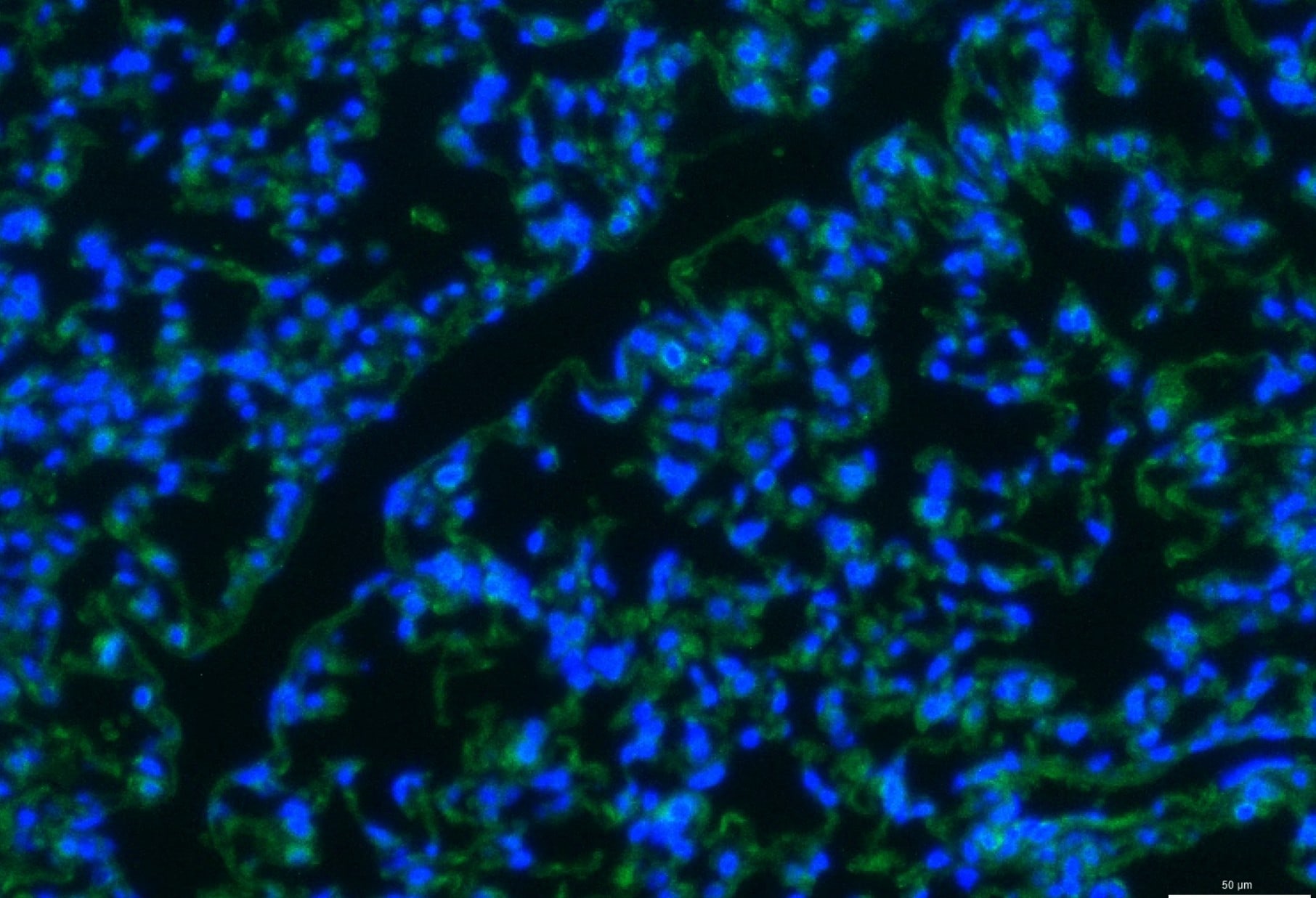
Application: ImmunofluorescenceSample Tested: Lung tissueSpecies: MouseVerified Customer | Posted 09/08/2025IF staining of FGF10 in mouse lungs (green) 10xFGF10 treated mice lungs staining
 Bio-Techne ResponseThis review reflects a new species or application tested on a primary antibody.
 -
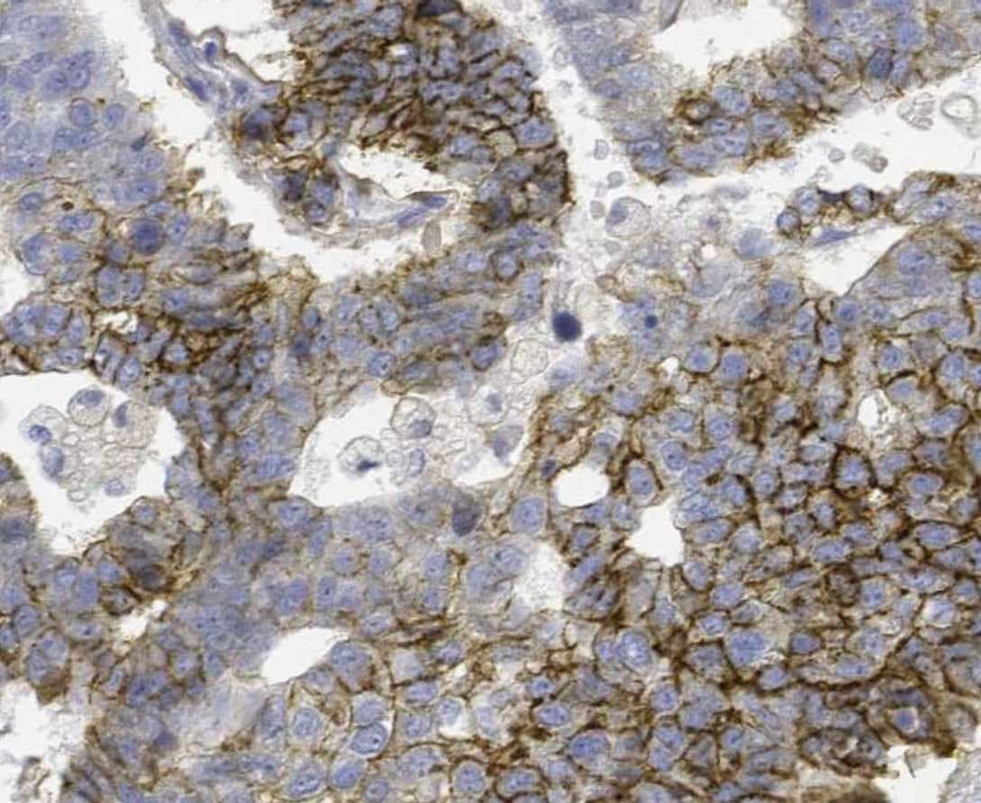
Application: ImmunohistochemistrySample Tested: Prostate cancer tissueSpecies: HumanVerified Customer | Posted 10/05/2021
-
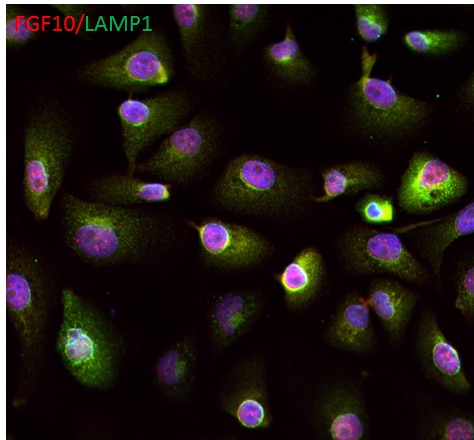
Application: Immunocytochemistry/ImmunofluorescenceSample Tested: iPSC derived epithelial cellsSpecies: HumanVerified Customer | Posted 09/27/2017
There are no reviews that match your criteria.
Protocols
Find general support by application which include: protocols, troubleshooting, illustrated assays, videos and webinars.
- Antigen Retrieval Protocol (PIER)
- Antigen Retrieval for Frozen Sections Protocol
- Appropriate Fixation of IHC/ICC Samples
- Cellular Response to Hypoxia Protocols
- Chromogenic IHC Staining of Formalin-Fixed Paraffin-Embedded (FFPE) Tissue Protocol
- Chromogenic Immunohistochemistry Staining of Frozen Tissue
- ClariTSA™ Fluorophore Kits
- Detection & Visualization of Antibody Binding
- ELISA Sample Preparation & Collection Guide
- ELISA Troubleshooting Guide
- Fluorescent IHC Staining of Frozen Tissue Protocol
- Graphic Protocol for Heat-induced Epitope Retrieval
- Graphic Protocol for the Preparation and Fluorescent IHC Staining of Frozen Tissue Sections
- Graphic Protocol for the Preparation and Fluorescent IHC Staining of Paraffin-embedded Tissue Sections
- Graphic Protocol for the Preparation of Gelatin-coated Slides for Histological Tissue Sections
- How to Run an R&D Systems DuoSet ELISA
- How to Run an R&D Systems Quantikine ELISA
- How to Run an R&D Systems Quantikine™ QuicKit™ ELISA
- IHC Sample Preparation (Frozen sections vs Paraffin)
- Immunofluorescent IHC Staining of Formalin-Fixed Paraffin-Embedded (FFPE) Tissue Protocol
- Immunohistochemistry (IHC) and Immunocytochemistry (ICC) Protocols
- Immunohistochemistry Frozen Troubleshooting
- Immunohistochemistry Paraffin Troubleshooting
- Preparing Samples for IHC/ICC Experiments
- Preventing Non-Specific Staining (Non-Specific Binding)
- Primary Antibody Selection & Optimization
- Protocol for Heat-Induced Epitope Retrieval (HIER)
- Protocol for Making a 4% Formaldehyde Solution in PBS
- Protocol for VisUCyte™ HRP Polymer Detection Reagent
- Protocol for the Preparation & Fixation of Cells on Coverslips
- Protocol for the Preparation and Chromogenic IHC Staining of Frozen Tissue Sections
- Protocol for the Preparation and Chromogenic IHC Staining of Frozen Tissue Sections - Graphic
- Protocol for the Preparation and Chromogenic IHC Staining of Paraffin-embedded Tissue Sections
- Protocol for the Preparation and Chromogenic IHC Staining of Paraffin-embedded Tissue Sections - Graphic
- Protocol for the Preparation and Fluorescent IHC Staining of Frozen Tissue Sections
- Protocol for the Preparation and Fluorescent IHC Staining of Paraffin-embedded Tissue Sections
- Protocol for the Preparation of Gelatin-coated Slides for Histological Tissue Sections
- Quantikine HS ELISA Kit Assay Principle, Alkaline Phosphatase
- Quantikine HS ELISA Kit Principle, Streptavidin-HRP Polymer
- R&D Systems Quality Control Western Blot Protocol
- Sandwich ELISA (Colorimetric) – Biotin/Streptavidin Detection Protocol
- Sandwich ELISA (Colorimetric) – Direct Detection Protocol
- TUNEL and Active Caspase-3 Detection by IHC/ICC Protocol
- The Importance of IHC/ICC Controls
- Troubleshooting Guide: ELISA
- Troubleshooting Guide: Immunohistochemistry
- Troubleshooting Guide: Western Blot Figures
- Western Blot Conditions
- Western Blot Protocol
- Western Blot Protocol for Cell Lysates
- Western Blot Troubleshooting
- Western Blot Troubleshooting Guide
- View all Protocols, Troubleshooting, Illustrated assays and Webinars


