
Key Product Details
Validated by
Biological Validation
Species Reactivity
Validated:
Human
Cited:
Human, Mouse
Applications
Validated:
Immunohistochemistry, Western Blot, Immunocytochemistry
Cited:
Immunohistochemistry, Immunohistochemistry-Paraffin, Immunohistochemistry-Frozen, Western Blot, Neutralization, Flow Cytometry, Immunocytochemistry, Binding Assay, Cell Culture
Label
Unconjugated
Antibody Source
Polyclonal Goat IgG
Loading...
Product Specifications
Immunogen
E. coli-derived recombinant human Galectin-9
Specificity
Detects human Galectin-9 in direct ELISAs and Western blots. In Western blots, approximately 10% cross-reactivity with recombinant human (rh) Galectin-3 and rhGalectin-4 is observed and less than 2% cross-reactivity with rhGalectin-1, rhGalectin-2, rhGalectin-7, and rhGalectin-8 is observed.
Clonality
Polyclonal
Host
Goat
Isotype
IgG
Scientific Data Images for Human Galectin-9 Antibody
Detection of Human Galectin‑9 by Western Blot.
Western blot shows conditioned media from HT-29 human colon adenocarcinoma cell line, human peripheral blood lymphocytes (PBL), and human dendritic cells untreated (-) or treated (+) with 10 µg/mL PHA for 6 days. PVDF membrane was probed with 1 µg/mL of Goat Anti-Human Galectin-9 Antigen Affinity-purified Polyclonal Antibody (Catalog # AF2045) followed by HRP-conjugated Anti-Goat IgG Secondary Antibody (Catalog # HAF109). Specific bands were detected for Galectin-9 at approximately 36 and 40 kDa. This experiment was conducted under reducing conditions and using Immunoblot Buffer Group 1.
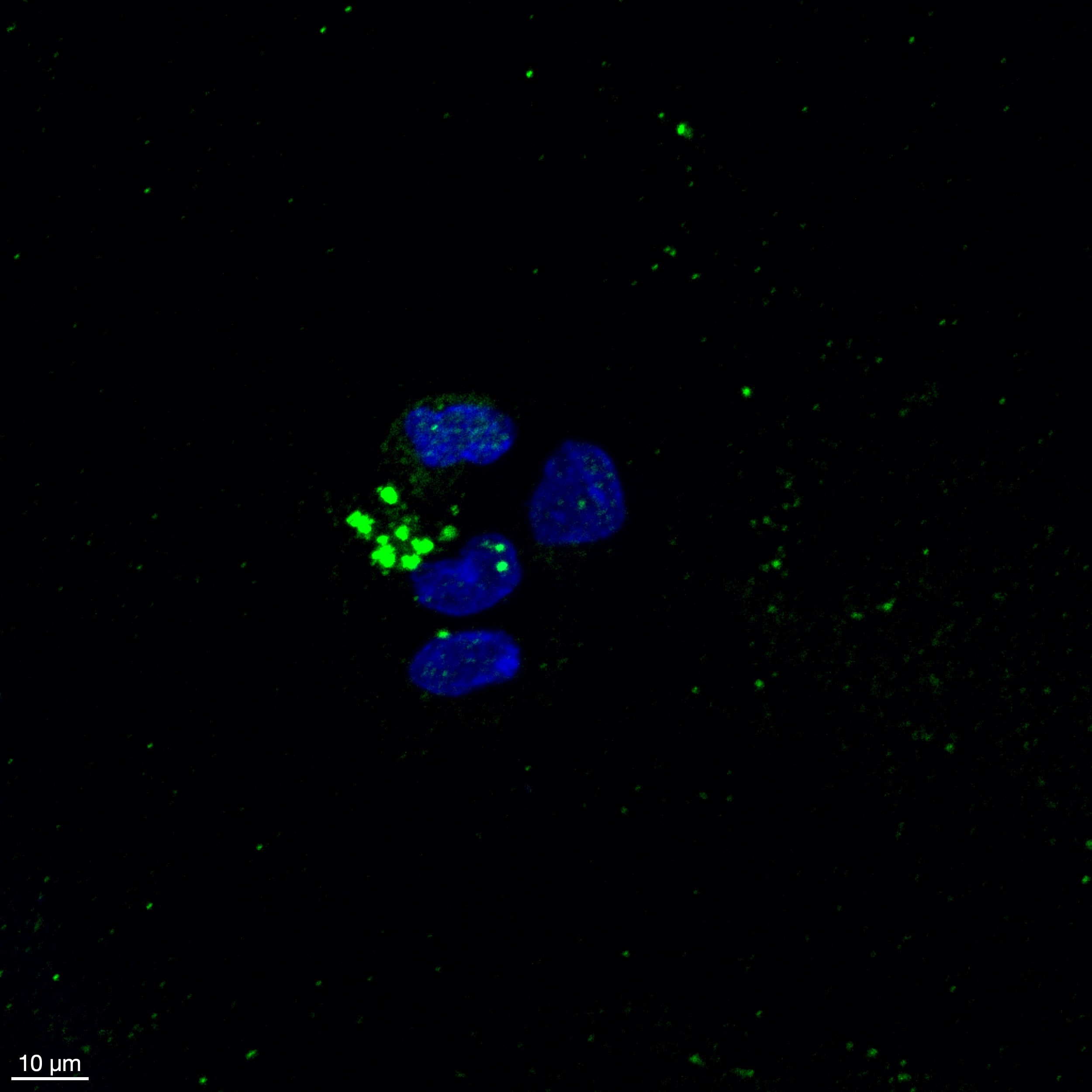
Galectin‑9 in Human PBMCs.
Galectin-9 was detected in immersion fixed human peripheral blood mononuclear cells (PBMCs) treated with Calcium Ionomycin and PMA using Goat Anti-Human Galectin-9 Antigen Affinity-purified Polyclonal Antibody (Catalog # AF2045) at 15 µg/mL for 3 hours at room temperature. Cells were stained using the NorthernLights™ 557-conjugated Anti-Goat IgG Secondary Antibody (red; Catalog # NL001) and counterstained with DAPI (blue). Specific staining was localized to cytoplasm. View our protocol for Fluorescent ICC Staining of Non-adherent Cells.
Galectin‑9 in Human Colon.
Galectin-9 was detected in immersion fixed paraffin-embedded sections of human colon using Goat Anti-Human Galectin-9 Antigen Affinity-purified Polyclonal Antibody (Catalog # AF2045) at 0.3 µg/mL for 1 hour at room temperature followed by incubation with the Anti-Goat IgG VisUCyte™ HRP Polymer Antibody (Catalog # VC004). Tissue was stained using DAB (brown) and counterstained with hematoxylin (blue). Specific staining was localized to epithelial cells. View our protocol for IHC Staining with VisUCyte HRP Polymer Detection Reagents.Applications for Human Galectin-9 Antibody
Application
Recommended Usage
Immunocytochemistry
5-15 µg/mL
Sample: Immersion fixed human peripheral blood mononuclear cells (PBMCs) treated with Calcium Ionomycin and PMA
Sample: Immersion fixed human peripheral blood mononuclear cells (PBMCs) treated with Calcium Ionomycin and PMA
Immunohistochemistry
0.3-15 µg/mL
Sample: Immersion fixed paraffin-embedded sections of human colon
Sample: Immersion fixed paraffin-embedded sections of human colon
Western Blot
1 µg/mL
Sample: HT‑29 human colon adenocarcinoma cell line, human peripheral blood lymphocytes (PBL), and human dendritic cells treated with PHA
Sample: HT‑29 human colon adenocarcinoma cell line, human peripheral blood lymphocytes (PBL), and human dendritic cells treated with PHA
Reviewed Applications
Read 4 reviews rated 4.5 using AF2045 in the following applications:
Formulation, Preparation, and Storage
Purification
Antigen Affinity-purified
Reconstitution
Reconstitute at 0.2 mg/mL in sterile PBS. For liquid material, refer to CoA for concentration.
Loading...
Formulation
Lyophilized from a 0.2 μm filtered solution in PBS with Trehalose. *Small pack size (SP) is supplied either lyophilized or as a 0.2 µm filtered solution in PBS.
Shipping
Lyophilized product is shipped at ambient temperature. Liquid small pack size (-SP) is shipped with polar packs. Upon receipt, store immediately at the temperature recommended below.
Stability & Storage
Use a manual defrost freezer and avoid repeated freeze-thaw cycles.
- 12 months from date of receipt, -20 to -70 °C as supplied.
- 1 month, 2 to 8 °C under sterile conditions after reconstitution.
- 6 months, -20 to -70 °C under sterile conditions after reconstitution.
Calculators
Background: Galectin-9
Alternate Names
Ecalectin, GAL9, Galectin9, LGALS9
Gene Symbol
LGALS9
Additional Galectin-9 Products
Product Documents for Human Galectin-9 Antibody
Certificate of Analysis
To download a Certificate of Analysis, please enter a lot or batch number in the search box below.
Note: Certificate of Analysis not available for kit components.
Product Specific Notices for Human Galectin-9 Antibody
For research use only
Related Research Areas
Citations for Human Galectin-9 Antibody
 Powered by Bioz
Customer Reviews for Human Galectin-9 Antibody (4)
4.5 out of 5
4 Customer Ratings
Have you used Human Galectin-9 Antibody?
Submit a review and receive an Amazon gift card!
$25/€18/£15/$25CAN/¥2500 Yen for a review with an image
$10/€7/£6/$10CAN/¥1110 Yen for a review without an image
Submit a review
Customer Images

Showing
1
-
4 of
4 reviews
Showing All
Filter By:
-
Application: ImmunocytochemistrySample Tested: ccd-841-conSpecies: HumanVerified Customer | Posted 04/18/2026Dapi (blue) and Galectin-9 (green)In vitro immunostaining of Galectin-9 (1:1000)

-
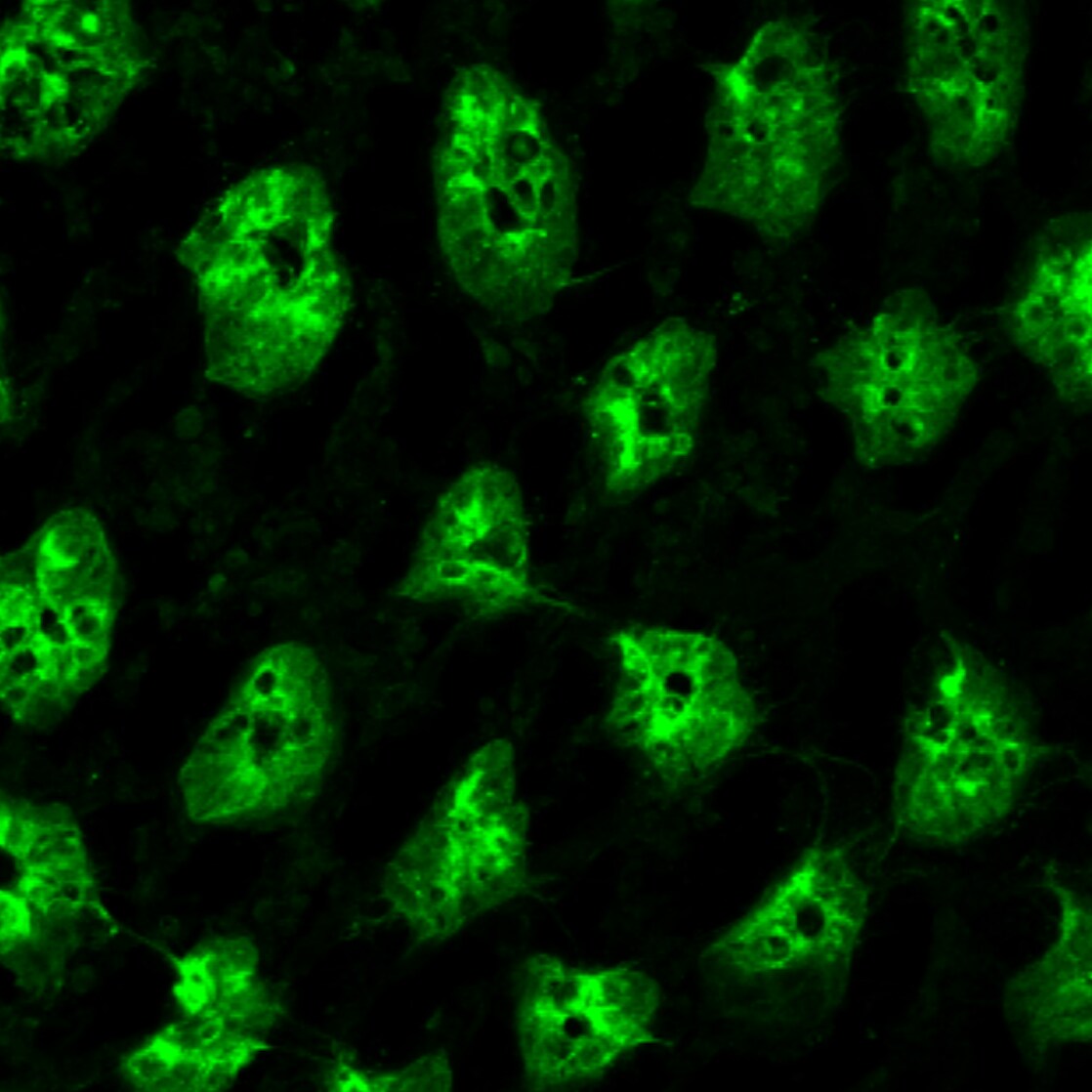
Application: Immunocytochemistry/ImmunofluorescenceVerified Customer | Posted 01/24/2024
-
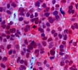
Application: Immunohistochemistry-ParaffinSample Tested: Breast cancer tissueSpecies: HumanVerified Customer | Posted 04/04/2020
-
Application: Western BlotSample Tested: SK-BR-3 human breast cancer cell lineSpecies: HumanVerified Customer | Posted 10/28/2018
There are no reviews that match your criteria.
Protocols
Find general support by application which include: protocols, troubleshooting, illustrated assays, videos and webinars.
- Antigen Retrieval Protocol (PIER)
- Antigen Retrieval for Frozen Sections Protocol
- Appropriate Fixation of IHC/ICC Samples
- Cellular Response to Hypoxia Protocols
- Chromogenic IHC Staining of Formalin-Fixed Paraffin-Embedded (FFPE) Tissue Protocol
- Chromogenic Immunohistochemistry Staining of Frozen Tissue
- ClariTSA™ Fluorophore Kits
- Detection & Visualization of Antibody Binding
- Fluorescent IHC Staining of Frozen Tissue Protocol
- Graphic Protocol for Heat-induced Epitope Retrieval
- Graphic Protocol for the Preparation and Fluorescent IHC Staining of Frozen Tissue Sections
- Graphic Protocol for the Preparation and Fluorescent IHC Staining of Paraffin-embedded Tissue Sections
- Graphic Protocol for the Preparation of Gelatin-coated Slides for Histological Tissue Sections
- ICC Cell Smear Protocol for Suspension Cells
- ICC Immunocytochemistry Protocol Videos
- ICC for Adherent Cells
- IHC Sample Preparation (Frozen sections vs Paraffin)
- Immunocytochemistry (ICC) Protocol
- Immunocytochemistry Troubleshooting
- Immunofluorescence of Organoids Embedded in Cultrex Basement Membrane Extract
- Immunofluorescent IHC Staining of Formalin-Fixed Paraffin-Embedded (FFPE) Tissue Protocol
- Immunohistochemistry (IHC) and Immunocytochemistry (ICC) Protocols
- Immunohistochemistry Frozen Troubleshooting
- Immunohistochemistry Paraffin Troubleshooting
- Preparing Samples for IHC/ICC Experiments
- Preventing Non-Specific Staining (Non-Specific Binding)
- Primary Antibody Selection & Optimization
- Protocol for Heat-Induced Epitope Retrieval (HIER)
- Protocol for Making a 4% Formaldehyde Solution in PBS
- Protocol for VisUCyte™ HRP Polymer Detection Reagent
- Protocol for the Fluorescent ICC Staining of Cell Smears - Graphic
- Protocol for the Fluorescent ICC Staining of Cultured Cells on Coverslips - Graphic
- Protocol for the Preparation & Fixation of Cells on Coverslips
- Protocol for the Preparation and Chromogenic IHC Staining of Frozen Tissue Sections
- Protocol for the Preparation and Chromogenic IHC Staining of Frozen Tissue Sections - Graphic
- Protocol for the Preparation and Chromogenic IHC Staining of Paraffin-embedded Tissue Sections
- Protocol for the Preparation and Chromogenic IHC Staining of Paraffin-embedded Tissue Sections - Graphic
- Protocol for the Preparation and Fluorescent ICC Staining of Cells on Coverslips
- Protocol for the Preparation and Fluorescent ICC Staining of Non-adherent Cells
- Protocol for the Preparation and Fluorescent ICC Staining of Stem Cells on Coverslips
- Protocol for the Preparation and Fluorescent IHC Staining of Frozen Tissue Sections
- Protocol for the Preparation and Fluorescent IHC Staining of Paraffin-embedded Tissue Sections
- Protocol for the Preparation of Gelatin-coated Slides for Histological Tissue Sections
- Protocol for the Preparation of a Cell Smear for Non-adherent Cell ICC - Graphic
- R&D Systems Quality Control Western Blot Protocol
- TUNEL and Active Caspase-3 Detection by IHC/ICC Protocol
- The Importance of IHC/ICC Controls
- Troubleshooting Guide: Immunohistochemistry
- Troubleshooting Guide: Western Blot Figures
- Western Blot Conditions
- Western Blot Protocol
- Western Blot Protocol for Cell Lysates
- Western Blot Troubleshooting
- Western Blot Troubleshooting Guide
- View all Protocols, Troubleshooting, Illustrated assays and Webinars
Loading...