Human HNF-4 alpha/NR2A1 Antibody (843716)
R&D Systems | Catalog # MAB4605

Key Product Details
Species Reactivity
Validated:
Human
Cited:
Human, Mouse
Applications
Validated:
Western Blot, Intracellular Staining by Flow Cytometry, Immunocytochemistry, Simple Western
Cited:
Immunohistochemistry
Label
Unconjugated
Antibody Source
Monoclonal Mouse IgG2B Clone # 843716
Loading...
Product Specifications
Immunogen
E. coli-derived recombinant human HNF‑4 alpha /NR2A1
Val130-Ser330
Accession # P41235
Val130-Ser330
Accession # P41235
Specificity
Detects human HNF‑4 alpha /NR2A1 in ELISAs and Western blots.
Clonality
Monoclonal
Host
Mouse
Isotype
IgG2B
Scientific Data Images for Human HNF-4 alpha/NR2A1 Antibody (843716)
Detection of Human HNF‑4 alpha /NR2A1 by Western Blot.
Western blot shows lysates of HepG2 human hepatocellular carcinoma cell line. Gels were loaded with 10 µg of cytoplasmic (Cyto) and 5 µg of nuclear (Nuc) extracts. PVDF membrane was probed with 1 µg/mL of Mouse Anti-Human HNF-4a/ NR2A1 Monoclonal Antibody (Catalog # MAB4605) followed by HRP-conjugated Anti-Mouse IgG Secondary Antibody (Catalog # HAF018). Specific bands were detected for HNF-4a/NR2A1 at approximately 50-55 kDa (as indicated). This experiment was conducted under reducing conditions and using Immunoblot Buffer Group 1.
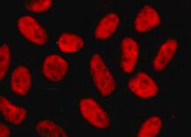
HNF-4 alpha /NR2A1 in HepG2 Human Cell Line.
HNF-4a/NR2A1 was detected in immersion fixed HepG2 human hepatocellular carcinoma cell line using Mouse Anti-Human HNF-4a/NR2A1 Monoclonal Antibody (Catalog # MAB4605) at 10 µg/mL for 3 hours at room temperature. Cells were stained using the NorthernLights™ 557-conjugated Anti-Mouse IgG Secondary Antibody (red, upper panel; Catalog # NL007) and counterstained with DAPI (blue, lower panel). Specific staining was localized to nuclei. View our protocol for Fluorescent ICC Staining of Cells on Coverslips.
Detection of Human HNF‑4 alpha /NR2A1 by Simple WesternTM.
Simple Western lane view shows lysates of human liver tissue, loaded at 0.2 mg/mL. A specific band was detected for HNF‑4 alpha /NR2A1 at approximately 62 kDa (as indicated) using 10 µg/mL of Mouse Anti-Human HNF‑4 alpha /NR2A1 Monoclonal Antibody (Catalog # MAB4605). This experiment was conducted under reducing conditions and using the 12-230 kDa separation system.Non-specific interaction with the 230 kDa Simple Western standard may be seen with this antibody.
Detection of HNF-4 alpha/NR2A1 in HepG2 Human Cell Line by Flow Cytometry.
HepG2 human hepatocellular carcinoma cell line was stained with Mouse Anti-Human HNF-4 alpha/NR2A1 Monoclonal Antibody (Catalog # MAB4605, filled histogram) or isotype control antibody (Catalog # MAB0041, open histogram) followed by PE-conjugated Anti-Mouse IgG Secondary Antibody (Catalog # F0102B). To facilitate intracellular staining, cells were fixed and permeabilized with FlowX FoxP3/Transcription Factor Fixation & Permeabilization Buffer Kit (Catalog # FC012). View our protocol for Staining Intracellular Molecules.
Detection of HNF-4 alpha/NR2A1 in Human Endodermal Cells by Flow Cytometry.
BG01V human embryonic stem cell line differentiated to endodermal cells (StemXVivo Endoderm Kit, Catalog # SC019B) was stained with Mouse Anti-Human HNF-4 alpha/NR2A1 Monoclonal Antibody (Catalog # MAB4605, filled histogram or isotype control antibody (Catalog # MAB0041, open histogram) followed by PE-conjugated Anti-Mouse IgG Secondary Antibody (Catalog # F0102B). To facilitate intracellular staining, cells were fixed and permeabilized with FlowX FoxP3/Transcription Factor Fixation & Permeabilization Buffer Kit (Catalog # FC012). View our protocol for Staining Intracellular Molecules.Applications for Human HNF-4 alpha/NR2A1 Antibody (843716)
Application
Recommended Usage
Immunocytochemistry
8-25 µg/mL
Sample: Immersion fixed HepG2 human hepatocellular carcinoma cell line
Sample: Immersion fixed HepG2 human hepatocellular carcinoma cell line
Intracellular Staining by Flow Cytometry
0.25 µg/106 cells
Sample: BG01V human embryonic stem cell line differentiated to endodermal cells (StemXVivo Endoderm Kit, Catalog # SC019B) and HepG2 human hepatocellular carcinoma cell line fixed and permeabilized with FlowX FoxP3/Transcription Factor Fixation & Permeabilization Buffer Kit (Catalog # FC012).
Sample: BG01V human embryonic stem cell line differentiated to endodermal cells (StemXVivo Endoderm Kit, Catalog # SC019B) and HepG2 human hepatocellular carcinoma cell line fixed and permeabilized with FlowX FoxP3/Transcription Factor Fixation & Permeabilization Buffer Kit (Catalog # FC012).
Simple Western
10 µg/mL
Sample: Human liver tissue
Sample: Human liver tissue
Western Blot
1 µg/mL
Sample: HepG2 human hepatocellular carcinoma cell line
Sample: HepG2 human hepatocellular carcinoma cell line
Reviewed Applications
Read 2 reviews rated 5 using MAB4605 in the following applications:
Flow Cytometry Panel Builder
Bio-Techne Knows Flow Cytometry
Save time and reduce costly mistakes by quickly finding compatible reagents using the Panel Builder Tool.
Advanced Features
- Spectra Viewer - Custom analysis of spectra from multiple fluorochromes
- Spillover Popups - Visualize the spectra of individual fluorochromes
- Antigen Density Selector - Match fluorochrome brightness with antigen density

Formulation, Preparation, and Storage
Purification
Protein A or G purified from hybridoma culture supernatant
Reconstitution
Sterile PBS to a final concentration of 0.5 mg/mL. For liquid material, refer to CoA for concentration.
Loading...
Formulation
Lyophilized from a 0.2 μm filtered solution in PBS with Trehalose. *Small pack size (SP) is supplied either lyophilized or as a 0.2 µm filtered solution in PBS.
Shipping
Lyophilized product is shipped at ambient temperature. Liquid small pack size (-SP) is shipped with polar packs. Upon receipt, store immediately at the temperature recommended below.
Stability & Storage
Use a manual defrost freezer and avoid repeated freeze-thaw cycles.
- 12 months from date of receipt, -20 to -70 °C as supplied.
- 1 month, 2 to 8 °C under sterile conditions after reconstitution.
- 6 months, -20 to -70 °C under sterile conditions after reconstitution.
Calculators
Background: HNF-4 alpha/NR2A1
Long Name
Hepatocyte Nuclear Factor-4, alpha
Alternate Names
HNF4 alpha, HNF4A, MODY1, NR2A1, TCF14
Entrez Gene IDs
3172 (Human)
Gene Symbol
HNF4A
UniProt
Additional HNF-4 alpha/NR2A1 Products
Product Documents for Human HNF-4 alpha/NR2A1 Antibody (843716)
Certificate of Analysis
To download a Certificate of Analysis, please enter a lot or batch number in the search box below.
Note: Certificate of Analysis not available for kit components.
Product Specific Notices for Human HNF-4 alpha/NR2A1 Antibody (843716)
For research use only
Related Research Areas
Citations for Human HNF-4 alpha/NR2A1 Antibody (843716)
 Powered by Bioz
Customer Reviews for Human HNF-4 alpha/NR2A1 Antibody (843716) (2)
5 out of 5
2 Customer Ratings
Have you used Human HNF-4 alpha/NR2A1 Antibody (843716)?
Submit a review and receive an Amazon gift card!
$25/€18/£15/$25CAN/¥2500 Yen for a review with an image
$10/€7/£6/$10CAN/¥1110 Yen for a review without an image
Submit a review
Customer Images
Showing
1
-
2 of
2 reviews
Showing All
Filter By:
-
Application: Immunocytochemistry/ImmunofluorescenceSample Tested: HepG2 Human Cell LineSpecies: HumanVerified Customer | Posted 02/10/2022

-
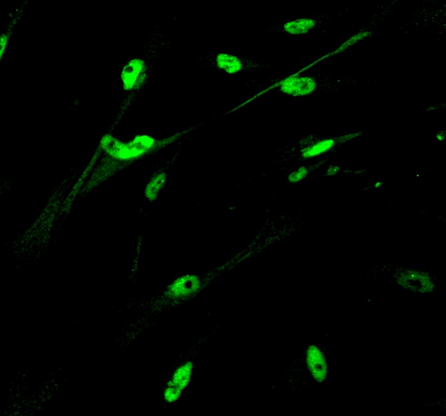
Application: ImmunocytochemistrySample Tested: Pig trabecular meshwork cellsSpecies: PigVerified Customer | Posted 12/05/2016Detection of HNF-4a in cultured pig trabecular meshwork cells using Human HNF-4a/NR2A1 Antibody (MAB4605) at 1:500 dilution overnight and donkey anti-mouse Alexa 488 (1:1000) for 1 hour.
There are no reviews that match your criteria.
Protocols
Find general support by application which include: protocols, troubleshooting, illustrated assays, videos and webinars.
- 7-Amino Actinomycin D (7-AAD) Cell Viability Flow Cytometry Protocol
- Appropriate Fixation of IHC/ICC Samples
- Cellular Response to Hypoxia Protocols
- ClariTSA™ Fluorophore Kits
- Detection & Visualization of Antibody Binding
- Extracellular Membrane Flow Cytometry Protocol
- Flow Cytometry Protocol for Cell Surface Markers
- Flow Cytometry Protocol for Staining Membrane Associated Proteins
- Flow Cytometry Staining Protocols
- Flow Cytometry Troubleshooting Guide
- ICC Cell Smear Protocol for Suspension Cells
- ICC Immunocytochemistry Protocol Videos
- ICC for Adherent Cells
- Immunocytochemistry (ICC) Protocol
- Immunocytochemistry Troubleshooting
- Immunofluorescence of Organoids Embedded in Cultrex Basement Membrane Extract
- Immunohistochemistry (IHC) and Immunocytochemistry (ICC) Protocols
- Intracellular Flow Cytometry Protocol Using Alcohol (Methanol)
- Intracellular Flow Cytometry Protocol Using Detergents
- Intracellular Nuclear Staining Flow Cytometry Protocol Using Detergents
- Intracellular Staining Flow Cytometry Protocol Using Alcohol Permeabilization
- Intracellular Staining Flow Cytometry Protocol Using Detergents to Permeabilize Cells
- Preparing Samples for IHC/ICC Experiments
- Preventing Non-Specific Staining (Non-Specific Binding)
- Primary Antibody Selection & Optimization
- Propidium Iodide Cell Viability Flow Cytometry Protocol
- Protocol for Liperfluo
- Protocol for VisUCyte™ HRP Polymer Detection Reagent
- Protocol for the Characterization of Human Th22 Cells
- Protocol for the Characterization of Human Th9 Cells
- Protocol for the Fluorescent ICC Staining of Cell Smears - Graphic
- Protocol for the Fluorescent ICC Staining of Cultured Cells on Coverslips - Graphic
- Protocol for the Preparation and Fluorescent ICC Staining of Cells on Coverslips
- Protocol for the Preparation and Fluorescent ICC Staining of Non-adherent Cells
- Protocol for the Preparation and Fluorescent ICC Staining of Stem Cells on Coverslips
- Protocol for the Preparation of a Cell Smear for Non-adherent Cell ICC - Graphic
- Protocol: Annexin V and PI Staining by Flow Cytometry
- Protocol: Annexin V and PI Staining for Apoptosis by Flow Cytometry
- R&D Systems Quality Control Western Blot Protocol
- TUNEL and Active Caspase-3 Detection by IHC/ICC Protocol
- The Importance of IHC/ICC Controls
- Troubleshooting Guide: Fluorokine Flow Cytometry Kits
- Troubleshooting Guide: Western Blot Figures
- Western Blot Conditions
- Western Blot Protocol
- Western Blot Protocol for Cell Lysates
- Western Blot Troubleshooting
- Western Blot Troubleshooting Guide
- View all Protocols, Troubleshooting, Illustrated assays and Webinars
Loading...
Associated Pathways
