Interleukin 10, also known as cytokine synthesis inhibitory factor (CSIF), is the charter member of the IL-10 family of alpha -helical cytokines that also includes IL-19, IL‑20, IL-22, IL-24, and IL-26/AK155 (1, 2). IL-10 is secreted by many activated hematopoietic cell types as well as hepatic stellate cells, keratinocytes, and placental cytotrophoblasts (2‑5). Mature human IL-10 shares 72%‑86% amino acid sequence identity with bovine, canine, equine, feline, mouse, ovine, porcine, and rat IL-10. Whereas human IL-10 is active on mouse cells, mouse IL-10 does not act on human cells (6, 7). IL-10 is a 178 amino acid molecule that contains two intrachain disulfide bridges and is expressed as a 36 kDa noncovalently associated homodimer (6, 8, 9). The IL-10 dimer binds to two IL-10 R alpha /IL-10 R1 chains, resulting in recruitment of two IL-10 R beta /IL-10 R2 chains and activation of a signaling cascade involving JAK1, TYK2, and STAT3 (10). IL-10 R beta does not bind IL-10 by itself but is required for signal transduction (1). IL-10 R beta also associates with IL‑20 R alpha, IL-22 R alpha, or IL-28 R alpha to form the receptor complexes for IL-22, IL-26, IL-28, and IL‑29 (11‑13). IL-10 is a critical molecule in the control of viral infections and allergic and autoimmune inflammation (14‑16). It promotes phagocytic uptake and Th2 responses but suppresses antigen presentation and Th1 proinflammatory responses (2).

Key Product Details
Species Reactivity
Validated:
Human
Cited:
Human, Primate - Macaca mulatta (Rhesus Macaque)
Applications
Validated:
Immunohistochemistry, Western Blot, Neutralization, Simple Western
Cited:
Immunohistochemistry, Immunohistochemistry-Paraffin, Western Blot, Neutralization, Assay Development, Functional Assay, Mass Cytometry, Imaging Mass Cytometry
Label
Unconjugated
Antibody Source
Polyclonal Goat IgG
Loading...
Product Specifications
Immunogen
S. frugiperda insect ovarian cell line Sf 21-derived recombinant human IL-10
Ser19-Asn178
Accession # P22301
Ser19-Asn178
Accession # P22301
Specificity
Detects human IL-10 in direct ELISAs and Western blots. In direct ELISAs, approximately 50% cross-reactivity with recombinant Epstein-Barr virus IL-10 is observed and less than 20% cross-reactivity with recombinant mouse IL‑10, recombinant feline IL-10, recombinant porcine IL‑10, recombinant equine IL-10, and recombinant rat IL‑10 is observed.
Clonality
Polyclonal
Host
Goat
Isotype
IgG
Endotoxin Level
<0.10 EU per 1 μg of the antibody by the LAL method.
Scientific Data Images for Human IL-10 Antibody
Detection of Human IL‑10 by Western Blot.
Western blot shows conditioned media of HEK293 human embryonic kidney cell line either mock transfected or transfected with human IL-10. PVDF membrane was probed with 1 µg/mL of Goat Anti-Human IL-10 Antigen Affinity-purified Polyclonal Antibody (Catalog # AF-217-NA) followed by HRP-conjugated Anti-Goat IgG Secondary Antibody (Catalog # HAF017). A specific band was detected for IL-10 at approximately 16 kDa (as indicated). This experiment was conducted under reducing conditions and using Immunoblot Buffer Group 1.
IL‑10 in Human Tonsil.
IL-10 was detected in immersion fixed paraffin-embedded sections of human tonsil using Goat Anti-Human IL-10 Antigen Affinity-purified Polyclonal Antibody (Catalog # AF-217-NA) at 15 µg/mL overnight at 4 °C. Tissue was stained using the Anti-Goat HRP-DAB Cell & Tissue Staining Kit (brown; Catalog # CTS008) and counterstained with hematoxylin (blue). View our protocol for Chromogenic IHC Staining of Paraffin-embedded Tissue Sections.
IL‑10 in Human Tonsil.
IL-10 was detected in immersion fixed paraffin-embedded sections of human tonsil using Goat Anti-Human IL-10 Antigen Affinity-purified Polyclonal Antibody (Catalog # AF-217-NA) at 15 µg/mL overnight at 4 °C. Tissue was stained using the Anti-Goat HRP-DAB Cell & Tissue Staining Kit (brown; Catalog # CTS008) and counterstained with hematoxylin (blue). Lower panel shows a lack of labeling if primary antibodies are omitted and tissue is stained only with secondary antibody followed by incubation with detection reagents. View our protocol for Chromogenic IHC Staining of Paraffin-embedded Tissue Sections.
Detection of Human IL‑10 by Simple WesternTM.
Simple Western lane view shows conditioned media of HEK293 human embryonic kidney cell line either mock transfected or transfected with human IL-10, loaded at 0.2 mg/mL. A specific band was detected for IL-10 at approximately 23 kDa (as indicated) using 10 µg/mL of Goat Anti-Human IL-10 Antigen Affinity-purified Polyclonal Antibody (Catalog # AF-217-NA) followed by 1:50 dilution of HRP-conjugated Anti-Goat IgG Secondary Antibody (Catalog # HAF109). This experiment was conducted under reducing conditions and using the 12-230 kDa separation system.
Cell Proliferation Induced by IL‑10 and Neutralization by Human IL‑10 Antibody.
Recombinant Human IL-10 (Catalog # 217-IL) stimulates proliferation in the MC/9-2 mouse mast cell line in a dose-dependent manner (orange line). Proliferation elicited by Recombinant Human IL-10 (5 ng/mL) is neutralized (green line) by increasing concentrations of Goat Anti-Human IL-10 Antigen Affinity-purified Polyclonal Antibody (Catalog # AF-217-NA). The ND50 is typically 0.2-1.0 µg/mL.Applications for Human IL-10 Antibody
Application
Recommended Usage
Immunohistochemistry
5-15 µg/mL
Sample: Immersion fixed paraffin-embedded sections of human tonsil
Sample: Immersion fixed paraffin-embedded sections of human tonsil
Simple Western
10 µg/mL
Sample: HEK293 human embryonic kidney cell line transfected with human IL-10
Sample: HEK293 human embryonic kidney cell line transfected with human IL-10
Western Blot
1 µg/mL
Sample: HEK293 human embryonic kidney cell line transfected with human IL-10
Sample: HEK293 human embryonic kidney cell line transfected with human IL-10
Neutralization
Measured by its ability to neutralize IL‑10-induced proliferation in the MC/9‑2 mouse mast cell line. The Neutralization Dose (ND50) is typically 0.2-1.0 µg/mL in the presence of 5 ng/mL Recombinant Human IL‑10.
Reviewed Applications
Read 2 reviews rated 4 using AF-217-NA in the following applications:
Formulation, Preparation, and Storage
Purification
Antigen Affinity-purified
Reconstitution
Reconstitute at 0.2 mg/mL in sterile PBS. For liquid material, refer to CoA for concentration.
Loading...
Formulation
Lyophilized from a 0.2 μm filtered solution in PBS with Trehalose. *Small pack size (SP) is supplied either lyophilized or as a 0.2 µm filtered solution in PBS.
Shipping
Lyophilized product is shipped at ambient temperature. Liquid small pack size (-SP) is shipped with polar packs. Upon receipt, store immediately at the temperature recommended below.
Stability & Storage
Use a manual defrost freezer and avoid repeated freeze-thaw cycles.
- 12 months from date of receipt, -20 to -70 °C as supplied.
- 1 month, 2 to 8 °C under sterile conditions after reconstitution.
- 6 months, -20 to -70 °C under sterile conditions after reconstitution.
Calculators
Background: IL-10
References
- Pestka, S. et al. (2004) Annu. Rev. Immunol. 22:929.
- O’Garra, A. and P. Vieira (2007) Nat. Rev. Immunol. 7:425.
- Mathurin, P. et al. (2002) Am. J. Physiol. Gastrointest. Liver Physiol. 282:G981.
- Grewe, M. et al. (1995) J. Invest. Dermatol. 104:3.
- Szony, B.J. et al. (1999) Mol. Hum. Reprod. 5:1059.
- Vieira, P. et al. (1991) Proc. Natl. Acad. Sci. 88:1172.
- Hsu, D.-H. et al. (1990) Science 250:830.
- Windsor, W.T. et al. (1993) Biochemistry 32:8807.
- Syto, R. et al. (1998) Biochemistry 37:16943.
- Kotenko, S.V. et al. (1997) EMBO J. 16:5894.
- Kotenko, S.V. et al. (2000) J. Biol. Chem. 276:2725.
- Hor, S. et al. (2004) J. Biol. Chem. 279:33343.
- Sheppard, P. et al. (2003) Nat. Immunol. 4:63.
- Fitzgerald, D.C. et al. (2007) Nat. Immunol. 8:1372.
- Wu, K. et al. (2007) Cell. Mol. Immunol. 4:269.
- Blackburn, S.D. and E.J. Wherry (2007) Trends Microbiol. 15:143.
Long Name
Interleukin 10
Alternate Names
CSIF, GVHDS, IL10, IL10A, TGIF
Entrez Gene IDs
Gene Symbol
IL10
UniProt
Additional IL-10 Products
Product Documents for Human IL-10 Antibody
Certificate of Analysis
To download a Certificate of Analysis, please enter a lot or batch number in the search box below.
Note: Certificate of Analysis not available for kit components.
Product Specific Notices for Human IL-10 Antibody
For research use only
Related Research Areas
Citations for Human IL-10 Antibody
 Powered by Bioz
Customer Reviews for Human IL-10 Antibody (2)
4 out of 5
2 Customer Ratings
Have you used Human IL-10 Antibody?
Submit a review and receive an Amazon gift card!
$25/€18/£15/$25CAN/¥2500 Yen for a review with an image
$10/€7/£6/$10CAN/¥1110 Yen for a review without an image
Submit a review
Customer Images
Showing
1
-
2 of
2 reviews
Showing All
Filter By:
-
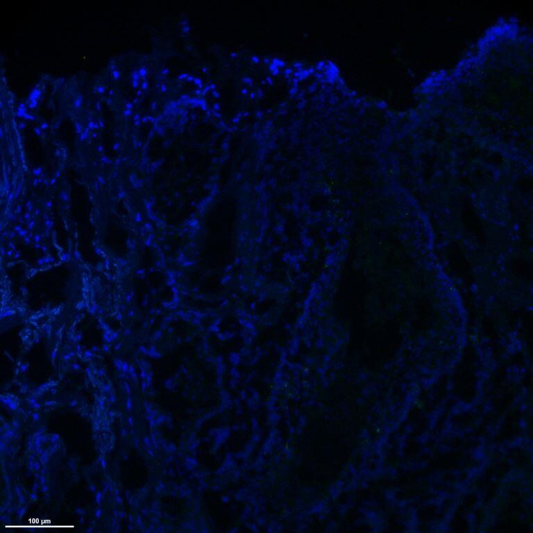
Application: Immunohistochemistry-FrozenSample Tested: Frozen human skin sectionsSpecies: HumanVerified Customer | Posted 12/14/2018Goat anti-IL10 (1:50, green) stained normal human skin, counterstained with DAPI (blue)Block with 5% BSA, incubated with antibody 1:50 in blocking solution at 37C for 1 hour

-
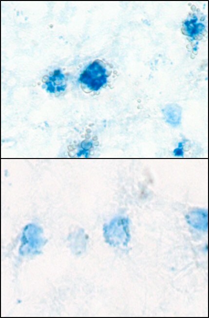
Application: ImmunohistochemistrySample Tested: Adult brainSpecies: HumanVerified Customer | Posted 04/13/2018Published in https://www.ncbi.nlm.nih.gov/pubmed/28169287 Used at 10ug/ml. Briefly, frozen brain sections were fixed in 4% PFA (Fisher Scientific), followed by antigen retrieval using heating in acid citric buffer (Vector, Burlingame, CA, USA). Endogenous avidin-biotin was blocked for 15 min (Vector). Sections were incubated with 10% horse serum in PBS (Biosera, Boussens, France) and Fc Receptor Blocking Solution was added (Human TruStain FcX Biolegend, London, UK). Primary antibody was added overnight at 4 °C and detected with donkey anti-goat-biotin (ab6578, Abcam), followed by streptavidin-alkaline phosphatase (SA-5100, Vector) and visualised with the Vector Blue Alkaline Phosphatase Substrate Kit III (Vector).
There are no reviews that match your criteria.
Protocols
Find general support by application which include: protocols, troubleshooting, illustrated assays, videos and webinars.
- Antigen Retrieval Protocol (PIER)
- Antigen Retrieval for Frozen Sections Protocol
- Appropriate Fixation of IHC/ICC Samples
- Cellular Response to Hypoxia Protocols
- Chromogenic IHC Staining of Formalin-Fixed Paraffin-Embedded (FFPE) Tissue Protocol
- Chromogenic Immunohistochemistry Staining of Frozen Tissue
- ClariTSA™ Fluorophore Kits
- Detection & Visualization of Antibody Binding
- Fluorescent IHC Staining of Frozen Tissue Protocol
- Graphic Protocol for Heat-induced Epitope Retrieval
- Graphic Protocol for the Preparation and Fluorescent IHC Staining of Frozen Tissue Sections
- Graphic Protocol for the Preparation and Fluorescent IHC Staining of Paraffin-embedded Tissue Sections
- Graphic Protocol for the Preparation of Gelatin-coated Slides for Histological Tissue Sections
- IHC Sample Preparation (Frozen sections vs Paraffin)
- Immunofluorescent IHC Staining of Formalin-Fixed Paraffin-Embedded (FFPE) Tissue Protocol
- Immunohistochemistry (IHC) and Immunocytochemistry (ICC) Protocols
- Immunohistochemistry Frozen Troubleshooting
- Immunohistochemistry Paraffin Troubleshooting
- Preparing Samples for IHC/ICC Experiments
- Preventing Non-Specific Staining (Non-Specific Binding)
- Primary Antibody Selection & Optimization
- Protocol for Heat-Induced Epitope Retrieval (HIER)
- Protocol for Making a 4% Formaldehyde Solution in PBS
- Protocol for VisUCyte™ HRP Polymer Detection Reagent
- Protocol for the Preparation & Fixation of Cells on Coverslips
- Protocol for the Preparation and Chromogenic IHC Staining of Frozen Tissue Sections
- Protocol for the Preparation and Chromogenic IHC Staining of Frozen Tissue Sections - Graphic
- Protocol for the Preparation and Chromogenic IHC Staining of Paraffin-embedded Tissue Sections
- Protocol for the Preparation and Chromogenic IHC Staining of Paraffin-embedded Tissue Sections - Graphic
- Protocol for the Preparation and Fluorescent IHC Staining of Frozen Tissue Sections
- Protocol for the Preparation and Fluorescent IHC Staining of Paraffin-embedded Tissue Sections
- Protocol for the Preparation of Gelatin-coated Slides for Histological Tissue Sections
- R&D Systems Quality Control Western Blot Protocol
- TUNEL and Active Caspase-3 Detection by IHC/ICC Protocol
- The Importance of IHC/ICC Controls
- Troubleshooting Guide: Immunohistochemistry
- Troubleshooting Guide: Western Blot Figures
- Western Blot Conditions
- Western Blot Protocol
- Western Blot Protocol for Cell Lysates
- Western Blot Troubleshooting
- Western Blot Troubleshooting Guide
- View all Protocols, Troubleshooting, Illustrated assays and Webinars
Loading...
Associated Pathways


