
Key Product Details
Validated by
Biological Validation
Species Reactivity
Validated:
Human
Cited:
Human
Applications
Validated:
Western Blot, Intracellular Staining by Flow Cytometry, Immunocytochemistry, CyTOF-ready
Cited:
Immunohistochemistry, Immunohistochemistry-Paraffin, Flow Cytometry, Competitive Binding Assay
Label
Unconjugated
Antibody Source
Monoclonal Mouse IgG1 Clone # 9906
Loading...
Product Specifications
Immunogen
S. frugiperda insect ovarian cell line Sf 21-derived recombinant human IL‑5
Ile20-Ser134
Accession # P05113
Ile20-Ser134
Accession # P05113
Specificity
Detects human IL-5 in direct ELISAs and Western blots. In direct ELISAs and Western blots, no cross-reactivity with recombinant mouse IL‑5 is observed.
Clonality
Monoclonal
Host
Mouse
Isotype
IgG1
Scientific Data Images for Human IL-5 Antibody (9906)
IL‑5 in Human Peripheral Blood Lymphocytes.
IL‑5 was detected in immersion fixed human peripheral blood lymphocytes using Mouse Anti-Human IL‑5 Monoclonal Antibody (Catalog # MAB605) at 5 µg/mL for 3 hours at room temperature. Cells were stained (red) and counterstained (green). Specific labeling was localized to the cytoplasm of PBMCs. View our protocol for Fluorescent ICC Staining of Non-adherent Cells.
Detection of IL‑5 in PMA and Ca2+ionomycin-treated Human PBMCs by Flow Cytometry.
Human PBMCs, untreated (light orange filled histogram) or activated with 50 ng/mL PMA and 500 ng/mL Ca2+ionomycin for 5 hours (dark orange filled histogram), were stained with Human IL-5 Monoclonal Antibody (Catalog # MAB605) or isotype control antibody (Catalog # MAB002, open histogram), followed by Fluorescein-conjugated Anti-Mouse IgG F(ab')2Secondary Antibody (Catalog # F0103B). To facilitate intracellular staining, cells were fixed with paraformaldehyde and permeabilized with saponin.
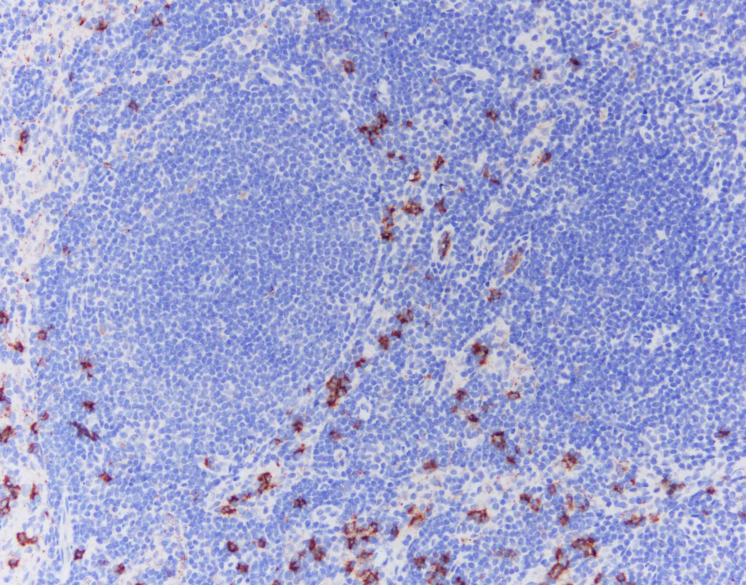
IL‑5 in Human Lymph Node Tissue.
pretreatment: basic buffer, heating Primary Ab: MAB605 x100 Detection: DAB Refine kit, standard protocol. Image from a verified customer review.Applications for Human IL-5 Antibody (9906)
Application
Recommended Usage
CyTOF-ready
Ready to be labeled using established conjugation methods. No BSA or other carrier proteins that could interfere with conjugation.
Immunocytochemistry
8-25 µg/mL
Sample: Immersion fixed human peripheral blood lymphocytes and activated T cells
Sample: Immersion fixed human peripheral blood lymphocytes and activated T cells
Intracellular Staining by Flow Cytometry
2.5 µg/106 cells
Sample: PMA and Ca2+ ionomycin‑treated human PBMCs, fixed with paraformaldehyde, and permeabilized with saponin
Sample: PMA and Ca2+ ionomycin‑treated human PBMCs, fixed with paraformaldehyde, and permeabilized with saponin
Western Blot
1 µg/mL
Sample: Recombinant Human IL‑5 (Catalog # 205-IL)
Sample: Recombinant Human IL‑5 (Catalog # 205-IL)
Reviewed Applications
Read 3 reviews rated 4.7 using MAB605 in the following applications:
Flow Cytometry Panel Builder
Bio-Techne Knows Flow Cytometry
Save time and reduce costly mistakes by quickly finding compatible reagents using the Panel Builder Tool.
Advanced Features
- Spectra Viewer - Custom analysis of spectra from multiple fluorochromes
- Spillover Popups - Visualize the spectra of individual fluorochromes
- Antigen Density Selector - Match fluorochrome brightness with antigen density

Formulation, Preparation, and Storage
Purification
Protein A or G purified from ascites
Reconstitution
Reconstitute at 0.5 mg/mL in sterile PBS. For liquid material, refer to CoA for concentration.
Loading...
Formulation
Lyophilized from a 0.2 μm filtered solution in PBS with Trehalose. *Small pack size (SP) is supplied either lyophilized or as a 0.2 µm filtered solution in PBS.
Shipping
Lyophilized product is shipped at ambient temperature. Liquid small pack size (-SP) is shipped with polar packs. Upon receipt, store immediately at the temperature recommended below.
Stability & Storage
Use a manual defrost freezer and avoid repeated freeze-thaw cycles.
- 12 months from date of receipt, -20 to -70 °C as supplied.
- 1 month, 2 to 8 °C under sterile conditions after reconstitution.
- 6 months, -20 to -70 °C under sterile conditions after reconstitution.
Calculators
Background: IL-5
References
- Rosenberg, H. F. et al. (2007) J. Allergy Clin. Immunol. 119:1303.
- Elsas, P.X. and M. I. G. Elsas (2007) Curr. Med. Chem. 14:1925.
- Martinez-Moczygemba, M. and D. P. Huston (2003) J. Allergy Clin. Immunol. 112:653.
- Minamitake, Y. et al. (1990) J. Biochem. 107:292.
- McKenzie, A. N. et al. (1991) Mol. Immunol. 28:155.
- Shakoory, B. et al. (2004) J. Interferon Cytokine Res. 24:271.
- Lalani, T. et al. (1999) Ann. Allergy Asthma Immunol. 82:317.
- Sakuishi, K. et al. (2007) J. Immunol. 179:3452.
- Clutterbuck, E. J. et al. (1989) Blood 73:1504.
- Cameron, L. et al. (2000) J. Immunol. 164:1538.
- Tavernier, J. et al. (1991) Cell 66:1175.
- Zaks-Zilberman, M. et al. (2008) J. Biol. Chem. 283:13398.
- Lipscombe, R. et al. (1998) J. Leukocyte Biol. 63:342.
- Tavernier, J. et al. (2000) Blood 95:1600.
- Kopf, M. et al. (1996) Immunity 4:15.
- Horikawa, K. and K. Takatsu (2006) Immunology 118:497.
- Denburg, J. A. et al. (1991) Blood 77:1462.
Long Name
Interleukin 5
Alternate Names
BCDF mu, BCGFII, EDF, Eo-CSF, IL5, TRF
Entrez Gene IDs
Gene Symbol
IL5
UniProt
Additional IL-5 Products
Product Documents for Human IL-5 Antibody (9906)
Certificate of Analysis
To download a Certificate of Analysis, please enter a lot or batch number in the search box below.
Note: Certificate of Analysis not available for kit components.
Product Specific Notices for Human IL-5 Antibody (9906)
For research use only
Citations for Human IL-5 Antibody (9906)
 Powered by Bioz
Customer Reviews for Human IL-5 Antibody (9906) (3)
4.7 out of 5
3 Customer Ratings
Have you used Human IL-5 Antibody (9906)?
Submit a review and receive an Amazon gift card!
$25/€18/£15/$25CAN/¥2500 Yen for a review with an image
$10/€7/£6/$10CAN/¥1110 Yen for a review without an image
Submit a review
Customer Images
Showing
1
-
3 of
3 reviews
Showing All
Filter By:
-
Application: ImmunohistochemistrySample Tested: Lymph node tissueSpecies: HumanVerified Customer | Posted 07/11/2025pretreatment: basic buffer, heating primary Ab: MAB605 x100 detection: DAB Refine kit, standard protocol
 Bio-Techne ResponseThis review reflects a new species or application tested on a primary antibody.
 -
Application: ImmunoprecipitationSample Tested: B16-F1 mouse melanoma cell lineSpecies: MouseVerified Customer | Posted 04/05/2018
-
Application: ELISASample Tested: A375 human melanoma cell lineSpecies: HumanVerified Customer | Posted 10/23/2017
There are no reviews that match your criteria.
Protocols
Find general support by application which include: protocols, troubleshooting, illustrated assays, videos and webinars.
- 7-Amino Actinomycin D (7-AAD) Cell Viability Flow Cytometry Protocol
- Appropriate Fixation of IHC/ICC Samples
- Cellular Response to Hypoxia Protocols
- ClariTSA™ Fluorophore Kits
- Detection & Visualization of Antibody Binding
- Extracellular Membrane Flow Cytometry Protocol
- Flow Cytometry Protocol for Cell Surface Markers
- Flow Cytometry Protocol for Staining Membrane Associated Proteins
- Flow Cytometry Staining Protocols
- Flow Cytometry Troubleshooting Guide
- ICC Cell Smear Protocol for Suspension Cells
- ICC Immunocytochemistry Protocol Videos
- ICC for Adherent Cells
- Immunocytochemistry (ICC) Protocol
- Immunocytochemistry Troubleshooting
- Immunofluorescence of Organoids Embedded in Cultrex Basement Membrane Extract
- Immunohistochemistry (IHC) and Immunocytochemistry (ICC) Protocols
- Intracellular Flow Cytometry Protocol Using Alcohol (Methanol)
- Intracellular Flow Cytometry Protocol Using Detergents
- Intracellular Nuclear Staining Flow Cytometry Protocol Using Detergents
- Intracellular Staining Flow Cytometry Protocol Using Alcohol Permeabilization
- Intracellular Staining Flow Cytometry Protocol Using Detergents to Permeabilize Cells
- Preparing Samples for IHC/ICC Experiments
- Preventing Non-Specific Staining (Non-Specific Binding)
- Primary Antibody Selection & Optimization
- Propidium Iodide Cell Viability Flow Cytometry Protocol
- Protocol for Liperfluo
- Protocol for VisUCyte™ HRP Polymer Detection Reagent
- Protocol for the Characterization of Human Th22 Cells
- Protocol for the Characterization of Human Th9 Cells
- Protocol for the Fluorescent ICC Staining of Cell Smears - Graphic
- Protocol for the Fluorescent ICC Staining of Cultured Cells on Coverslips - Graphic
- Protocol for the Preparation and Fluorescent ICC Staining of Cells on Coverslips
- Protocol for the Preparation and Fluorescent ICC Staining of Non-adherent Cells
- Protocol for the Preparation and Fluorescent ICC Staining of Stem Cells on Coverslips
- Protocol for the Preparation of a Cell Smear for Non-adherent Cell ICC - Graphic
- Protocol: Annexin V and PI Staining by Flow Cytometry
- Protocol: Annexin V and PI Staining for Apoptosis by Flow Cytometry
- R&D Systems Quality Control Western Blot Protocol
- TUNEL and Active Caspase-3 Detection by IHC/ICC Protocol
- The Importance of IHC/ICC Controls
- Troubleshooting Guide: Fluorokine Flow Cytometry Kits
- Troubleshooting Guide: Western Blot Figures
- Western Blot Conditions
- Western Blot Protocol
- Western Blot Protocol for Cell Lysates
- Western Blot Troubleshooting
- Western Blot Troubleshooting Guide
- View all Protocols, Troubleshooting, Illustrated assays and Webinars


