Irisin (also known as FNDC5) is a 12 kDa glycosylated polypeptide hormone that regulates energy metabolism, stem cell differentiation, and neuronal development (1, 2). Human Irisin is synthesized as a 212 amino acid (aa) precursor encoding a type 1 transmembrane protein with a 121 aa extracellular domain (ECD), a 21 aa transmembrane domain, and a 39 aa cytoplasmic domain. The ECD of Irisin contains a fibronectin type III domain and multiple glycosylation sites. The ECD is proteolytically cleaved to release the 112 aa soluble Irisin hormone into circulation (2-5). Mature human, mouse, and rat Irisin share 100% sequence identity. Expression of Irisin is induced in skeletal muscle and subcutaneous adipose tissue during and shortly after exercise (2, 6). Irisin induces expression of peroxisome proliferator-activated receptor gamma co-activator 1 alpha (PGC1 alpha ) and uncoupling protein-1 (UCP1), mitochondrial-associated metabolic proteins (7, 8). Irisin induces the transition of white adipose tissue into more metabolically active beige adipose tissue. In mice, expression of Irisin has been shown to regulate obesity and diabetes (1, 2). A similar function in humans is suggested (9). Irisin also regulates neuronal cell differentiation and neurite outgrowth in the brain and is involved in the differentiation of osteoblasts (10-14).
Human Irisin/FNDC5 Antibody (936434)
R&D Systems | Catalog # MAB9420

Key Product Details
Species Reactivity
Validated:
Cited:
Applications
Validated:
Cited:
Label
Antibody Source
Product Specifications
Immunogen
Asp32-Glu143
Accession # Q8NAU1
Specificity
Clonality
Host
Isotype
Scientific Data Images for Human Irisin/FNDC5 Antibody (936434)
Detection of Irisin/FNDC5 in HepG2 Human Cell Line by Flow Cytometry.
HepG2 human hepatocarcinoma cell line was stained with Mouse Anti-Human Irisin/FNDC5 Monoclonal Antibody (Catalog # MAB9420, filled histogram) or isotype control antibody (MAB0041, open histogram), followed by APC-conjugated Anti-Mouse IgG F(ab')2 Secondary Antibody (F0101B). View our protocol for Staining Membrane-associated Proteins.
Irisin/FNDC5 in Rat Skeletal Muscle.
Irisin/FNDC5 was detected in perfusion fixed frozen sections of rat skeletal muscle using Mouse Anti-Human Irisin/FNDC5 Monoclonal Antibody (Catalog # MAB9420) at 25 µg/mL overnight at 4 °C. Tissue was stained using the NorthernLights™ 557-conjugated Anti-Mouse IgG Secondary Antibody (red; NL007) and counterstained with DAPI (blue). Specific staining was localized to plasma membrane and cytoplasm of muscle cells. View our protocol for Fluorescent IHC Staining of Frozen Tissue Sections.
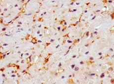
Immersion fixed paraffin-embedded sections of human skeletal muscle
Irisin/FNDC5 was detected in immersion fixed paraffin-embedded sections of human skeletal muscle using Mouse Anti-Human Irisin/FNDC5 Monoclonal Antibody (Catalog # MAB9420) at 5 µg/ml overnight at 4 °C. Before incubation with the primary antibody, tissue was subjected to heat-induced epitope retrieval using VisUCyte Antigen Retrieval Reagent-Basic (VCTS021). Tissue was stained using the HRP-conjugated Anti-Mouse IgG Secondary Antibody (HAF007) and counterstained with hematoxylin (blue). Specific staining was localized to cytoplasm. View our protocol for Chromogenic IHC Staining of Paraffin-embedded Tissue Sections.Applications for Human Irisin/FNDC5 Antibody (936434)
CyTOF-ready
ELISA
This antibody functions as an ELISA detection antibody when paired with Mouse Anti-Human Irisin/FNDC5 Monoclonal Antibody (Catalog # MAB8880).
This product is intended for assay development on various assay platforms requiring antibody pairs.
Flow Cytometry
Sample: HepG2 cell line
Immunohistochemistry
Sample:
Perfusion fixed frozen sections of rat skeletal muscle and Immersion fixed paraffin-embedded sections of human skeletal muscle
Reviewed Applications
Read 1 review rated 5 using MAB9420 in the following applications:
Flow Cytometry Panel Builder
Bio-Techne Knows Flow Cytometry
Save time and reduce costly mistakes by quickly finding compatible reagents using the Panel Builder Tool.
Advanced Features
- Spectra Viewer - Custom analysis of spectra from multiple fluorochromes
- Spillover Popups - Visualize the spectra of individual fluorochromes
- Antigen Density Selector - Match fluorochrome brightness with antigen density

Formulation, Preparation, and Storage
Purification
Reconstitution
Reconstitute at 0.5 mg/mL in sterile PBS. For liquid material, refer to CoA for concentration.
Formulation
*Small pack size (-SP) is supplied either lyophilized or as a 0.2 µm filtered solution in PBS.
Shipping
Stability & Storage
- 12 months from date of receipt, -20 to -70 °C as supplied.
- 1 month, 2 to 8 °C under sterile conditions after reconstitution.
- 6 months, -20 to -70 °C under sterile conditions after reconstitution.
Calculators
Background: Irisin/FNDC5
References
Long Name
Alternate Names
Gene Symbol
UniProt
Additional Irisin/FNDC5 Products
Product Documents for Human Irisin/FNDC5 Antibody (936434)
Certificate of Analysis
To download a Certificate of Analysis, please enter a lot or batch number in the search box below.
Note: Certificate of Analysis not available for kit components.
Product Specific Notices for Human Irisin/FNDC5 Antibody (936434)
For research use only
Citations for Human Irisin/FNDC5 Antibody (936434)
 Powered by Bioz
Customer Reviews for Human Irisin/FNDC5 Antibody (936434) (1)
Have you used Human Irisin/FNDC5 Antibody (936434)?
Submit a review and receive an Amazon gift card!
$25/€18/£15/$25CAN/¥2500 Yen for a review with an image
$10/€7/£6/$10CAN/¥1110 Yen for a review without an image
Submit a review
Customer Images
-
Application: ImmunohistochemistrySample Tested: Skeletal muscle tissueSpecies: HumanVerified Customer | Posted 06/28/2022

There are no reviews that match your criteria.
Protocols
Find general support by application which include: protocols, troubleshooting, illustrated assays, videos and webinars.
- 7-Amino Actinomycin D (7-AAD) Cell Viability Flow Cytometry Protocol
- Antigen Retrieval Protocol (PIER)
- Antigen Retrieval for Frozen Sections Protocol
- Appropriate Fixation of IHC/ICC Samples
- Cellular Response to Hypoxia Protocols
- Chromogenic IHC Staining of Formalin-Fixed Paraffin-Embedded (FFPE) Tissue Protocol
- Chromogenic Immunohistochemistry Staining of Frozen Tissue
- ClariTSA™ Fluorophore Kits
- Detection & Visualization of Antibody Binding
- ELISA Sample Preparation & Collection Guide
- ELISA Troubleshooting Guide
- Extracellular Membrane Flow Cytometry Protocol
- Flow Cytometry Protocol for Cell Surface Markers
- Flow Cytometry Protocol for Staining Membrane Associated Proteins
- Flow Cytometry Staining Protocols
- Flow Cytometry Troubleshooting Guide
- Fluorescent IHC Staining of Frozen Tissue Protocol
- Graphic Protocol for Heat-induced Epitope Retrieval
- Graphic Protocol for the Preparation and Fluorescent IHC Staining of Frozen Tissue Sections
- Graphic Protocol for the Preparation and Fluorescent IHC Staining of Paraffin-embedded Tissue Sections
- Graphic Protocol for the Preparation of Gelatin-coated Slides for Histological Tissue Sections
- How to Run an R&D Systems DuoSet ELISA
- How to Run an R&D Systems Quantikine ELISA
- How to Run an R&D Systems Quantikine™ QuicKit™ ELISA
- IHC Sample Preparation (Frozen sections vs Paraffin)
- Immunofluorescent IHC Staining of Formalin-Fixed Paraffin-Embedded (FFPE) Tissue Protocol
- Immunohistochemistry (IHC) and Immunocytochemistry (ICC) Protocols
- Immunohistochemistry Frozen Troubleshooting
- Immunohistochemistry Paraffin Troubleshooting
- Intracellular Flow Cytometry Protocol Using Alcohol (Methanol)
- Intracellular Flow Cytometry Protocol Using Detergents
- Intracellular Nuclear Staining Flow Cytometry Protocol Using Detergents
- Intracellular Staining Flow Cytometry Protocol Using Alcohol Permeabilization
- Intracellular Staining Flow Cytometry Protocol Using Detergents to Permeabilize Cells
- Preparing Samples for IHC/ICC Experiments
- Preventing Non-Specific Staining (Non-Specific Binding)
- Primary Antibody Selection & Optimization
- Propidium Iodide Cell Viability Flow Cytometry Protocol
- Protocol for Heat-Induced Epitope Retrieval (HIER)
- Protocol for Liperfluo
- Protocol for Making a 4% Formaldehyde Solution in PBS
- Protocol for VisUCyte™ HRP Polymer Detection Reagent
- Protocol for the Characterization of Human Th22 Cells
- Protocol for the Characterization of Human Th9 Cells
- Protocol for the Preparation & Fixation of Cells on Coverslips
- Protocol for the Preparation and Chromogenic IHC Staining of Frozen Tissue Sections
- Protocol for the Preparation and Chromogenic IHC Staining of Frozen Tissue Sections - Graphic
- Protocol for the Preparation and Chromogenic IHC Staining of Paraffin-embedded Tissue Sections
- Protocol for the Preparation and Chromogenic IHC Staining of Paraffin-embedded Tissue Sections - Graphic
- Protocol for the Preparation and Fluorescent IHC Staining of Frozen Tissue Sections
- Protocol for the Preparation and Fluorescent IHC Staining of Paraffin-embedded Tissue Sections
- Protocol for the Preparation of Gelatin-coated Slides for Histological Tissue Sections
- Protocol: Annexin V and PI Staining by Flow Cytometry
- Protocol: Annexin V and PI Staining for Apoptosis by Flow Cytometry
- Quantikine HS ELISA Kit Assay Principle, Alkaline Phosphatase
- Quantikine HS ELISA Kit Principle, Streptavidin-HRP Polymer
- Sandwich ELISA (Colorimetric) – Biotin/Streptavidin Detection Protocol
- Sandwich ELISA (Colorimetric) – Direct Detection Protocol
- TUNEL and Active Caspase-3 Detection by IHC/ICC Protocol
- The Importance of IHC/ICC Controls
- Troubleshooting Guide: ELISA
- Troubleshooting Guide: Fluorokine Flow Cytometry Kits
- Troubleshooting Guide: Immunohistochemistry
- View all Protocols, Troubleshooting, Illustrated assays and Webinars