Human ISG15/UCRP Antibody (539442)
R&D Systems | Catalog # MAB4845

Key Product Details
Species Reactivity
Validated:
Human
Cited:
Human
Applications
Validated:
Immunohistochemistry, Western Blot, Simple Western
Cited:
Western Blot
Label
Unconjugated
Antibody Source
Monoclonal Mouse IgG2B Clone # 539442
Loading...
Product Specifications
Immunogen
E. coli-derived recombinant human ISG15/UCRP
Gly2-Gly157
Accession # ABW03539
Gly2-Gly157
Accession # ABW03539
Specificity
Detects human ISG15/UCRP in direct ELISAs and Western blots.
Clonality
Monoclonal
Host
Mouse
Isotype
IgG2B
Scientific Data Images for Human ISG15/UCRP Antibody (539442)
Detection of Human ISG15/UCRP by Western Blot.
Western blot shows lysates of MCF-7 human breast cancer cell line and HT-29 human colon adenocarcinoma cell line. PVDF membrane was probed with 0.5 µg/mL of Mouse Anti-Human ISG15/UCRP Monoclonal Antibody (Catalog # MAB4845) followed by HRP-conjugated Anti-Mouse IgG Secondary Antibody (Catalog # HAF007). A specific band was detected for ISG15/UCRP at approximately 15 kDa (as indicated). This experiment was conducted under reducing conditions and using Immunoblot Buffer Group 2.
Detection of ISG15/UCRP by Western Blot
ISG15 is necessary for autocrine IFNAR1-mediated control of DV replication. (C, D) A549 infected with 20 DV PFU. At 36 hpi, harvested, cell lysates analyzed by immunoblotting (C). Error bars represent mean ± SD. Results are representative of two independent experiments. Data was analyzed by unpaired t test. Image collected & cropped by CiteAb from the following open publication (https://www.frontiersin.org/articles/10.3389/fimmu.2024.1331731/full), licensed under a CC-BY license. Not internally tested by R&D Systems.
Detection of Human ISG15/UCRP by Simple WesternTM.
Simple Western lane view shows lysates of MCF‑7 human breast cancer cell line and HT‑29 human colon adenocarcinoma cell line, loaded at 0.2 mg/mL. A specific band was detected for ISG15/UCRP at approximately 20 kDa (as indicated) using 5 µg/mL of Mouse Anti-Human ISG15/UCRP Monoclonal Antibody (Catalog # MAB4845). This experiment was conducted under reducing conditions and using the 12-230 kDa separation system.
Detection of ISG15/UCRP by Western Blot
ISG15 is necessary for autocrine IFNAR1-mediated control of DV replication. (A) A549 primed with IFN alpha 2b (100 IU/ml) for 12 h, washed three times with DPBS & allowed to rest. harvested at the indicated time point & cell lysates analyzed by WB with the corresponding antibodies. Results are representative of two independent experiments. Image collected & cropped by CiteAb from the following open publication (https://www.frontiersin.org/articles/10.3389/fimmu.2024.1331731/full), licensed under a CC-BY license. Not internally tested by R&D Systems.
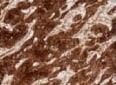
Detection of ISG15/UCRP in Human Endometrial Cancer.
ISG15/UCRP was detected in immersion fixed paraffin-embedded sections of Human Endometrial Cancer using Mouse Anti-Human ISG15/UCRP Monoclonal Antibody (Catalog # MAB4845) at 0.5 µg/mL for 1 hour at room temperature followed by incubation with the Anti-Mouse IgG VisUCyte™ HRP Polymer Antibody (Catalog # VC001). Before incubation with the primary antibody, tissue was subjected to heat-induced epitope retrieval using VisUCyte Antigen Retrieval Reagent-Basic (Catalog # VCTS021). Tissue was stained using DAB (brown) and counterstained with hematoxylin (blue). Specific staining was localized to cytoplasm in cancer cells. View our protocol for IHC Staining with VisUCyte HRP Polymer Detection Reagents.
Detection of ISG15/UCRP by Western Blot
ISG15 counteracts DV IFN-I evasion. (A) A549 WT and ISG15 KO were infected with 20 DV PFU. Cells were harvested at the indicated time points after infection (hpi) and cell lysates were analyzed by Western blot using STAT2 and Actin antibodies. (B–D) A549 WT and ISG15 KO immunofluorescence assay (IFA) 36 hpi for cellular IFIT3 and flavivirus E protein expression (D). Percentage of IFIT3 (B) and DV (C) positive cells per foci were quantified by ImageJ software and analyzed using unpaired t test with Welch’s correction when appropriate. Displayed images were acquired with a Leica DMI6000 B microscope. (E) A549 cells were infected with DV at MOI 0.01. At 36 hpi, cells were fixed, permeabilized and stained for flavivirus E protein. Cell lysates were analyzed by Western blot with the indicated antibodies before (upper panel) and after (bottom panel) cells were sorted by fluorescence-activated cell sorting (FACS) based on E protein expression. U, uninfected; B, bystander; I, infected. Error bars represent mean ± SD. Results are representative of two independent experiments. Statistical analyses were performed using Prism 8 (GraphPad Software). p values ***<0.001. Image collected and cropped by CiteAb from the following open publication (https://www.frontiersin.org/articles/10.3389/fimmu.2024.1331731/full), licensed under a CC-BY license. Not internally tested by R&D Systems.
Detection of ISG15/UCRP by Western Blot
PKR deletion does not affect the innate immune response or cell viability. A549 WT or PKR−/− cells infected with DENV4 (MOI 2) or ZIKV (MOI 3) and harvested at 24 h.p.i. for analysis. (A) Quantification by RT-qPCR of IFN beta, IFN lambda, ISG15, and TNF-alpha gene expression. Relative expression calculated by 2− delta delta Ct methods using mock cells as a reference. (B) Immunoblot analysis of cell extracts resolved in denaturing SDS-PAGE. Representative image of two independent experiments. (C) Flow cytometry analysis for quantification of living cells by staining with Zombie NIR viability dye. Mock WT cells set as 100% reference. In the column charts, bars represent the means ± standard error of the mean from three independent experiments. Statistical analysis was performed by paired t test comparing the two cell lineages under the same conditions. *P ≤ 0.05. ns/no markup, no statistical difference. Image collected and cropped by CiteAb from the following open publication (https://pubmed.ncbi.nlm.nih.gov/37732809), licensed under a CC-BY license. Not internally tested by R&D Systems.
Detection of ISG15/UCRP by Western Blot
ISGylation is not sufficient to restrict DV spread. (A) ISGylation profile of A549 WT and HERC5 KO cells by Western blot. Cells were primed with IFN alpha 2b (100 IU/ml) for 24 h and cell lysates were analyzed with an ISG15 antibody. (*) indicates antibody unspecific band. (B, C) A549 cells were infected with 20 DV PFUs. At 36 hpi cells were fixed, permeabilized and stained for the flavivirus E protein. DV relative foci area (B) and the number of infected cells per foci (C) quantified by ImageJ software and analyzed by one-way ANOVA. Images were acquired with an Olympus IX83 inverted microscope. Error bars represent mean ± SD. Results are representative of three or more independent experiments. Statistical analyses were performed using Prism 8 (GraphPad Software). p values ****<0.0001. Image collected and cropped by CiteAb from the following open publication (https://www.frontiersin.org/articles/10.3389/fimmu.2024.1331731/full), licensed under a CC-BY license. Not internally tested by R&D Systems.Applications for Human ISG15/UCRP Antibody (539442)
Application
Recommended Usage
Immunohistochemistry
8-25 µg/mL
Sample: Immersion fixed paraffin-embedded sections of human endometrial cancer tissue
Sample: Immersion fixed paraffin-embedded sections of human endometrial cancer tissue
Simple Western
5 µg/mL
Sample: MCF‑7 human breast cancer cell line and HT‑29 human colon adenocarcinoma cell line
Sample: MCF‑7 human breast cancer cell line and HT‑29 human colon adenocarcinoma cell line
Western Blot
0.5 µg/mL
Sample: MCF-7 human breast cancer cell line and HT-29 human colon adenocarcinoma cell line
Sample: MCF-7 human breast cancer cell line and HT-29 human colon adenocarcinoma cell line
Reviewed Applications
Read 1 review rated 5 using MAB4845 in the following applications:
Formulation, Preparation, and Storage
Purification
Protein A or G purified from hybridoma culture supernatant
Reconstitution
Reconstitute at 0.5 mg/mL in sterile PBS. For liquid material, refer to CoA for concentration.
Loading...
Formulation
Lyophilized from a 0.2 μm filtered solution in PBS with Trehalose. *Small pack size (SP) is supplied either lyophilized or as a 0.2 µm filtered solution in PBS.
Shipping
Lyophilized product is shipped at ambient temperature. Liquid small pack size (-SP) is shipped with polar packs. Upon receipt, store immediately at the temperature recommended below.
Stability & Storage
Use a manual defrost freezer and avoid repeated freeze-thaw cycles.
- 12 months from date of receipt, -20 to -70 °C as supplied.
- 1 month, 2 to 8 °C under sterile conditions after reconstitution.
- 6 months, -20 to -70 °C under sterile conditions after reconstitution.
Calculators
Background: ISG15/UCRP
Long Name
ISG15 Ubiquitin-like Modifier
Alternate Names
G1P2, IFI15, IP17, UCRP
Gene Symbol
ISG15
UniProt
Additional ISG15/UCRP Products
Product Documents for Human ISG15/UCRP Antibody (539442)
Certificate of Analysis
To download a Certificate of Analysis, please enter a lot or batch number in the search box below.
Note: Certificate of Analysis not available for kit components.
Product Specific Notices for Human ISG15/UCRP Antibody (539442)
For research use only
Related Research Areas
Citations for Human ISG15/UCRP Antibody (539442)
 Powered by Bioz
Customer Reviews for Human ISG15/UCRP Antibody (539442) (1)
5 out of 5
1 Customer Rating
Have you used Human ISG15/UCRP Antibody (539442)?
Submit a review and receive an Amazon gift card!
$25/€18/£15/$25CAN/¥2500 Yen for a review with an image
$10/€7/£6/$10CAN/¥1110 Yen for a review without an image
Submit a review
Customer Images
Showing
1
-
1 of
1 review
Showing All
Filter By:
-
Application: ImmunohistochemistrySample Tested: Cheek sectionSpecies: HumanVerified Customer | Posted 11/15/2021

There are no reviews that match your criteria.
Protocols
Find general support by application which include: protocols, troubleshooting, illustrated assays, videos and webinars.
- Antigen Retrieval Protocol (PIER)
- Antigen Retrieval for Frozen Sections Protocol
- Appropriate Fixation of IHC/ICC Samples
- Cellular Response to Hypoxia Protocols
- Chromogenic IHC Staining of Formalin-Fixed Paraffin-Embedded (FFPE) Tissue Protocol
- Chromogenic Immunohistochemistry Staining of Frozen Tissue
- ClariTSA™ Fluorophore Kits
- Detection & Visualization of Antibody Binding
- Fluorescent IHC Staining of Frozen Tissue Protocol
- Graphic Protocol for Heat-induced Epitope Retrieval
- Graphic Protocol for the Preparation and Fluorescent IHC Staining of Frozen Tissue Sections
- Graphic Protocol for the Preparation and Fluorescent IHC Staining of Paraffin-embedded Tissue Sections
- Graphic Protocol for the Preparation of Gelatin-coated Slides for Histological Tissue Sections
- IHC Sample Preparation (Frozen sections vs Paraffin)
- Immunofluorescent IHC Staining of Formalin-Fixed Paraffin-Embedded (FFPE) Tissue Protocol
- Immunohistochemistry (IHC) and Immunocytochemistry (ICC) Protocols
- Immunohistochemistry Frozen Troubleshooting
- Immunohistochemistry Paraffin Troubleshooting
- Preparing Samples for IHC/ICC Experiments
- Preventing Non-Specific Staining (Non-Specific Binding)
- Primary Antibody Selection & Optimization
- Protocol for Heat-Induced Epitope Retrieval (HIER)
- Protocol for Making a 4% Formaldehyde Solution in PBS
- Protocol for VisUCyte™ HRP Polymer Detection Reagent
- Protocol for the Preparation & Fixation of Cells on Coverslips
- Protocol for the Preparation and Chromogenic IHC Staining of Frozen Tissue Sections
- Protocol for the Preparation and Chromogenic IHC Staining of Frozen Tissue Sections - Graphic
- Protocol for the Preparation and Chromogenic IHC Staining of Paraffin-embedded Tissue Sections
- Protocol for the Preparation and Chromogenic IHC Staining of Paraffin-embedded Tissue Sections - Graphic
- Protocol for the Preparation and Fluorescent IHC Staining of Frozen Tissue Sections
- Protocol for the Preparation and Fluorescent IHC Staining of Paraffin-embedded Tissue Sections
- Protocol for the Preparation of Gelatin-coated Slides for Histological Tissue Sections
- R&D Systems Quality Control Western Blot Protocol
- TUNEL and Active Caspase-3 Detection by IHC/ICC Protocol
- The Importance of IHC/ICC Controls
- Troubleshooting Guide: Immunohistochemistry
- Troubleshooting Guide: Western Blot Figures
- Western Blot Conditions
- Western Blot Protocol
- Western Blot Protocol for Cell Lysates
- Western Blot Troubleshooting
- Western Blot Troubleshooting Guide
- View all Protocols, Troubleshooting, Illustrated assays and Webinars
Loading...