Human Kallikrein 3/PSA Antibody (181823)
R&D Systems | Catalog # MAB1344

Key Product Details
Species Reactivity
Validated:
Human
Cited:
Human
Applications
Validated:
Immunohistochemistry, Western Blot, Immunocytochemistry, Immunoprecipitation
Cited:
ELISA Microarray Development
Label
Unconjugated
Antibody Source
Monoclonal Mouse IgG2B Clone # 181823
Loading...
Product Specifications
Immunogen
Mouse myeloma cell line NS0-derived recombinant human Kallikrein 3/PSA
Ala18-Pro261
Accession # P07288
Ala18-Pro261
Accession # P07288
Specificity
Detects human Kallikrein 3/PSA in direct ELISAs and Western blots. In direct ELISAs, no cross-reactivity with recombinant human Kallikrein 5 or 11 is observed.
Clonality
Monoclonal
Host
Mouse
Isotype
IgG2B
Scientific Data Images for Human Kallikrein 3/PSA Antibody (181823)
Detection of Human Kallikrein 3/PSA by Western Blot.
Western blot shows lysates of human prostate tissue. PVDF membrane was probed with 2 µg/mL of Mouse Anti-Human Kallikrein 3/PSA Monoclonal Antibody (Catalog # MAB1344) followed by HRP-conjugated Anti-Mouse IgG Secondary Antibody (Catalog # HAF018). A specific band was detected for Kallikrein 3/PSA at approximately 30 kDa (as indicated). This experiment was conducted under reducing conditions and using Immunoblot Buffer Group 1.
Kallikrein 3/PSA in LNCaP and MCF-7 Human Cell Lines.
Kallikrein 3/PSA was detected in immersion fixed LNCaP human prostate cancer cell line (positive control, left panel) and MCF-7 human breast cancer cell line (negative control, right panel) using Mouse Anti-Human Kallikrein 3/PSA Monoclonal Antibody (Catalog # MAB1344) at 8 µg/mL for 3 hours at room temperature. Cells were stained using the NorthernLights™ 557-conjugated Anti-Mouse IgG Secondary Antibody (red; Catalog # NL007) and counterstained with DAPI (blue). Specific staining was localized to cytoplasm in LNCaP cells. View our protocol for Fluorescent ICC Staining of Cells on Coverslips.
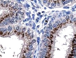
Kallikrein 3/PSA in Prostate Cancer.
Kallikrein 3/PSA was detected in immersion fixed paraffin-embedded sections of prostate cancer tissue using Mouse Anti-Human Kallikrein 3/PSA Monoclonal Antibody (Catalog # MAB1344) at 1.7 µg/mL overnight at 4 °C. Tissue was stained using the Anti-Mouse HRP-DAB Cell & Tissue Staining Kit (brown; Catalog # CTS002) and counterstained with hematoxylin (blue). Specific staining was localized to the apical plasma membrane. View our protocol for Chromogenic IHC Staining of Paraffin-embedded Tissue Sections.Applications for Human Kallikrein 3/PSA Antibody (181823)
Application
Recommended Usage
Immunocytochemistry
8-25 µg/mL
Sample: Immersion fixed LNCaP human prostate cancer cell line
Sample: Immersion fixed LNCaP human prostate cancer cell line
Immunohistochemistry
8-25 µg/mL
Sample: Immersion fixed paraffin-embedded prostate cancer tissue
Sample: Immersion fixed paraffin-embedded prostate cancer tissue
Immunoprecipitation
25 µg/mL
Sample: Conditioned cell culture medium spiked with Recombinant Human Kallikrein 3/PSA (Catalog # 1344-SE), see our available Western blot detection antibodies
Sample: Conditioned cell culture medium spiked with Recombinant Human Kallikrein 3/PSA (Catalog # 1344-SE), see our available Western blot detection antibodies
Western Blot
2 µg/mL
Sample: Human prostate tissue
Sample: Human prostate tissue
Reviewed Applications
Read 1 review rated 5 using MAB1344 in the following applications:
Formulation, Preparation, and Storage
Purification
Protein A or G purified from hybridoma culture supernatant
Reconstitution
Reconstitute at 0.5 mg/mL in sterile PBS. For liquid material, refer to CoA for concentration.
Loading...
Formulation
Lyophilized from a 0.2 μm filtered solution in PBS with Trehalose. *Small pack size (SP) is supplied either lyophilized or as a 0.2 µm filtered solution in PBS.
Shipping
Lyophilized product is shipped at ambient temperature. Liquid small pack size (-SP) is shipped with polar packs. Upon receipt, store immediately at the temperature recommended below.
Stability & Storage
Use a manual defrost freezer and avoid repeated freeze-thaw cycles.
- 12 months from date of receipt, -20 to -70 °C as supplied.
- 1 month, 2 to 8 °C under sterile conditions after reconstitution.
- 6 months, -20 to -70 °C under sterile conditions after reconstitution.
Calculators
Background: Kallikrein 3/PSA
References
- Yousef, G.M. and E.P. Diamandis (2001) Endocrine Rev. 22:184.
- Ward, A.M. et al. (2001) Ann. Clin. Biochem. 38:633.
- Jain, S. et al. (2002) Postgrad. Med. J. 78:646.
- Lilja H. (2003) Urology 62:270.
- Takayama, T.K. et al. (1997) J. Biol. Chem. 272:21582.
Alternate Names
KLK3, PSA
Entrez Gene IDs
354 (Human)
Gene Symbol
KLK3
UniProt
Additional Kallikrein 3/PSA Products
Product Documents for Human Kallikrein 3/PSA Antibody (181823)
Certificate of Analysis
To download a Certificate of Analysis, please enter a lot or batch number in the search box below.
Note: Certificate of Analysis not available for kit components.
Product Specific Notices for Human Kallikrein 3/PSA Antibody (181823)
For research use only
Citations for Human Kallikrein 3/PSA Antibody (181823)
 Powered by Bioz
Customer Reviews for Human Kallikrein 3/PSA Antibody (181823) (1)
5 out of 5
1 Customer Rating
Have you used Human Kallikrein 3/PSA Antibody (181823)?
Submit a review and receive an Amazon gift card!
$25/€18/£15/$25CAN/¥2500 Yen for a review with an image
$10/€7/£6/$10CAN/¥1110 Yen for a review without an image
Submit a review
Customer Images
Showing
1
-
1 of
1 review
Showing All
Filter By:
-
Application: ImmunohistochemistrySample Tested: Prostate cancerSpecies: HumanVerified Customer | Posted 03/11/2022

There are no reviews that match your criteria.
Protocols
Find general support by application which include: protocols, troubleshooting, illustrated assays, videos and webinars.
- Antigen Retrieval Protocol (PIER)
- Antigen Retrieval for Frozen Sections Protocol
- Appropriate Fixation of IHC/ICC Samples
- Cellular Response to Hypoxia Protocols
- Chromogenic IHC Staining of Formalin-Fixed Paraffin-Embedded (FFPE) Tissue Protocol
- Chromogenic Immunohistochemistry Staining of Frozen Tissue
- ClariTSA™ Fluorophore Kits
- Detection & Visualization of Antibody Binding
- Fluorescent IHC Staining of Frozen Tissue Protocol
- Graphic Protocol for Heat-induced Epitope Retrieval
- Graphic Protocol for the Preparation and Fluorescent IHC Staining of Frozen Tissue Sections
- Graphic Protocol for the Preparation and Fluorescent IHC Staining of Paraffin-embedded Tissue Sections
- Graphic Protocol for the Preparation of Gelatin-coated Slides for Histological Tissue Sections
- ICC Cell Smear Protocol for Suspension Cells
- ICC Immunocytochemistry Protocol Videos
- ICC for Adherent Cells
- IHC Sample Preparation (Frozen sections vs Paraffin)
- Immunocytochemistry (ICC) Protocol
- Immunocytochemistry Troubleshooting
- Immunofluorescence of Organoids Embedded in Cultrex Basement Membrane Extract
- Immunofluorescent IHC Staining of Formalin-Fixed Paraffin-Embedded (FFPE) Tissue Protocol
- Immunohistochemistry (IHC) and Immunocytochemistry (ICC) Protocols
- Immunohistochemistry Frozen Troubleshooting
- Immunohistochemistry Paraffin Troubleshooting
- Immunoprecipitation Protocol
- Preparing Samples for IHC/ICC Experiments
- Preventing Non-Specific Staining (Non-Specific Binding)
- Primary Antibody Selection & Optimization
- Protocol for Heat-Induced Epitope Retrieval (HIER)
- Protocol for Making a 4% Formaldehyde Solution in PBS
- Protocol for VisUCyte™ HRP Polymer Detection Reagent
- Protocol for the Fluorescent ICC Staining of Cell Smears - Graphic
- Protocol for the Fluorescent ICC Staining of Cultured Cells on Coverslips - Graphic
- Protocol for the Preparation & Fixation of Cells on Coverslips
- Protocol for the Preparation and Chromogenic IHC Staining of Frozen Tissue Sections
- Protocol for the Preparation and Chromogenic IHC Staining of Frozen Tissue Sections - Graphic
- Protocol for the Preparation and Chromogenic IHC Staining of Paraffin-embedded Tissue Sections
- Protocol for the Preparation and Chromogenic IHC Staining of Paraffin-embedded Tissue Sections - Graphic
- Protocol for the Preparation and Fluorescent ICC Staining of Cells on Coverslips
- Protocol for the Preparation and Fluorescent ICC Staining of Non-adherent Cells
- Protocol for the Preparation and Fluorescent ICC Staining of Stem Cells on Coverslips
- Protocol for the Preparation and Fluorescent IHC Staining of Frozen Tissue Sections
- Protocol for the Preparation and Fluorescent IHC Staining of Paraffin-embedded Tissue Sections
- Protocol for the Preparation of Gelatin-coated Slides for Histological Tissue Sections
- Protocol for the Preparation of a Cell Smear for Non-adherent Cell ICC - Graphic
- R&D Systems Quality Control Western Blot Protocol
- TUNEL and Active Caspase-3 Detection by IHC/ICC Protocol
- The Importance of IHC/ICC Controls
- Troubleshooting Guide: Immunohistochemistry
- Troubleshooting Guide: Western Blot Figures
- Western Blot Conditions
- Western Blot Protocol
- Western Blot Protocol for Cell Lysates
- Western Blot Troubleshooting
- Western Blot Troubleshooting Guide
- View all Protocols, Troubleshooting, Illustrated assays and Webinars
Loading...