
Key Product Details
Species Reactivity
Validated:
Human
Cited:
Human, Mouse
Applications
Validated:
Immunohistochemistry, Western Blot, Immunocytochemistry
Cited:
Western Blot
Label
Unconjugated
Antibody Source
Monoclonal Mouse IgG2A Clone # 919007
Loading...
Product Specifications
Immunogen
E. coli-derived recombinant human Lamin B1
Arg418-Ser582
Accession # P20700
Arg418-Ser582
Accession # P20700
Specificity
Detects human Lamin B1 in direct ELISAs.
Clonality
Monoclonal
Host
Mouse
Isotype
IgG2A
Scientific Data Images for Human Lamin B1 Antibody
Detection of Human Lamin B1 by Western Blot.
Western blot shows lysates of Jurkat human acute T cell leukemia cell line and MOLT-4 human acute lymphoblastic leukemia cell line. PVDF membrane was probed with 2 µg/mL of Mouse Anti-Human Lamin B1 Monoclonal Antibody (Catalog # MAB8525) followed by HRP-conjugated Anti-Mouse IgG Secondary Antibody (Catalog # HAF018). A specific band was detected for Lamin B1 at approximately 68 kDa (as indicated). This experiment was conducted under reducing conditions and using Immunoblot Buffer Group 1.
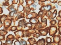
Lamin B1 in Human Liver.
Lamin B1 was detected in immersion fixed paraffin-embedded sections of human liver using Mouse Anti-Human Lamin B1 Monoclonal Antibody (Catalog # MAB8525) at 3 µg/mL for 1 hour at room temperature followed by incubation with the Anti-Mouse IgG VisUCyte™ HRP Polymer Antibody (Catalog # VC001). Tissue was stained using DAB (brown) and counterstained with hematoxylin (blue). Specific staining was localized to nuclear membrane. View our protocol for IHC Staining with VisUCyte HRP Polymer Detection Reagents.
Lamin B1 in HeLa Human Cell Line.
Lamin B1 was detected in immersion fixed HeLa human cervical epithelial carcinoma cell line using Mouse Anti-Human Lamin B1 Monoclonal Antibody (Catalog # MAB8525) at 8 µg/mL for 3 hours at room temperature. Cells were stained using the NorthernLights™ 557-conjugated Anti-Mouse IgG Secondary Antibody (red; Catalog # NL007) and counterstained with DAPI (blue). Specific staining was localized to nuclear membrane. View our protocol for Fluorescent ICC Staining of Cells on Coverslips.
Detection of Lamin B1 by Western Blot
LCZ696 suppresses LPS-induced activation of the TLR4/Myd88 pathway in HUVECs.Cells were stimulated with LPS (1 μg/mL) in the presence or absence of LCZ696 (20 μM) for 24 hours. (A). The levels of TLR4 and Myd88 were measured using Western blot; (B). Nuclear levels of NF-kappa B p65 (***P < 0.005 vs. control group; ##, ###P < 0.01, 0.005 vs. LPS group). Image collected and cropped by CiteAb from the following open publication (https://pubmed.ncbi.nlm.nih.gov/33839697), licensed under a CC-BY license. Not internally tested by R&D Systems.
Detection of Lamin B1 by Western Blot
Pristimerin suppresses endothelial cellular motilities required for angiogenesis through inactivating Shh/Gli1 signaling pathway. A Pristimerin suppressed nucleus translocation of Gli1 in endothelial cells. After incubating with Shh (100 ng/mL) and pristimerin (125, 250, and 500 nM) for 24 h, the HUVECs and HMEC-1 cells were stained with Gli1 antibody and Alexa Fluor 488 Donkey anti-Rabbit IgG. DAPI was used to label the nucleus. The cells were observed and photographed with a laser scanning confocal microscope. Scale bar: 20 μm. b, c Pristimerin inhibits Gli1 distribution in endothelial cells. The HUVECs and HMEC-1 cells were incubated with Shh and pristimerin for 24 h, and then lysed with a NE-PERTM Nucleus and Cytoplasmic Extraction Kit. After that, proteins were subjected to western blotting assay. beta -actin and Lamin B were set as loading control. The quantitative data were presented as ratios of Gli1/ beta -actin and Gli1/Lamin B. The representative blot and quantitative data were shown in B&C, respectively. d, e Pristimerin inactivated Shh/Gli1 and its downstream signaling pathway. The cells were treated with Shh and pristimerin for 24 h and lysed using RIPA buffer. Total proteins were subjected to western blotting assay. The representative blot and quantitative data were shown in d, e, respectively. The data are presented as mean ± SEM, n = 3. ***P < 0.001 compared with the control group; #P < 0.05 and ###P < 0.001 compared with the SHH-treated group. Image collected and cropped by CiteAb from the following open publication (https://pubmed.ncbi.nlm.nih.gov/32286274), licensed under a CC-BY license. Not internally tested by R&D Systems.Applications for Human Lamin B1 Antibody
Application
Recommended Usage
Immunocytochemistry
8-25 µg/mL
Sample: Immersion fixed HeLa human cervical epithelial carcinoma cell line
Sample: Immersion fixed HeLa human cervical epithelial carcinoma cell line
Immunohistochemistry
3-25 µg/mL
Sample: Immersion fixed paraffin-embedded sections of human liver
Sample: Immersion fixed paraffin-embedded sections of human liver
Western Blot
2 µg/mL
Sample: Jurkat human acute T cell leukemia cell line and MOLT‑4 human acute lymphoblastic leukemia cell line
Sample: Jurkat human acute T cell leukemia cell line and MOLT‑4 human acute lymphoblastic leukemia cell line
Reviewed Applications
Read 1 review rated 5 using MAB8525 in the following applications:
Formulation, Preparation, and Storage
Purification
Protein A or G purified from hybridoma culture supernatant
Reconstitution
Reconstitute at 0.5 mg/mL in sterile PBS. For liquid material, refer to CoA for concentration.
Loading...
Formulation
Lyophilized from a 0.2 μm filtered solution in PBS with Trehalose. *Small pack size (SP) is supplied either lyophilized or as a 0.2 µm filtered solution in PBS.
Shipping
Lyophilized product is shipped at ambient temperature. Liquid small pack size (-SP) is shipped with polar packs. Upon receipt, store immediately at the temperature recommended below.
Stability & Storage
Use a manual defrost freezer and avoid repeated freeze-thaw cycles.
- 12 months from date of receipt, -20 to -70 °C as supplied.
- 1 month, 2 to 8 °C under sterile conditions after reconstitution.
- 6 months, -20 to -70 °C under sterile conditions after reconstitution.
Calculators
Background: Lamin B1
Alternate Names
ADLD, LMN2, LMNB1
Gene Symbol
LMNB1
UniProt
Additional Lamin B1 Products
Product Documents for Human Lamin B1 Antibody
Certificate of Analysis
To download a Certificate of Analysis, please enter a lot or batch number in the search box below.
Note: Certificate of Analysis not available for kit components.
Product Specific Notices for Human Lamin B1 Antibody
For research use only
Related Research Areas
Citations for Human Lamin B1 Antibody
 Powered by Bioz
Customer Reviews for Human Lamin B1 Antibody (1)
5 out of 5
1 Customer Rating
Have you used Human Lamin B1 Antibody?
Submit a review and receive an Amazon gift card!
$25/€18/£15/$25CAN/¥2500 Yen for a review with an image
$10/€7/£6/$10CAN/¥1110 Yen for a review without an image
Submit a review
Customer Images
Showing
1
-
1 of
1 review
Showing All
Filter By:
-
Application: ImmunohistochemistrySample Tested: Pituitary gland tissueSpecies: HumanVerified Customer | Posted 02/10/2022

There are no reviews that match your criteria.
Protocols
Find general support by application which include: protocols, troubleshooting, illustrated assays, videos and webinars.
- Antigen Retrieval Protocol (PIER)
- Antigen Retrieval for Frozen Sections Protocol
- Appropriate Fixation of IHC/ICC Samples
- Cellular Response to Hypoxia Protocols
- Chromogenic IHC Staining of Formalin-Fixed Paraffin-Embedded (FFPE) Tissue Protocol
- Chromogenic Immunohistochemistry Staining of Frozen Tissue
- ClariTSA™ Fluorophore Kits
- Detection & Visualization of Antibody Binding
- Fluorescent IHC Staining of Frozen Tissue Protocol
- Graphic Protocol for Heat-induced Epitope Retrieval
- Graphic Protocol for the Preparation and Fluorescent IHC Staining of Frozen Tissue Sections
- Graphic Protocol for the Preparation and Fluorescent IHC Staining of Paraffin-embedded Tissue Sections
- Graphic Protocol for the Preparation of Gelatin-coated Slides for Histological Tissue Sections
- ICC Cell Smear Protocol for Suspension Cells
- ICC Immunocytochemistry Protocol Videos
- ICC for Adherent Cells
- IHC Sample Preparation (Frozen sections vs Paraffin)
- Immunocytochemistry (ICC) Protocol
- Immunocytochemistry Troubleshooting
- Immunofluorescence of Organoids Embedded in Cultrex Basement Membrane Extract
- Immunofluorescent IHC Staining of Formalin-Fixed Paraffin-Embedded (FFPE) Tissue Protocol
- Immunohistochemistry (IHC) and Immunocytochemistry (ICC) Protocols
- Immunohistochemistry Frozen Troubleshooting
- Immunohistochemistry Paraffin Troubleshooting
- Preparing Samples for IHC/ICC Experiments
- Preventing Non-Specific Staining (Non-Specific Binding)
- Primary Antibody Selection & Optimization
- Protocol for Heat-Induced Epitope Retrieval (HIER)
- Protocol for Making a 4% Formaldehyde Solution in PBS
- Protocol for VisUCyte™ HRP Polymer Detection Reagent
- Protocol for the Fluorescent ICC Staining of Cell Smears - Graphic
- Protocol for the Fluorescent ICC Staining of Cultured Cells on Coverslips - Graphic
- Protocol for the Preparation & Fixation of Cells on Coverslips
- Protocol for the Preparation and Chromogenic IHC Staining of Frozen Tissue Sections
- Protocol for the Preparation and Chromogenic IHC Staining of Frozen Tissue Sections - Graphic
- Protocol for the Preparation and Chromogenic IHC Staining of Paraffin-embedded Tissue Sections
- Protocol for the Preparation and Chromogenic IHC Staining of Paraffin-embedded Tissue Sections - Graphic
- Protocol for the Preparation and Fluorescent ICC Staining of Cells on Coverslips
- Protocol for the Preparation and Fluorescent ICC Staining of Non-adherent Cells
- Protocol for the Preparation and Fluorescent ICC Staining of Stem Cells on Coverslips
- Protocol for the Preparation and Fluorescent IHC Staining of Frozen Tissue Sections
- Protocol for the Preparation and Fluorescent IHC Staining of Paraffin-embedded Tissue Sections
- Protocol for the Preparation of Gelatin-coated Slides for Histological Tissue Sections
- Protocol for the Preparation of a Cell Smear for Non-adherent Cell ICC - Graphic
- R&D Systems Quality Control Western Blot Protocol
- TUNEL and Active Caspase-3 Detection by IHC/ICC Protocol
- The Importance of IHC/ICC Controls
- Troubleshooting Guide: Immunohistochemistry
- Troubleshooting Guide: Western Blot Figures
- Western Blot Conditions
- Western Blot Protocol
- Western Blot Protocol for Cell Lysates
- Western Blot Troubleshooting
- Western Blot Troubleshooting Guide
- View all Protocols, Troubleshooting, Illustrated assays and Webinars
Loading...