Matrix metalloproteinases are a family of zinc and calcium dependent endopeptidases with the combined ability to degrade all the components of the extracellular matrix. MMP-1 (interstitial collagenase), can degrade a broad range of substrates including types I, II, III, VII, VIII, and X collagens as well as casein, gelatin,
alpha -1 antitrypsin, myelin basic protein, L-Selectin, pro-TNF, IL-1 beta, IGFBP-3, IGFBP-5, pro-MMP-2, and pro-MMP-9. A significant role of MMP-1 is the degradation of fibrillar collagens in extracellular matrix remodeling, characterized by the cleavage of the interstitial collagen triple helix into ¾, ¼ fragments. However, as the list of substrates above illustrates, the role of MMP-1 is more diverse than originally envisaged, and may involve enzyme cascades, cytokine regulation, and cell surface molecule modulation. MMP-1 is expressed by fibroblasts, keratinocytes, endothelial cells, monocytes, and macrophages. Structurally, MMP-1 may be divided into several distinct domains; a pro-domain which is cleaved upon activation; a catalytic domain containing the zinc binding site; a short hinge region and a carboxyl terminal (hemopexin-like) domain.

Key Product Details
Validated by
Knockout/Knockdown
Species Reactivity
Validated:
Human
Cited:
Human
Applications
Validated:
Knockout Validated, Immunohistochemistry, Western Blot, ELISA Capture (Matched Antibody Pair), Simple Western
Cited:
Immunohistochemistry, Western Blot, ELISA Development, ELISA Microarray Development
Label
Unconjugated
Antibody Source
Polyclonal Goat IgG
Loading...
Product Specifications
Immunogen
Mouse myeloma cell line NS0-derived recombinant human MMP-1
Phe20-Asn469
Accession # P03956
Phe20-Asn469
Accession # P03956
Specificity
Detects human MMP-1 in ELISAs and Western blots. In sandwich immunoassays, less than 0.5% cross-reactivity with recombinant human (rh) MMP‑2, rhMMP-3, rhMMP-7, rhMMP-8, rhMMP-9, rhMMP-10, rhMMP-12, rhMMP-13, and rhMMP-16 is observed.
Clonality
Polyclonal
Host
Goat
Isotype
IgG
Scientific Data Images for Human MMP-1 Antibody
Detection of Human MMP‑1 by Western Blot.
Western blot shows lysates of PC-3 human prostate cancer cell line and HEK001 human epidermal keratinocyte cell line. PVDF membrane was probed with 1 µg/mL of Goat Anti-Human MMP-1 Antigen Affinity-purified Polyclonal Antibody (Catalog # AF901) followed by HRP-conjugated Anti-Goat IgG Secondary Antibody (Catalog # HAF017). A specific band was detected for MMP-1 at approximately 50 kDa (as indicated). This experiment was conducted under reducing conditions and using Immunoblot Buffer Group 1.
Detection of Human MMP‑1 by Simple WesternTM.
Left: Simple Western lane view shows lysates of MDA‑MB‑231 human breast cancer cell line, loaded at 0.1 mg/ml. A specific band was detected for MMP‑1 at approximately 55 kDa (as indicated) using both 10 µg/ml and 50 µg/ml of Goat Anti-Human MMP‑1 Antigen Affinity-purified Polyclonal Antibody (Catalog # AF901) followed by HRP-conjugated Donkey Anti-Goat Secondary Antibody (Catalog # 043-522-2). This experiment was conducted under reducing conditions and using the 12-230kDa separation system. Right: Simple Western electropherogram showing the same Goat Anti-Human MMP‑1 Antigen Affinity-purified Polyclonal Antibody (Catalog # AF901) tested at 10 µg/ml (blue line) and 50 µg/ml (green line) in the MDA‑MB‑231 human breast cancer cell line.
MMP‑1 in Human Ovarian Cancer Tissue.
MMP-1 was detected in immersion fixed paraffin-embedded sections of human ovarian cancer tissue using 15 µg/mL Goat Anti-Human MMP-1 Antigen Affinity-purified Polyclonal Antibody (Catalog # AF901) overnight at 4 °C. Tissue was stained with the Anti-Goat HRP-DAB Cell & Tissue Staining Kit (brown; Catalog # CTS008) and counterstained with hematoxylin (blue). View our protocol for Chromogenic IHC Staining of Paraffin-embedded Tissue Sections.
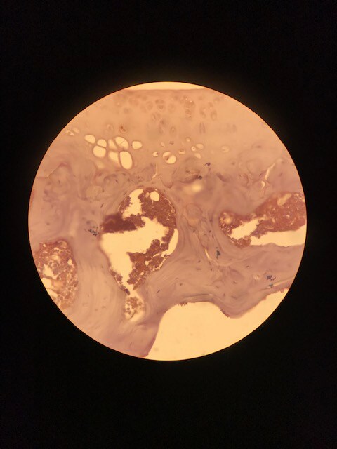
MMP‑1 in Human Prostate Tissue.
MMP-1 was detected in immersion fixed paraffin-embedded sections of human prostate tissue using Goat Anti-Human MMP-1 Antigen Affinity-purified Polyclonal Antibody (Catalog # AF901) at 10 µg/mL overnight at 4 °C. Tissue was stained using the Anti-Goat HRP-DAB Cell & Tissue Staining Kit (brown; Catalog # CTS008) and counterstained with hematoxylin (blue). View our protocol for Chromogenic IHC Staining of Paraffin-embedded Tissue Sections.
Western Blot Shows Human MMP-1 Specificity by Using Knockout Cell Line.
Western blot shows lysates of PC-3 human prostate cancer parental cell line and MMP-1 knockout PC-3 cell line (KO). PVDF membrane was probed with 1 µg/mL of Goat Anti-Human MMP-1 Antigen Affinity-purified Polyclonal Antibody (Catalog # AF901) followed by HRP-conjugated Anti-Goat IgG Secondary Antibody (Catalog # HAF017). A specific band was detected for MMP-1 at approximately 50 kDa (as indicated) in the parental PC-3 cell line, but is not detectable in knockout PC-3 cell line. GAPDH (Catalog # AF5718) is shown as a loading control. This experiment was conducted under reducing conditions and using Immunoblot Buffer Group 1.Applications for Human MMP-1 Antibody
Application
Recommended Usage
Immunohistochemistry
5-15 µg/mL
Sample: Immersion fixed paraffin-embedded sections of human ovarian cancer tissue and immersion fixed paraffin-embedded sections of human prostate
Sample: Immersion fixed paraffin-embedded sections of human ovarian cancer tissue and immersion fixed paraffin-embedded sections of human prostate
Knockout Validated
MMP-1 is specifically detected in PC‑3 human prostate cancer parental cell line but is not detectable in MMP-1 knockout PC‑3 cell line.
Simple Western
10-50 µg/mL
Sample: MDA-MB-231 human breast cancer cell line
Sample: MDA-MB-231 human breast cancer cell line
Western Blot
1 µg/mL
Sample: PC‑3 human prostate cancer cell line and HEK001 human epidermal keratinocyte cell line
Sample: PC‑3 human prostate cancer cell line and HEK001 human epidermal keratinocyte cell line
Human MMP-1 Sandwich Immunoassay
Please Note: Optimal dilutions of this antibody should be experimentally determined.
Reviewed Applications
Read 2 reviews rated 4 using AF901 in the following applications:
Formulation, Preparation, and Storage
Purification
Antigen Affinity-purified
Reconstitution
Reconstitute at 0.2 mg/mL in sterile PBS. For liquid material, refer to CoA for concentration.
Loading...
Formulation
Lyophilized from a 0.2 μm filtered solution in PBS with Trehalose. See Certificate of Analysis for details.
*Small pack size (-SP) is supplied either lyophilized or as a 0.2 µm filtered solution in PBS.
*Small pack size (-SP) is supplied either lyophilized or as a 0.2 µm filtered solution in PBS.
Shipping
Lyophilized product is shipped at ambient temperature. Liquid small pack size (-SP) is shipped with polar packs. Upon receipt, store immediately at the temperature recommended below.
Stability & Storage
Use a manual defrost freezer and avoid repeated freeze-thaw cycles.
- 12 months from date of receipt, -20 to -70 °C as supplied.
- 1 month, 2 to 8 °C under sterile conditions after reconstitution.
- 6 months, -20 to -70 °C under sterile conditions after reconstitution.
Calculators
Background: MMP-1
References
- Cawston, T.E. (2004) in Interstitial Collagenase. Barrett, A.J. et al. (eds): Handbook of Proteolytic Enzymes, San Diego: Academic Press, p. 472.
Long Name
Matrix Metalloproteinase 1
Alternate Names
MMP1
Gene Symbol
MMP1
UniProt
Additional MMP-1 Products
Product Documents for Human MMP-1 Antibody
Certificate of Analysis
To download a Certificate of Analysis, please enter a lot or batch number in the search box below.
Note: Certificate of Analysis not available for kit components.
Product Specific Notices for Human MMP-1 Antibody
For research use only
Related Research Areas
Citations for Human MMP-1 Antibody
 Powered by Bioz
Customer Reviews for Human MMP-1 Antibody (2)
4 out of 5
2 Customer Ratings
Have you used Human MMP-1 Antibody?
Submit a review and receive an Amazon gift card!
$25/€18/£15/$25CAN/¥2500 Yen for a review with an image
$10/€7/£6/$10CAN/¥1110 Yen for a review without an image
Submit a review
Customer Images
Showing
1
-
2 of
2 reviews
Showing All
Filter By:
-
Application: ImmunohistochemistrySample Tested: Cartilage tissue and Bone ExtractsSpecies: HumanVerified Customer | Posted 07/12/2019These samples were IHC-P, and the antibody was diluted 1:100 in BSA blocking solution. We used a HRP conjugate.

-
Application: ELISASample Tested: Serum and PlasmaSpecies: HumanVerified Customer | Posted 11/09/2017The antibody AF901 was used as both the capture and detection molecule in an ELISA to measure MMP-1 in human plasma samples.
There are no reviews that match your criteria.
Protocols
Find general support by application which include: protocols, troubleshooting, illustrated assays, videos and webinars.
- Antigen Retrieval Protocol (PIER)
- Antigen Retrieval for Frozen Sections Protocol
- Appropriate Fixation of IHC/ICC Samples
- Cellular Response to Hypoxia Protocols
- Chromogenic IHC Staining of Formalin-Fixed Paraffin-Embedded (FFPE) Tissue Protocol
- Chromogenic Immunohistochemistry Staining of Frozen Tissue
- ClariTSA™ Fluorophore Kits
- Detection & Visualization of Antibody Binding
- Fluorescent IHC Staining of Frozen Tissue Protocol
- Graphic Protocol for Heat-induced Epitope Retrieval
- Graphic Protocol for the Preparation and Fluorescent IHC Staining of Frozen Tissue Sections
- Graphic Protocol for the Preparation and Fluorescent IHC Staining of Paraffin-embedded Tissue Sections
- Graphic Protocol for the Preparation of Gelatin-coated Slides for Histological Tissue Sections
- IHC Sample Preparation (Frozen sections vs Paraffin)
- Immunofluorescent IHC Staining of Formalin-Fixed Paraffin-Embedded (FFPE) Tissue Protocol
- Immunohistochemistry (IHC) and Immunocytochemistry (ICC) Protocols
- Immunohistochemistry Frozen Troubleshooting
- Immunohistochemistry Paraffin Troubleshooting
- Preparing Samples for IHC/ICC Experiments
- Preventing Non-Specific Staining (Non-Specific Binding)
- Primary Antibody Selection & Optimization
- Protocol for Heat-Induced Epitope Retrieval (HIER)
- Protocol for Making a 4% Formaldehyde Solution in PBS
- Protocol for VisUCyte™ HRP Polymer Detection Reagent
- Protocol for the Preparation & Fixation of Cells on Coverslips
- Protocol for the Preparation and Chromogenic IHC Staining of Frozen Tissue Sections
- Protocol for the Preparation and Chromogenic IHC Staining of Frozen Tissue Sections - Graphic
- Protocol for the Preparation and Chromogenic IHC Staining of Paraffin-embedded Tissue Sections
- Protocol for the Preparation and Chromogenic IHC Staining of Paraffin-embedded Tissue Sections - Graphic
- Protocol for the Preparation and Fluorescent IHC Staining of Frozen Tissue Sections
- Protocol for the Preparation and Fluorescent IHC Staining of Paraffin-embedded Tissue Sections
- Protocol for the Preparation of Gelatin-coated Slides for Histological Tissue Sections
- R&D Systems Quality Control Western Blot Protocol
- TUNEL and Active Caspase-3 Detection by IHC/ICC Protocol
- The Importance of IHC/ICC Controls
- Troubleshooting Guide: Immunohistochemistry
- Troubleshooting Guide: Western Blot Figures
- Western Blot Conditions
- Western Blot Protocol
- Western Blot Protocol for Cell Lysates
- Western Blot Troubleshooting
- Western Blot Troubleshooting Guide
- View all Protocols, Troubleshooting, Illustrated assays and Webinars
Loading...