EphB2, also known as Cek5, Nuk, Erk, Qek2, Tyro5, Sek3, Hek5, and Drt (1), is a member of the Eph receptor family which binds members of the ephrin ligand family. There are two classes of receptors, designated A and B. Both the A and B class receptors have an extracellular region consisting of a globular domain, a cysteine-rich domain, and two fibronectin type III domains. This is followed by the transmembrane region and the cytoplasmic region. The cytoplasmic region contains a juxtamembrane motif with two tyrosine residues which are the major autophosphorylation sites, a kinase domain, and a conserved sterile alpha motif (SAM) in the carboxy tail which contains one conserved tyrosine residue. Activation of kinase activity occurs after ligand recognition and binding. EphB2 has been shown to bind ephrin-B1, ephrin-B2, and ephrin-B3 (2, 3). The extracellular domains of human and mouse EphB2 share 99% amino acid identity. Only membrane-bound or
Fc‑clustered ligands are capable of activating the receptor in vitro. Soluble monomeric ligands bind the receptor but do not induce receptor autophosphorylation and activation (2). In vivo, the ligands and receptors display reciprocal expression (3). It has been found that nearly all the receptors and ligands are expressed in developing and adult neural tissue (3). The ephrin/Eph families also appear to play a role in angiogenesis (3).

Key Product Details
Validated by
Biological Validation
Species Reactivity
Validated:
Human, Mouse
Cited:
Human, Mouse, Rat, Frog - Xenopus (African Clawed Frog), Transgenic Mouse
Applications
Validated:
Immunohistochemistry, Western Blot, Flow Cytometry, Immunocytochemistry, Simple Western, CyTOF-ready
Cited:
Immunohistochemistry, Immunohistochemistry-Paraffin, Immunohistochemistry-Frozen, Western Blot, Neutralization, Immunofluorescence, Immunocytochemistry, Simple Western, Immunoprecipitation, Proximity Ligation Assay, In vivo assay, Functional Assay
Label
Unconjugated
Antibody Source
Polyclonal Goat IgG
Loading...
Product Specifications
Immunogen
Mouse myeloma cell line NS0-derived recombinant mouse EphB2
Val27-Lys548
Accession # P54763
Val27-Lys548
Accession # P54763
Specificity
Detects mouse and human EphB2 in direct ELISAs and Western blots. In Western blots, approximately 5% cross-reactivity with recombinant rat (rr) EphB1, recombinant mouse (rm) EphA8, rmEphA6, rmEphB6, rmEphA3, rmEphA4, rmEphA7, rrEphA5, recombinant human EphA1, rmEphA2 and rmEphB3 is observed.
Clonality
Polyclonal
Host
Goat
Isotype
IgG
Scientific Data Images for EphB2 Antibody
Detection of Recombinant Human and Mouse EphB2 by Western Blot.
Western blot shows 25 ng of Recombinant Human EphB2 Fc Chimera (Catalog # 5189-B2) and Recombinant Mouse EphB2 Fc Chimera (Catalog # 467-B2). PVDF Membrane was probed with 0.1 µg/mL of Goat Anti-Human/Mouse EphB2 Antigen Affinity-purified Polyclonal Antibody (Catalog # AF467) followed by HRP-conjugated Anti-Goat IgG Secondary Antibody (Catalog # HAF109). A specific band was detected for EphB2 at approximately 120 kDa (as indicated). This experiment was conducted under reducing conditions and using Immunoblot Buffer Group 3.
Detection of EphB2 in COLO 205 Human Cell Line by Flow Cytometry.
COLO 205 human colorectal adenocarcinoma cell line was stained with Goat Anti-Human/Mouse EphB2 Antigen Affinity-purified Polyclonal Antibody (Catalog # AF467, filled histogram) or isotype control antibody (Catalog # AB-108-C, open histogram), followed by Phycoerythrin-conjugated Anti-Goat IgG Secondary Antibody (Catalog # F0107). View our protocol for Staining Membrane-associated Proteins.
Detection of EphB2 in D3 Mouse Cell Line by Flow Cytometry.
D3 mouse embryonic stem cell line was stained with Goat Anti-Human/Mouse EphB2 Antigen Affinity-purified Polyclonal Antibody (Catalog # AF467, filled histogram) or isotype control antibody (Catalog # AB-108-C, open histogram), followed by Phycoerythrin-conjugated Anti-Goat IgG Secondary Antibody (Catalog # F0107). View our protocol for Staining Membrane-associated Proteins.
EphB2 in MBA‑MB‑468 Human Cell Line.
EphB2 was detected in immersion fixed MBA-MB-468 human breast cancer cell line using Goat Anti-Human/Mouse EphB2 Antigen Affinity-purified Polyclonal Antibody (Catalog # AF467) at 5 µg/mL for 3 hours at room temperature. Cells were stained using the NorthernLights™ 557-conjugated Anti-Goat IgG Secondary Antibody (red; Catalog # NL001) and counterstained with DAPI (blue). Specific staining was localized to cytoplasm. View our protocol for Fluorescent ICC Staining of Cells on Coverslips.
EphB2 in Embryonic Mouse Brain.
EphB2 was detected in immersion fixed frozen sections of embryonic mouse brain (15 d.p.c.) using 15 µg/mL Goat Anti-Mouse EphB2 Antigen Affinity-purified Polyclonal Antibody (Catalog # AF467) overnight at 4 °C. Tissue was stained with the NorthernLights™ 557-conjugated Anti-Goat IgG Secondary Antibody (red; Catalog # NL001) and counterstained (green). View our protocol for Fluorescent IHC Staining of Frozen Tissue Sections.
EphB2 in Human Esophageal Squamous Cell Carcinoma.
EphB2 was detected in immersion fixed paraffin-embedded sections of human esophageal squamous cell carcinoma using Goat Anti-Human/Mouse EphB2 Antigen Affinity-purified Polyclonal Antibody (Catalog # AF467) at 3 µg/mL for 1 hour at room temperature followed by incubation with the Anti-Goat IgG VisUCyte™ HRP Polymer Antibody (Catalog # VC004). Tissue was stained using DAB (brown) and counterstained with hematoxylin (blue). Specific staining was localized to cytoplasm in cancer cells. View our protocol for IHC Staining with VisUCyte HRP Polymer Detection Reagents.
EphB2 in Mouse Embryo.
EphB2 was detected in immersion fixed frozen sections of mouse embryo (13 d.p.c.) using Goat Anti-Human/Mouse EphB2 Antigen Affinity-purified Polyclonal Antibody (Catalog # AF467) at 1.7 µg/mL for 1 hour at room temperature followed by incubation with the Anti-Goat IgG VisUCyte™ HRP Polymer Antibody (Catalog # VC004). Tissue was stained using DAB (brown) and counterstained with hematoxylin (blue). Specific staining was localized to developing brain. View our protocol for IHC Staining with VisUCyte HRP Polymer Detection Reagents.
Detection of Human EphB2 by Simple WesternTM.
Simple Western lane view shows lysates of Exosome Standards (COLO1) (NBP2-49845) and COLO 205 human colorectal adenocarcinoma cell line, loaded at 0.5 mg/ml. A specific band was detected for EphB2 at approximately 135 kDa (as indicated) using 1 µg/ml of Goat Anti-Human/Mouse EphB2 Antigen Affinity-purified Polyclonal Antibody (Catalog # AF467) followed by HRP-conjugated Donkey Anti-Goat Secondary Antibody (Catalog # 042-206). This experiment was conducted under reducing conditions and using the 12-230kDa separation system.
Detection of Human EphB2 by Simple WesternTM.
Simple Western lane view shows lysates of COLO 205 human colorectal adenocarcinoma cell line and MBA-MB-468 human breast cancer cell line, loaded at 0.2 mg/mL. A specific band was detected for EphB2 at approximately 139 and 146 kDa (as indicated) using 20 µg/mL of Goat Anti-Human/Mouse EphB2 Antigen Affinity-purified Polyclonal Antibody (Catalog # AF467) followed by 1:50 dilution of HRP-conjugated Anti-Goat IgG Secondary Antibody (Catalog # HAF109). This experiment was conducted under reducing conditions and using the 12-230 kDa separation system.
Detection of Human EphB2 by Immunocytochemistry/Immunofluorescence
Colorectal tumours maintain tissue organisation similar to normal colon.Detection of EPHB2 (green) and ERBB3 (red, A and B) by co-immunofluorescence in normal colon (A) and colorectal cancer (B) (DAPI, blue). Scale bar, 50μm. Image collected and cropped by CiteAb from the following publication (https://pubmed.ncbi.nlm.nih.gov/26367378), licensed under a CC-BY license. Not internally tested by R&D Systems.
Detection of Mouse EphB2 by Immunocytochemistry/Immunofluorescence
Expression of EphB2 increases and is activated on HSCs after chronic CCl4 -induced liver injury. (a) Isolated cell fractions from livers of mice subjected to chronic CCl4 injections were analyzed for EphB2, Ephrin-B1, Ephrin-B2 and Ephrin-B3 mRNA levels using RT-qPCR. Results are shown as fold change compared to liver cell fractions obtained from vehicle-treated controls. Error bars represent mean ± SEM.; n = 6 animals; CD11b = macrophages, LSEC = Liver sinusoidal endothelial cells, HEP = Hepatocytes and HSCs = Hepatic stellate cells. (b) OCT liver sections from C57BL/6 J mice chronically injected with CCl4 or vehicle (oil) controls were stained with EphB2 (red), alpha SMA (green) and DAPI/DNA (blue) and analyzed using confocal microscopy. Scale bar = 100 µm, “C” denotes the central vein. All images are representative of 5 mice per group. (c) OCT liver sections from C57BL/6 J mice chronically injected with CCl4 or vehicle controls were stained with phospho-EphB1/EphB2-Y594 (red), PDGFR beta (green) and DAPI/DNA (blue) and analyzed using confocal microscopy. Scale bar = 50 µm. All images are representative of 5 mice per group. Image collected and cropped by CiteAb from the following publication (https://pubmed.ncbi.nlm.nih.gov/29416088), licensed under a CC-BY license. Not internally tested by R&D Systems.
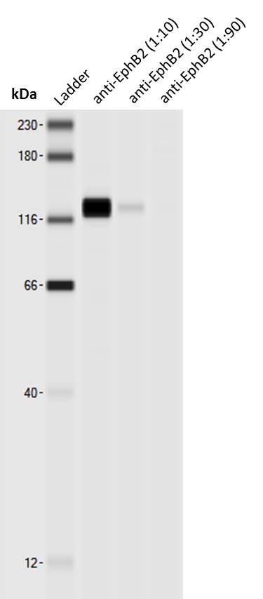
Detection of Human EphB2 by Western Blot
Increased cell-cell adhesion within one cell population is required for the formation of tightly packed cell clusters.A) Simulation of cell-cell segregation using the same adhesion term in both cell types (Aeph = Aephrin = 100, left panel) vs. increased adhesion only in the green cell population (Aeph = 110, Aephrin = 100, right panel). Both simulations started with the same number of Eph (green) and ephrin (black) expressing cells. In the “Equal adhesion” case, an ‘Islands-in-a-sea’ pattern is less apparent. B) Representative images from segregation assays of unlabelled ephrin-B1 cells co-cultured with Cell Tracker-green labelled (green staining) EphB2 cells, without (left) or with E-cadherin-cherry expression (red staining, right); scale bar, 75 µm. C) Quantitation of cell densities in the cell clusters shown in B (n = 10). D) Western blot analysis of lysates from parental and E-cadherin-cherry-transduced cells, using the indicated antibodies. Image collected and cropped by CiteAb from the following publication (https://dx.plos.org/10.1371/journal.pone.0111803), licensed under a CC-BY license. Not internally tested by R&D Systems.Applications for EphB2 Antibody
Application
Recommended Usage
CyTOF-ready
Ready to be labeled using established conjugation methods. No BSA or other carrier proteins that could interfere with conjugation.
Flow Cytometry
0.25 µg/106 cells
Sample: COLO 205 human colorectal adenocarcinoma cell line and D3 mouse embryonic stem cell line
Sample: COLO 205 human colorectal adenocarcinoma cell line and D3 mouse embryonic stem cell line
Immunocytochemistry
5-15 µg/mL
Sample: Immersion fixed MDA-MB-468 human breast cancer cell line
Sample: Immersion fixed MDA-MB-468 human breast cancer cell line
Immunohistochemistry
1-15 µg/mL
Sample: Immersion fixed frozen sections of embryonic mouse brain (15 d.p.c.), immersion fixed paraffin-embedded sections of human esophageal squamous cell carcinoma, and immersion fixed frozen sections of mouse embryo (13 d.p.c.)
Sample: Immersion fixed frozen sections of embryonic mouse brain (15 d.p.c.), immersion fixed paraffin-embedded sections of human esophageal squamous cell carcinoma, and immersion fixed frozen sections of mouse embryo (13 d.p.c.)
Simple Western
1-20 µg/mL
Sample: Exosome Standards (COLO1) (Catalog # NBP2-49845), COLO 205 human colorectal adenocarcinoma cell line and MBA‑MB‑468 human breast cancer cell line
Sample: Exosome Standards (COLO1) (Catalog # NBP2-49845), COLO 205 human colorectal adenocarcinoma cell line and MBA‑MB‑468 human breast cancer cell line
Western Blot
0.1 µg/mL
Sample: Recombinant Human EphB2 Fc Chimera (Catalog # 5189-B2) and Recombinant Mouse EphB2 Fc Chimera (Catalog # 467-B2).
Sample: Recombinant Human EphB2 Fc Chimera (Catalog # 5189-B2) and Recombinant Mouse EphB2 Fc Chimera (Catalog # 467-B2).
Reviewed Applications
Read 9 reviews rated 4.3 using AF467 in the following applications:
Flow Cytometry Panel Builder
Bio-Techne Knows Flow Cytometry
Save time and reduce costly mistakes by quickly finding compatible reagents using the Panel Builder Tool.
Advanced Features
- Spectra Viewer - Custom analysis of spectra from multiple fluorochromes
- Spillover Popups - Visualize the spectra of individual fluorochromes
- Antigen Density Selector - Match fluorochrome brightness with antigen density

Formulation, Preparation, and Storage
Purification
Antigen Affinity-purified
Reconstitution
Reconstitute at 0.2 mg/mL in sterile PBS. For liquid material, refer to CoA for concentration.
Loading...
Formulation
Lyophilized from a 0.2 μm filtered solution in PBS with Trehalose. See Certificate of Analysis for details.
*Small pack size (-SP) is supplied either lyophilized or as a 0.2 µm filtered solution in PBS.
*Small pack size (-SP) is supplied either lyophilized or as a 0.2 µm filtered solution in PBS.
Shipping
Lyophilized product is shipped at ambient temperature. Liquid small pack size (-SP) is shipped with polar packs. Upon receipt, store immediately at the temperature recommended below.
Stability & Storage
Use a manual defrost freezer and avoid repeated freeze-thaw cycles.
- 12 months from date of receipt, -20 to -70 °C as supplied.
- 1 month, 2 to 8 °C under sterile conditions after reconstitution.
- 6 months, -20 to -70 °C under sterile conditions after reconstitution.
Calculators
Background: EphB2
References
- Eph Nomenclature Committee [letter] (1997) Cell 90:403.
- Flanagan, J.G. and P. Vanderhaeghen (1998) Annu. Rev. Neurosci. 21:309.
- Pasquale, E.B. (1997) Curr. Opin. Cell Biol. 9:608.
Long Name
Eph Receptor B2
Alternate Names
Cek5, Drt, Erk, Hek5, Nuk, Qek2, Sek3, Tyro5
Gene Symbol
EPHB2
UniProt
Additional EphB2 Products
Product Documents for EphB2 Antibody
Certificate of Analysis
To download a Certificate of Analysis, please enter a lot or batch number in the search box below.
Note: Certificate of Analysis not available for kit components.
Product Specific Notices for EphB2 Antibody
For research use only
Citations for EphB2 Antibody
 Powered by Bioz
Customer Reviews for EphB2 Antibody (9)
4.3 out of 5
9 Customer Ratings
Have you used EphB2 Antibody?
Submit a review and receive an Amazon gift card!
$25/€18/£15/$25CAN/¥2500 Yen for a review with an image
$10/€7/£6/$10CAN/¥1110 Yen for a review without an image
Submit a review
Customer Images

Showing
1
-
5 of
9 reviews
Showing All
Filter By:
-
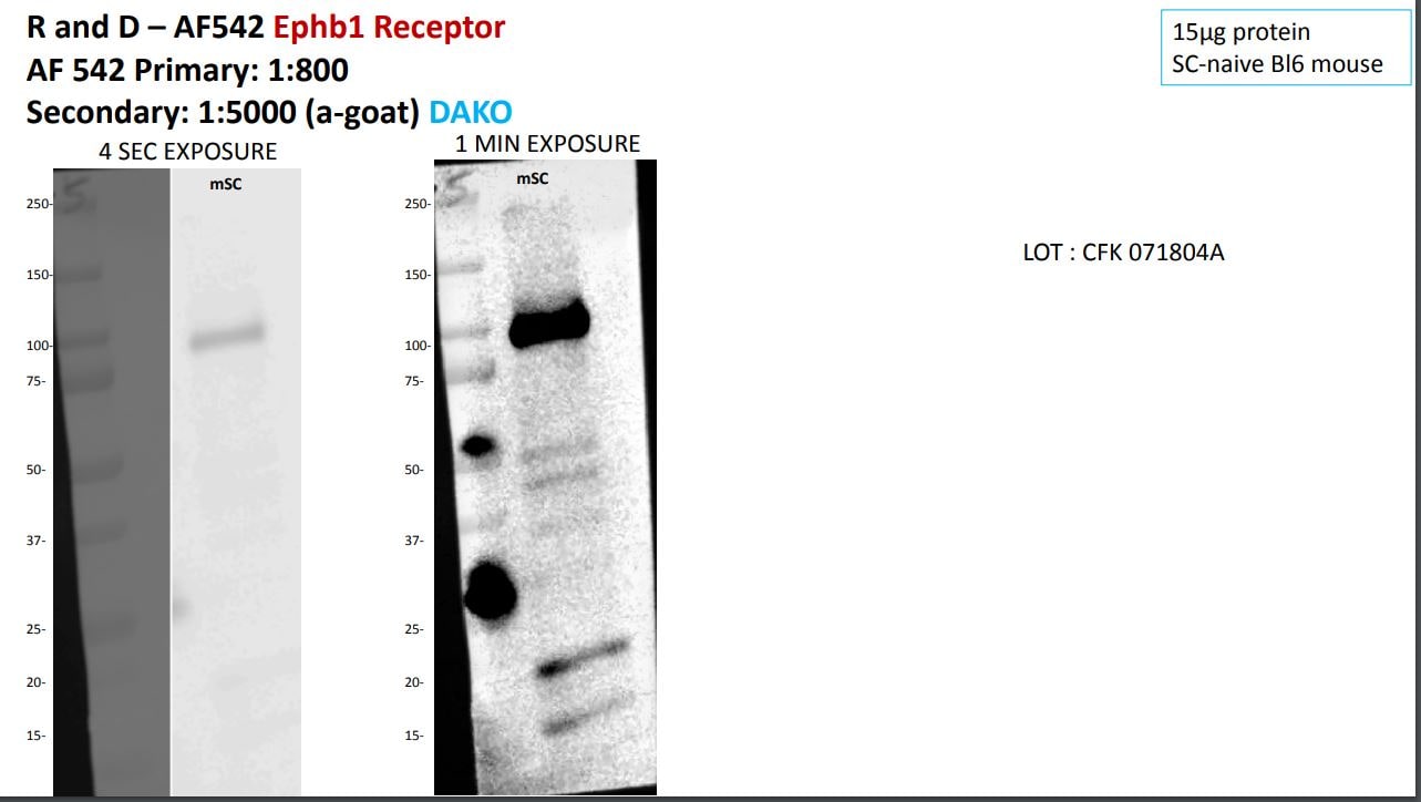
Application: Western BlotSample Tested: brain and spinal cordSpecies: MouseVerified Customer | Posted 07/05/2018Lane1: Precision Plus Protein™ Dual Color Standards7ul Lane2:Ephb1 receptor (SC tissue from naive C57BL6j mouse)Blot Loading • ANTIBODY: Human/Mouse EphB2Antibody cat noAF542 Lane1: Precision Plus Protein™ Dual Color Standards7ul Lane2:Ephb1 receptor (SC tissue from naive C57BL6j mouse) Western blot experiment details Gel:4-20% SDS page Tranfer apparatus: Trans-Blot® Turbo™ Transfer System Membrane: midi size nitrocellulose Time: 25min at 75-125V Primary ab:Goat –rat EphB1 Ab 1mg for 20h Secondary ab: rabbit a-goat 1:5,000 for 1h Substrate: HRP for 15min

-
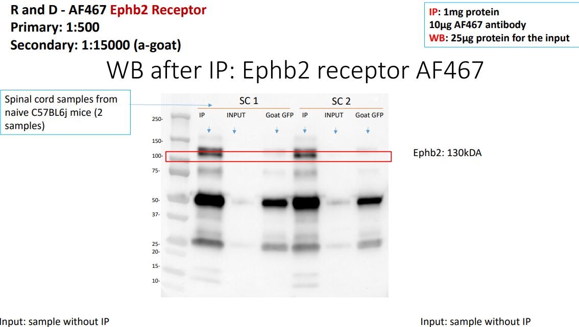
Application: ImmunoprecipitationSample Tested: brain and spinal cordSpecies: MouseVerified Customer | Posted 04/16/2018Blot Loading • ANTIBODY: Human/Mouse EphB2Antibody cat noAF467 Lane1: PreĐision Plus Protein™ Dual Color Standards8ul Lane2:Ephb2 receptors IP 1mg protein (SC tissue) Lane3:Ephb2 receptors no IP 25ug (SC tissue) Lane4:Goat GFP IP (negative control instead of goat IgG)1mg (SC tissue) Lane5:Ephb2 receptors IP 1mg (SC tissue) Lane6:Ephb2 receptors no IP 25ug (SC tissue) Lane7:Goat GFP IP (negative control instead of goat IgG)1mg (SC tissue) Western blot experiment details Gel:4-20% SDS page Tranfer apparatus: Trans-Blot® Turďo™ Transfer System Membrane: midi size nitrocellulose Time: 25min at 75-125V Primary ab:Goat –m/h EphB2 Ab 1mg for 20h Secondary ab: donkey a-goat 1:15,000 for 1h Substrate: HRP for 15min
-
Application: Western BlotSample Tested: Mouse iPSSpecies: MouseVerified Customer | Posted 01/17/2018
-
Application: Simple WesternSample Tested: Adult lungSpecies: MouseVerified Customer | Posted 01/12/2018
-
Application: Western BlotSample Tested: Embryonic stem cellsSpecies: HumanVerified Customer | Posted 12/21/2017
-
Application: Western BlotSample Tested: Lung tissueSpecies: MouseVerified Customer | Posted 11/10/2017
-
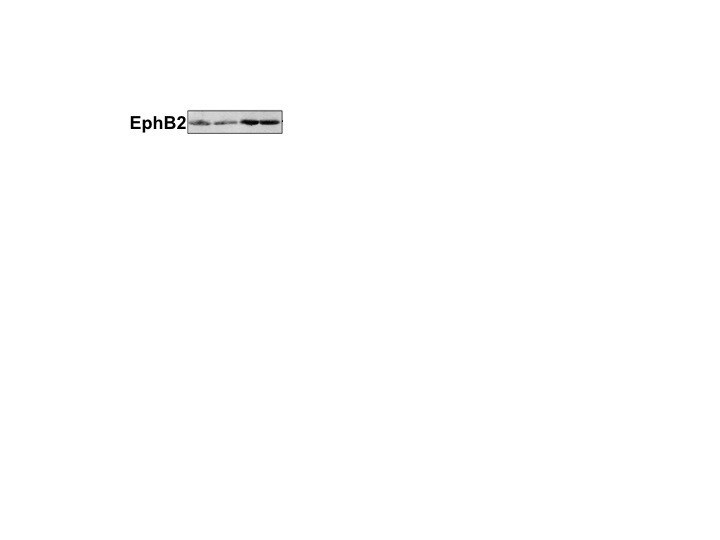
Application: Simple WesternSample Tested: mouse brain lysateSpecies: MouseVerified Customer | Posted 07/12/2017Simple Western: anti-EphB2 antibody [biotechne AF467] Simple Western lane view shows a specific 128 kDa signal for EphB2 in 0.5 mg/ml of mouse brain lysate (lysis buffer from ARY014 proteome profiler kit). This run was performed under reducing conditions using the 12-230 kDa separation system in combination with a secondary anti-goat-HRP antibody (biotechne, HAF109, 1:50).
-
Application: Western BlotSample Tested: See PMID 21276420Species: MouseVerified Customer | Posted 01/08/2015
-
Application: Western BlotSample Tested: See PMID 22144690Species: OtherVerified Customer | Posted 01/08/2015
There are no reviews that match your criteria.
Protocols
Find general support by application which include: protocols, troubleshooting, illustrated assays, videos and webinars.
- 7-Amino Actinomycin D (7-AAD) Cell Viability Flow Cytometry Protocol
- Antigen Retrieval Protocol (PIER)
- Antigen Retrieval for Frozen Sections Protocol
- Appropriate Fixation of IHC/ICC Samples
- Cellular Response to Hypoxia Protocols
- Chromogenic IHC Staining of Formalin-Fixed Paraffin-Embedded (FFPE) Tissue Protocol
- Chromogenic Immunohistochemistry Staining of Frozen Tissue
- ClariTSA™ Fluorophore Kits
- Detection & Visualization of Antibody Binding
- Extracellular Membrane Flow Cytometry Protocol
- Flow Cytometry Protocol for Cell Surface Markers
- Flow Cytometry Protocol for Staining Membrane Associated Proteins
- Flow Cytometry Staining Protocols
- Flow Cytometry Troubleshooting Guide
- Fluorescent IHC Staining of Frozen Tissue Protocol
- Graphic Protocol for Heat-induced Epitope Retrieval
- Graphic Protocol for the Preparation and Fluorescent IHC Staining of Frozen Tissue Sections
- Graphic Protocol for the Preparation and Fluorescent IHC Staining of Paraffin-embedded Tissue Sections
- Graphic Protocol for the Preparation of Gelatin-coated Slides for Histological Tissue Sections
- ICC Cell Smear Protocol for Suspension Cells
- ICC Immunocytochemistry Protocol Videos
- ICC for Adherent Cells
- IHC Sample Preparation (Frozen sections vs Paraffin)
- Immunocytochemistry (ICC) Protocol
- Immunocytochemistry Troubleshooting
- Immunofluorescence of Organoids Embedded in Cultrex Basement Membrane Extract
- Immunofluorescent IHC Staining of Formalin-Fixed Paraffin-Embedded (FFPE) Tissue Protocol
- Immunohistochemistry (IHC) and Immunocytochemistry (ICC) Protocols
- Immunohistochemistry Frozen Troubleshooting
- Immunohistochemistry Paraffin Troubleshooting
- Intracellular Flow Cytometry Protocol Using Alcohol (Methanol)
- Intracellular Flow Cytometry Protocol Using Detergents
- Intracellular Nuclear Staining Flow Cytometry Protocol Using Detergents
- Intracellular Staining Flow Cytometry Protocol Using Alcohol Permeabilization
- Intracellular Staining Flow Cytometry Protocol Using Detergents to Permeabilize Cells
- Preparing Samples for IHC/ICC Experiments
- Preventing Non-Specific Staining (Non-Specific Binding)
- Primary Antibody Selection & Optimization
- Propidium Iodide Cell Viability Flow Cytometry Protocol
- Protocol for Heat-Induced Epitope Retrieval (HIER)
- Protocol for Liperfluo
- Protocol for Making a 4% Formaldehyde Solution in PBS
- Protocol for VisUCyte™ HRP Polymer Detection Reagent
- Protocol for the Characterization of Human Th22 Cells
- Protocol for the Characterization of Human Th9 Cells
- Protocol for the Fluorescent ICC Staining of Cell Smears - Graphic
- Protocol for the Fluorescent ICC Staining of Cultured Cells on Coverslips - Graphic
- Protocol for the Preparation & Fixation of Cells on Coverslips
- Protocol for the Preparation and Chromogenic IHC Staining of Frozen Tissue Sections
- Protocol for the Preparation and Chromogenic IHC Staining of Frozen Tissue Sections - Graphic
- Protocol for the Preparation and Chromogenic IHC Staining of Paraffin-embedded Tissue Sections
- Protocol for the Preparation and Chromogenic IHC Staining of Paraffin-embedded Tissue Sections - Graphic
- Protocol for the Preparation and Fluorescent ICC Staining of Cells on Coverslips
- Protocol for the Preparation and Fluorescent ICC Staining of Non-adherent Cells
- Protocol for the Preparation and Fluorescent ICC Staining of Stem Cells on Coverslips
- Protocol for the Preparation and Fluorescent IHC Staining of Frozen Tissue Sections
- Protocol for the Preparation and Fluorescent IHC Staining of Paraffin-embedded Tissue Sections
- Protocol for the Preparation of Gelatin-coated Slides for Histological Tissue Sections
- Protocol for the Preparation of a Cell Smear for Non-adherent Cell ICC - Graphic
- Protocol: Annexin V and PI Staining by Flow Cytometry
- Protocol: Annexin V and PI Staining for Apoptosis by Flow Cytometry
- R&D Systems Quality Control Western Blot Protocol
- TUNEL and Active Caspase-3 Detection by IHC/ICC Protocol
- The Importance of IHC/ICC Controls
- Troubleshooting Guide: Fluorokine Flow Cytometry Kits
- Troubleshooting Guide: Immunohistochemistry
- Troubleshooting Guide: Western Blot Figures
- Western Blot Conditions
- Western Blot Protocol
- Western Blot Protocol for Cell Lysates
- Western Blot Troubleshooting
- Western Blot Troubleshooting Guide
- View all Protocols, Troubleshooting, Illustrated assays and Webinars
Loading...